Keratosis Pilaris
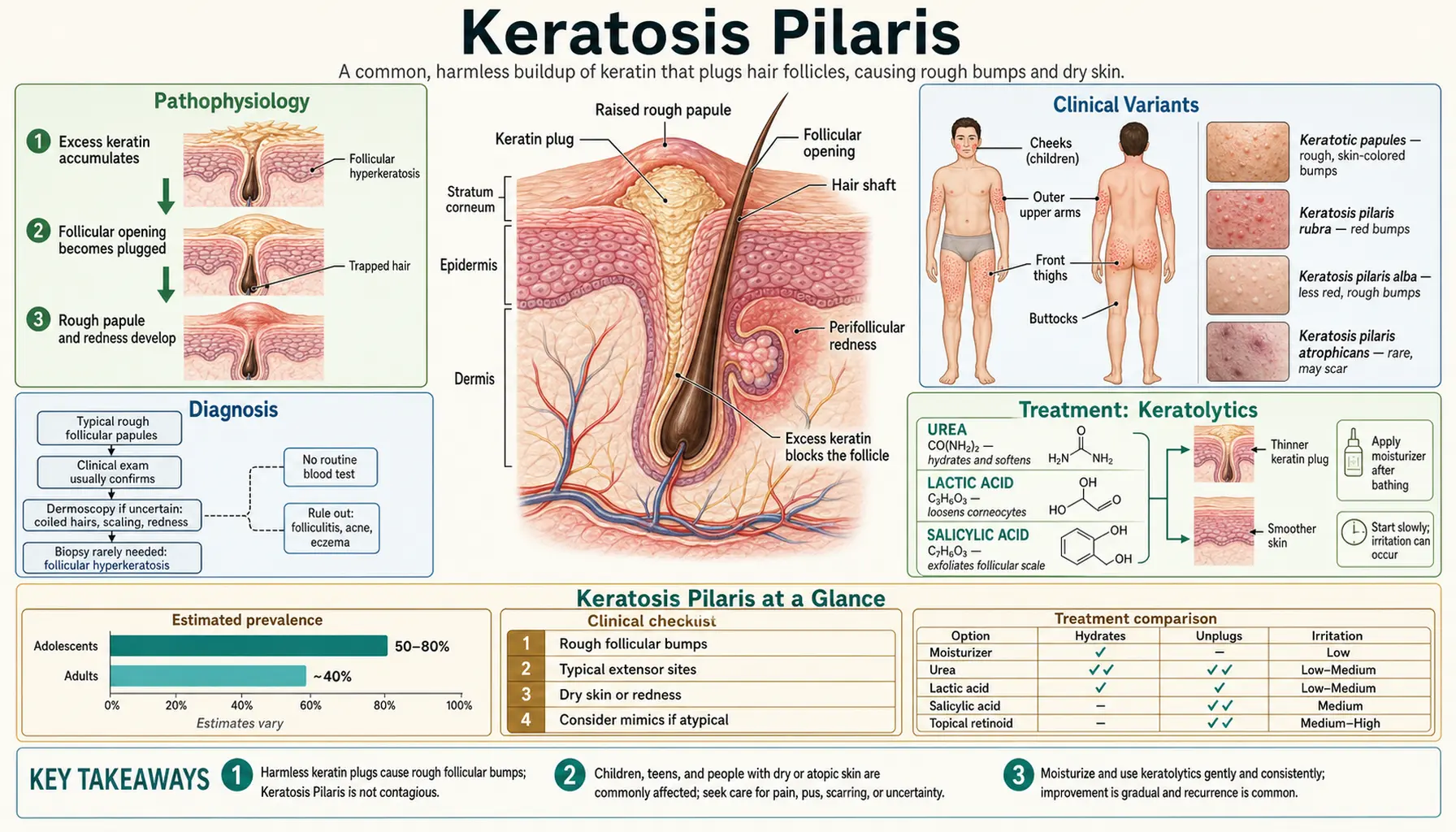
Keratosis pilaris (KP) is an extremely common, benign genetic skin condition affecting 50–80% of adolescents and 40% of adults worldwide — caused by abnormal cornification of hair follicles that produces the characteristic rough, sandpaper-like bumps most often found on the upper arms, thighs, and buttocks.
Table of Contents
1. Pathophysiology
Keratosis pilaris arises from a defect in the normal process of follicular cornification — the orderly shedding of dead skin cells from hair follicle openings. At the molecular level, the condition is associated with loss-of-function variants in the filaggrin gene (FLG), the same gene implicated in atopic dermatitis and ichthyosis vulgaris — although a 2015 study of 20 people with KP found filaggrin mutations in only 35% of them, so these variants only partly account for the condition.
Filaggrin and the Skin Barrier
Filaggrin is synthesized as a large precursor protein called profilaggrin, stored in keratohyalin granules of the stratum granulosum. During terminal keratinocyte differentiation, profilaggrin is cleaved into individual filaggrin monomers that cross-link keratin filaments into tight bundles within corneocytes — giving the stratum corneum its structural integrity and mechanical resilience. Filaggrin is then further degraded into small hygroscopic amino acids and their derivatives (urocanic acid, pyrrolidone carboxylic acid) that collectively constitute the natural moisturizing factor (NMF), which retains water within the cornified layer and maintains skin surface pH.
When FLG is mutated or underexpressed, three downstream problems emerge:
- Barrier defect: Cornified cells fail to pack tightly, creating microscopic gaps that allow water to escape (transepidermal water loss, TEWL) and allergens, irritants, and microbes to penetrate.
- Reduced NMF: Less filaggrin means fewer hygroscopic breakdown products, resulting in a drier, more brittle stratum corneum — explaining why KP worsens in low-humidity environments.
- Microbiome dysbiosis: A compromised barrier and altered pH shift the skin microbiome toward pro-inflammatory species, amplifying perifollicular inflammation.
Follicular Plugging Mechanism
The characteristic bumps of KP are follicular keratinous plugs formed within the infundibulum (upper segment) of vellus hair follicles. Excess keratin accumulates at the follicular ostium, trapping the fine vellus hair within a compact plug. The trapped hair may coil beneath the surface or perforate the follicular wall, triggering a perifollicular inflammatory response. This inflammation generates the surrounding erythema — an important distinction from infectious folliculitis: KP inflammation is sterile, not caused by bacterial or fungal invasion.
Genetics
KP follows an autosomal dominant inheritance pattern with incomplete penetrance, meaning not everyone who carries a predisposing variant expresses it clinically. Filaggrin mutations explain only part of KP: in a 2015 study, 35% of people with KP carried one. Concordance studies in twins show high heritability. Many patients have a parent who recalls similar "chicken skin" on their own arms, though it may have improved by adulthood. The strong genetic overlap with atopic dermatitis and ichthyosis vulgaris means these three conditions frequently coexist — clinicians commonly look for eczema and ichthyosis features in people with KP.
Biopsy Findings
Skin biopsy is rarely needed for diagnosis but is occasionally performed when the presentation is atypical. Histology shows dilated follicular ostia occluded by compact lamellar keratinous plugs, sometimes containing a coiled vellus hair. Mild perifollicular lymphocytic infiltrate is present in erythematous variants. A 2015 study comparing 20 KP patients with 20 matched controls found a striking absence of sebaceous glands in KP lesions (but not in unaffected skin), which its authors suggest is an early step in how KP develops.
2. Clinical Variants
KP encompasses a spectrum of clinical presentations that share the same underlying follicular plugging mechanism but differ in location, degree of inflammation, and risk of scarring.
Keratosis Pilaris Rubra
The most visually striking variant, KP rubra features pronounced perifollicular erythema — a red halo surrounding each follicular plug — giving the skin a diffusely flushed, inflamed appearance. It most commonly involves the cheeks (particularly in children), upper outer arms, and anterior thighs. The erythema can persist even when the follicular plugs themselves are reduced with treatment, making this variant the most frustrating to manage cosmetically.
KP Alba
In KP alba, erythema is minimal or absent. The follicular papules are skin-colored or slightly whitish, reflecting predominantly keratinous scale with little inflammatory component. This variant is the most prevalent form in adults and is more amenable to keratolytic treatment alone.
KP Atrophicans Faciei (Ulerythema Ophryogenes)
This rare, scarring variant affects the lateral eyebrows and adjacent cheeks, causing erythema, follicular plugging, and progressive scarring alopecia of the outer third of the eyebrows. The eyebrow loss can be permanent. It is associated with systemic syndromes — particularly Noonan syndrome and cardio-facio-cutaneous (CFC) syndrome, both involving RAS-MAPK pathway mutations. Because of this association, children with ulerythema ophryogenes are commonly evaluated for these syndromes.
Atrophoderma Vermiculata
An exceptionally rare variant producing a reticulated, pitted, "worm-eaten" texture on the cheeks due to atrophic follicular scarring. It typically manifests in mid-childhood and progresses through adolescence. Unlike ordinary KP, the scarring is largely irreversible, and treatment is primarily aimed at halting progression rather than restoration.
KP with Ichthyosis
In patients carrying two loss-of-function FLG alleles (compound heterozygotes or homozygotes), KP commonly co-presents with ichthyosis vulgaris — fine white scaling on the trunk and limbs resembling fish scales — and atopic dermatitis. This triad (ichthyosis vulgaris + KP + atopic dermatitis) is recognized as a distinct clinical phenotype with a more severe barrier defect and higher inflammatory burden.
Distribution Rules
Regardless of variant, KP has characteristic predilection sites: the posterior upper arms are the most common location in all age groups, followed by the anterior thighs, buttocks, and (in children) cheeks. The face involvement decreases as children age. KP never involves the palms or soles — follicular plugging on plantar surfaces suggests an alternative diagnosis.
Seasonal Variation
KP follows a predictable seasonal pattern: worsening in winter and dry climates (low humidity drives TEWL and disrupts the barrier) and improving in summer (higher ambient humidity, increased sun exposure — ultraviolet (UV) may have a mild immunomodulatory effect on perifollicular inflammation). Patients who relocate to humid climates often report dramatic improvement.
3. Diagnosis
KP is a clinical diagnosis made at the bedside based on the characteristic appearance and distribution of lesions. Laboratory studies and biopsy are not required in typical presentations.
Clinical Features
The classic presentation is multiple discrete, rough, follicular papules 1–2 mm in diameter distributed symmetrically over the posterior upper arms or anterior thighs. Individual papules may be skin-colored, white, or surrounded by erythematous halos. Running a fingertip over affected skin produces a sandpaper-like sensation. Pruritus is variable — mild in most patients, absent in many, occasionally bothersome in inflamed variants. There is no vesiculation, exudate, or crusting in uncomplicated KP.
Biopsy (When Needed)
Punch biopsy to 4 mm depth is occasionally performed when the diagnosis is uncertain. Histological findings: dilated follicular ostia with compact lamellar keratinous plugs; mild perifollicular lymphocytic infiltrate; no significant dermal inflammation or vascular changes; sebaceous glands absent from KP lesions in a 2015 controlled study. A trapped coiled vellus hair within the plug is a specific histological sign.
Differential Diagnosis
- Milia: Deeper, pearly-white, subepidermal cysts containing keratin; found on the face around eyes; no follicular association; firm on palpation; distinct histology (epidermal-lined cysts).
- Acne vulgaris: Involves open/closed comedones and inflammatory lesions (papules, pustules, nodules) within sebaceous follicles predominantly on the face, chest, and back; not follicular vellus-hair-unit based; sebum production is a key driver.
- Lichen planopilaris: Violaceous, pruritic, scarring follicular condition; leads to permanent alopecia on scalp; skin lesions have wickham striae; autoimmune etiology.
- Phrynoderma (vitamin A deficiency): Follicular hyperkeratosis with similar-looking plugs, but occurs in the context of nutritional deficiency (rare in high-income countries); systemic features of vitamin A deficiency present (night blindness, xerophthalmia).
- Folliculitis (bacterial/fungal): Pustular and tender; lesions are inflammatory papulopustules rather than firm keratinous plugs; culture may be positive for Staphylococcus aureus, Pseudomonas (hot tub folliculitis), or Malassezia (pityrosporum folliculitis).
- Pityriasis rubra pilaris (PRP): Orange-red follicular papules coalescing into plaques with islands of normal skin; hallmark waxy palmoplantar keratoderma; salmon-colored face; nail changes; distinguished by involvement of palms/soles (never in KP).
- Eruptive vellus hair cysts: Asymptomatic skin-colored to brown follicular papules on the chest and abdomen of children and young adults; contain multiple vellus hairs on histology; may resolve spontaneously.
4. Treatment: Keratolytics
There is no cure for KP. Treatment aims to soften and dissolve follicular plugs, reduce perifollicular inflammation, and restore the skin barrier. Management is chronic — consistent daily care is required to maintain results, and discontinuing treatment leads to recurrence within weeks.
Moisturization and Gentle Cleansing (Cornerstone)
The foundation of KP management is restoring the compromised skin barrier through consistent moisturization. Ceramide-containing emollients are widely used because ceramides are the predominant lipid in the lamellar bodies that seal the intercorneocyte spaces — precisely the structural element deficient in FLG-mutant skin. Ceramide creams made without fragrance, dyes, or comedogenic ingredients are widely sold.
Commonly described moisturizing practice: applying within 3 minutes of bathing ("soak and seal" technique) to trap transepidermal moisture; lukewarm rather than hot water (hot water strips lipids and worsens the barrier defect); fragrance-free, soap-free cleansers; no loofah scrubbing (micro-abrasion worsens perifollicular inflammation); moisturizer at least twice daily.
Alpha-Hydroxy Acids (AHAs)
Ammonium lactate 12% lotion is widely considered first-line pharmacological therapy for KP. Lactic acid is an AHA that loosens the corneodesmosomes holding corneocytes together, allowing the keratinous plug to shed more readily. It also acts as a humectant, drawing water into the stratum corneum. Applied once or twice daily, ammonium lactate typically produces noticeable smoothing within 4–8 weeks. Mild stinging on broken skin is expected and usually diminishes with continued use.
Glycolic acid at 6–12% concentration is an alternative AHA available in lotions, creams, and chemical peels. It has smaller molecular weight than lactic acid and penetrates the follicular infundibulum more readily, which may make it more effective for stubborn cases. Both AHAs irritate the eyes and open skin.
Urea
Urea is both a keratolytic and a humectant — at lower concentrations (10–20%) it primarily acts as a humectant, while at higher concentrations (20–40%) it disrupts the hydrogen bonds holding keratin fibers together, dissolving plugs. Urea 20–40% cream is particularly effective for thick, stubborn KP plaques and may be used in combination with lactic acid. It can sting on excoriated or eroded skin. Urea-containing products are used cautiously in children and generally not on the face.
Salicylic Acid
Salicylic acid 2–6% in wash or lotion formulations exploits its lipophilicity to penetrate the lipid-rich follicular canal — making it uniquely "folliculotropic" compared to water-soluble AHAs. It disrupts corneocyte cohesion within the plug and has mild anti-inflammatory properties. Salicylic acid washes (e.g., 2% in a body wash) are convenient for large body-surface-area involvement. Not used in children under 2 years old — systemic salicylate absorption through compromised skin can cause salicylism (tinnitus, metabolic acidosis).
5. Treatment: Topical Retinoids
Topical retinoids address the root cause of KP by normalizing follicular keratinization at the molecular level — reducing the excess keratin production in the infundibulum that forms the plug in the first place. They are particularly useful for patients who respond inadequately to keratolytics alone or who have the KP rubra variant with prominent erythema.
Tretinoin
Tretinoin 0.025–0.05% cream is the most studied retinoid for KP. It binds retinoic acid receptors (RARs) in keratinocytes, downregulating genes responsible for excess keratin production and normalizing the differentiation program of follicular epithelium. This prevents new plug formation while lactic acid or urea dissolves existing plugs — making combination therapy more effective than either alone.
Initiation protocol: apply a pea-sized amount to affected areas on alternate evenings for the first 2 weeks, then advance to every evening as tolerated. The retinoid dermatitis (dryness, flaking, erythema) is expected during the first 4–6 weeks and diminishes as skin adapts. A moisturizer is commonly applied 20–30 minutes after tretinoin, or tretinoin is layered in the "sandwich technique" (moisturizer → tretinoin → moisturizer). Retinoids can cause birth defects: oral tretinoin is teratogenic, and the topical tretinoin label reports birth defects in animal studies and 30 human cases of birth defects reported during its use, without an established causal link.
Tazarotene
Tazarotene (0.05–0.1% cream or gel) is a more potent, receptor-selective retinoid that may be more effective for thick, hyperkeratotic KP plaques but carries a higher risk of retinoid dermatitis. It is typically reserved for patients who have tolerated tretinoin without adequate response. Like tretinoin, it is teratogenic.
Adapalene
Adapalene 0.1% gel (now sold over the counter in the US) is the best-tolerated retinoid option, with lower receptor-binding affinity and less irritant potential than tretinoin or tazarotene. It is often used as a starting point for people with sensitive skin or for adolescents who are new to retinoid therapy. Initiation: apply 3 times per week, increasing to nightly as tolerated over 4–6 weeks. Over-the-counter availability makes it accessible without a prescription; improvement takes 8–12 weeks of consistent use.
Combination Strategy
A commonly described regimen for moderate-to-severe KP combines moisturization with a morning keratolytic and an evening retinoid:
- Morning: Gentle soap-free cleanser → ceramide moisturizer → ammonium lactate 12% lotion or glycolic acid cream
- Evening: Lukewarm shower → pat dry → ceramide moisturizer (wait 20 min) → adapalene or tretinoin cream to affected areas
This approach addresses both plug dissolution (AHA/urea) and prevention of new plug formation (retinoid) while supporting barrier repair (ceramides). Maintenance is indefinite — this is a management strategy, not a cure.
6. Treatments for Erythema
The perifollicular erythema of KP rubra is the component most distressing to patients — particularly on visible areas like cheeks and upper arms — and is also the hardest to treat. Keratolytics can smooth the skin texture but often leave persistent redness, requiring targeted vascular or anti-inflammatory therapy.
Vascular Laser and Light Therapies
The pulsed dye laser (PDL) operating at 585 nm or 595 nm targets oxyhemoglobin in the dilated perifollicular capillaries without damaging surrounding tissue (selective photothermolysis). Treatment sessions typically involve multiple passes with a 10 mm spot and standard fluences; no anesthesia is required for most patients though a cooling spray is applied. Erythema reduction is visible after 2–3 sessions, with full benefit at 4–6 sessions spaced 4–6 weeks apart. Effects can be durable (months to years) but are not permanent.
Intense pulsed light (IPL) uses a broad-spectrum filtered light source (typically 560–1200 nm) to achieve similar vascular targeting with a larger treatment footprint — useful for extensive involvement of the upper arms or thighs. Efficacy for erythema is comparable to PDL, though individual response varies. Fractional laser (non-ablative 1550 nm or ablative CO2) can be combined with PDL in the same session to address both erythema and surface texture simultaneously — an approach gaining traction in published case series.
Topical Anti-Inflammatory Agents
Azelaic acid 10–20% has a dual mechanism relevant to KP: it normalizes aberrant keratinization in the follicular epithelium and exerts direct anti-inflammatory effects by inhibiting reactive oxygen species in neutrophils. At 15–20% concentration it provides meaningful erythema reduction over 8–12 weeks of twice-daily application. It is well tolerated, minimally absorbed through the skin (its label states that use in pregnancy is not expected to expose the fetus), and can be combined with lactic acid or a retinoid.
Low-potency topical corticosteroids (e.g., hydrocortisone 1% or desonide 0.05%) may transiently reduce perifollicular erythema during acute flares, but are not appropriate for maintenance use: prolonged application causes skin atrophy, telangiectasia, and tachyphylaxis. Courses are typically limited to 1–2 weeks on acutely inflamed areas, and they are generally not applied to children's faces.
Calcineurin Inhibitors
Tacrolimus 0.1% ointment and pimecrolimus 1% cream are off-label options for the inflammatory component of KP rubra, particularly on facial lesions (cheeks in children) where the atrophy risk of topical corticosteroids is especially unwanted. They suppress T-lymphocyte-mediated perifollicular inflammation without causing skin atrophy or tachyphylaxis — which is why they are used on sensitive areas. Their U.S. Food and Drug Administration (FDA) boxed warning states that long-term safety has not been established and that rare cases of malignancy (skin cancer and lymphoma) have been reported, although a causal relationship has not been established; the tacrolimus label states that continuous long-term use in any age group should be avoided.
7. Special Populations and Natural History
Infants and Young Children
KP is common in infancy, presenting most notably on the cheeks as rough, red papules that alarm parents who fear acne or a rash. The cheek involvement in toddlers can be striking but is entirely benign and typically improves through childhood without treatment. First-line management in this age group is limited to gentle soap-free cleansing with fragrance-free wash and twice-daily ceramide moisturizer. Salicylic acid is not used under 2 years; retinoids are generally not used under 12 years; urea is used cautiously. Parental reassurance is as important as any topical therapy.
Adolescents
Prevalence peaks during adolescence (up to 80%), coinciding with hormonal changes that increase keratinocyte proliferation and sebum production. For teenagers, KP carries a disproportionate psychosocial burden: self-consciousness about wearing short sleeves or shorts, avoidance of swimming pools or beach activities, and social comparison amplified by social media. Adolescents are also the group most likely to have come across online KP "cures" — dietary changes, supplements, and home remedies that people report trying. KP is a chronic genetic condition managed (not cured) by consistent skincare. Adapalene 0.1%, sold over the counter, is a commonly used first retinoid for teenagers who want treatment beyond moisturizer.
Pregnancy
KP may flare during pregnancy due to hormonal influences on keratinocyte turnover. Management options are limited by safety constraints: the tazarotene label lists pregnancy as a contraindication, systemic retinoids cause birth defects, and the topical tretinoin label reports birth defects in animal studies and case reports of birth defects in humans without an established causal link. Lactic acid 12% and ceramide moisturizers are considered safe for topical use in pregnancy. Urea has limited safety data in pregnancy and is used conservatively and not on large body surface areas. The azelaic acid label states that it is minimally absorbed through the skin and that use in pregnancy is not expected to expose the fetus. For the topical calcineurin inhibitors, there are no adequate and well-controlled studies in pregnant women, and the tacrolimus ointment label states that experience in pregnancy is too limited to assess its safety.
Adults
Approximately 40% of adults carry KP, though prevalence declines with age as barrier function evolves. Many adults self-manage with over-the-counter lactic acid lotions without ever seeking medical advice. Those who present to a dermatologist are often motivated by cosmetic concerns about the erythematous variant or by years of home remedies that did not help them. Adult patients may respond better to vascular laser for erythema than younger patients, as cumulative sun exposure provides some background photodamage that laser treatment can address simultaneously.
8. Prognosis and Outlook
Natural Course
KP is a benign, non-contagious condition with no risk of malignant transformation, systemic complications, or spread beyond the characteristic distribution. In the one natural-history study (a 1994 questionnaire of 49 patients aged 18 months to 25 years), KP had improved with age in 35%, stayed the same in 43%, and worsened in 22%; the mean age of improvement was 16 years. Improvement is not the same as resolution — a subset of patients retain KP throughout their adult life, and environmental factors (dry climates, harsh soaps, stress) can trigger recurrences even in those who had previously improved.
Environmental Modifiers
Environmental factors that affect KP include:
- Climate and humidity: Dry winter air and low-humidity climates (arid regions, heated indoor air) reliably worsen KP. A humidifier in the bedroom during winter months can produce noticeable improvement. Air travel (very low cabin humidity) often triggers visible flares.
- Swimming pools: Chlorinated water is both a barrier irritant and a desiccant. Showering and moisturizing soon after swimming are commonly described ways of limiting this.
- Hot showers and baths: Hot water dissolves the surface lipid film and disrupts barrier function. Switching to lukewarm showers is one of the most impactful low-cost interventions.
- Friction and tight clothing: Repeated friction from tight fabrics (jeans on affected thighs, sleeve hems on upper arms) can worsen perifollicular inflammation. Loose, breathable clothing reduces this friction during flares.
What to Expect From Treatment
The main facts about treatment:
- There is no cure for KP — the genetic basis cannot be altered by any current therapy.
- Improvement with treatment takes weeks (4–8 weeks for keratolytics; 8–12 weeks for retinoids; 3–6 months for full benefit of laser).
- Stopping treatment leads to regression within weeks to months — maintenance is indefinite.
- The texture (follicular plugs) responds better to treatment than the redness (erythema), which often requires laser therapy for significant improvement.
- People report trying home remedies such as oil cleansing, dietary changes, and diluted apple cider vinegar for KP. Undiluted acids and aggressive physical scrubbing can worsen KP by disrupting the barrier.
Overlapping Conditions
Approximately 40% of KP patients have concurrent atopic dermatitis, driven by the same FLG barrier defect. When eczema is present, KP care is usually folded into a broader atopic skin regimen — this often means barrier repair first and no topical retinoids on actively inflamed eczematous patches. Some clinicians look for food allergies, particularly egg and peanut, in young children with severe, early-onset KP (especially with cheek involvement and associated eczema), on the view that these allergies can drive barrier inflammation. Contact dermatitis can complicate KP when patients experiment aggressively with multiple keratolytic products simultaneously — patch testing is sometimes used in people who worsen despite a seemingly logical regimen.
Key Research Papers
- Gruber R, Sugarman JL, Crumrine D, et al. "Sebaceous gland, hair shaft, and epidermal barrier abnormalities in keratosis pilaris with and without filaggrin deficiency." Am J Pathol. 2015;185(4):1012–1021. — PubMed PMID: 25660180
- Poskitt L, Wilkinson JD. "Natural history of keratosis pilaris." Br J Dermatol. 1994;130(6):711–713. — PubMed PMID: 8011494
- Brown SJ, McLean WH. "One remarkable molecule: filaggrin." J Invest Dermatol. 2012;132(3 Pt 2):751–762. — PubMed PMID: 22158554
- Mevorah B, et al. "The prevalence of accentuated palmoplantar markings and keratosis pilaris in atopic dermatitis, autosomal dominant ichthyosis and control dermatological patients." Br J Dermatol. 1985;112(6):679–685. — PubMed PMID: 4005167
- Maghfour J, Ly S, Haidari W, Taylor SL, Feldman SR. "Treatment of keratosis pilaris and its variants: a systematic review." J Dermatolog Treat. 2022;33(3):1231–1242. — PubMed PMID: 32886029
- Chamlin SL, et al. "Ceramide-dominant barrier repair lipids alleviate childhood atopic dermatitis: changes in barrier function provide a sensitive indicator of disease activity." J Am Acad Dermatol. 2002;47(2):198–208. — PubMed PMID: 12140465
- Hwang S, Schwartz RA. "Keratosis pilaris: A common follicular hyperkeratosis." Cutis. 2008;82(3):177–180. — PubMed PMID: 18856156