Patent Foramen Ovale (PFO)
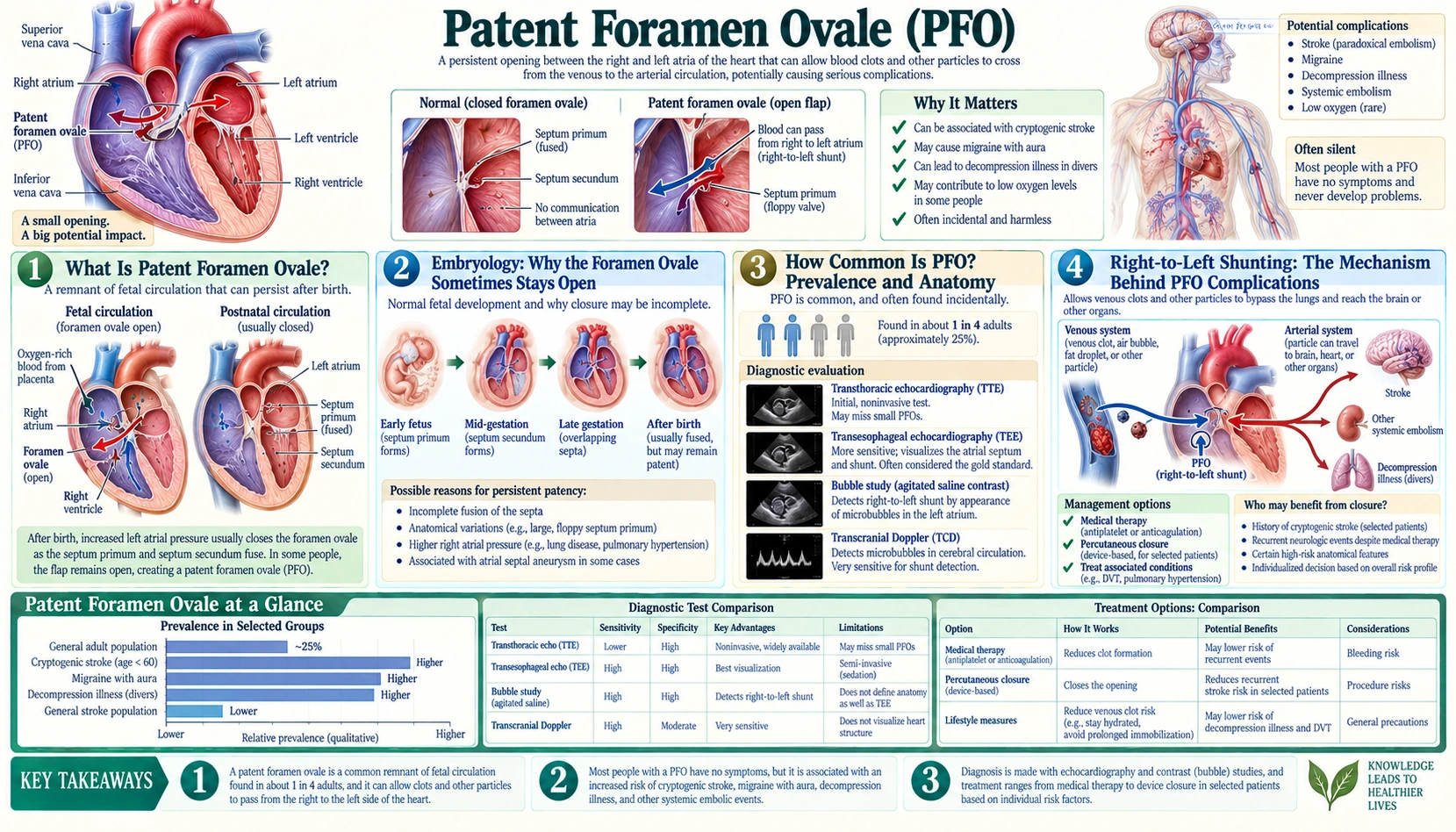
A patent foramen ovale (PFO) is a small opening between the upper chambers of the heart that failed to close after birth. The 2020 American Academy of Neurology practice advisory describes it as present in about one in four adults, and in most people it causes no symptoms. However, in certain situations — particularly unexplained stroke in younger adults — a PFO can allow blood clots or other material to pass directly from the venous to the arterial circulation, bypassing the lungs entirely. Understanding of when a PFO matters, and how it is managed, changed with a set of randomized trials published in 2017 and 2018.
Table of Contents
- What Is Patent Foramen Ovale?
- Embryology: Why the Foramen Ovale Sometimes Stays Open
- How Common Is PFO? Prevalence and Anatomy
- Right-to-Left Shunting: The Mechanism Behind PFO Complications
- Clinical Associations: Stroke, Migraine, and Diving Risks
- Diagnosis: Echo, Bubble Studies, and TCD
- PFO Closure: The Pivotal Trials and Guideline Statements
- What to Expect: The Closure Procedure and Recovery
- Key Research Papers
- Connections
- Featured Videos
What Is Patent Foramen Ovale?
The word patent simply means open. The foramen ovale is a natural opening in the wall between the heart's two upper chambers — the right and left atria — that every human fetus needs in order to survive. In most people this opening seals shut within the first few months after birth. In about one in four adults, it never fully closes, leaving a flap-like channel that can intermittently allow blood to pass from the right atrium to the left atrium without first going to the lungs.
On its own, a PFO is not a disease. The vast majority of people who have one will live their entire lives without ever knowing it is there. A PFO only becomes medically relevant when it creates a pathway for material — a blood clot, air bubble, or fat droplet — to bypass the lungs and travel directly into the arterial circulation, a phenomenon called paradoxical embolism.
In an autopsy study of 965 normal hearts, the open foramen ovale measured 1 to 19 mm in maximal potential diameter (mean 4.9 mm), and the size tended to increase with age (Hagen 1984, PubMed PMID: 6694427).
Researchers use PFO features to define "high-risk" anatomy. The DEFENSE-PFO trial, for example, defined a high-risk PFO as one with an atrial septal aneurysm (ASA, a redundant, mobile interatrial septum), hypermobility of the septum (excursion of 10 mm or more), or a PFO size of 2 mm or more (Lee 2018, PubMed PMID: 29544871).
Back to Table of ContentsEmbryology: Why the Foramen Ovale Sometimes Stays Open
To understand why a PFO can persist into adulthood, it helps to understand why the foramen ovale exists in the first place. During fetal life, the lungs are collapsed and filled with amniotic fluid — they perform no gas exchange. Instead, the placenta oxygenates the blood. The fetal heart faces an unusual challenge: it must route the richest, most oxygenated blood directly to the brain and body while bypassing the non-functional lungs almost entirely.
The foramen ovale is nature's elegant solution. The interatrial septum develops from two overlapping sheets of tissue — the septum primum (the first partition to grow, arriving from the top of the atrium) and the septum secundum (a thicker, crescent-shaped ridge that grows alongside it). The septum secundum develops a gap — the foramen ovale — and the thinner septum primum acts as a one-way flap over this gap. Because right atrial pressure is higher than left atrial pressure in the fetus (the lungs are not pumping blood back to the left side), oxygenated blood returning from the placenta flows through the inferior vena cava, bounces off a ridge of tissue called the Eustachian valve, and is funneled through the foramen ovale into the left atrium, then into the left ventricle and out to the brain and body.
At birth, everything changes in an instant. The infant takes its first breath, the lungs expand and fill with air, pulmonary vascular resistance drops dramatically, and blood begins flooding the pulmonary circulation. Left atrial pressure rises and exceeds right atrial pressure. This pressure reversal presses the septum primum flap against the foramen ovale, sealing it shut — first functionally, within minutes of birth, and then anatomically, as the two layers fuse together over the following weeks to months.
In a minority of people, this fusion never completes. The septum primum flap remains unsealed, leaving a potential channel — the patent foramen ovale — that can open whenever right atrial pressure momentarily exceeds left atrial pressure.
Back to Table of ContentsHow Common Is PFO? Prevalence and Anatomy
PFO is strikingly common. In the autopsy study of 965 normal hearts, 27.3% had a patent foramen ovale; the rate declined with age, from 34.3% in the first three decades of life to 25.4% in the 4th through 8th decades and 20.2% in the 9th and 10th (Hagen 1984, PubMed PMID: 6694427). In a French multicentre study of 581 young adults with cryptogenic stroke, 45.9% had a PFO on transesophageal echocardiography (Lamy 2002, PubMed PMID: 11872892) — one reason PFO has attracted so much research interest as a potential stroke cause.
Several anatomical features influence how clinically significant a PFO is likely to be:
- Tunnel length: Some PFOs are simple slits; others form long tunnels between the two septal layers. Longer tunnels are more likely to allow sustained shunting.
- Shunt grade: The number of bubbles that cross from right to left during a bubble study reflects the functional size of the opening; trials such as CLOSE enrolled patients with a large interatrial shunt (Mas 2017, PubMed PMID: 28902593).
- Atrial septal aneurysm (ASA): In some people with PFO, the interatrial septum is unusually mobile and redundant, bulging back and forth with the heartbeat. The CLOSE trial enrolled patients whose PFO came with an atrial septal aneurysm or a large shunt, and in the PFO-ASA study a PFO with an atrial septal aneurysm was more strongly associated with migraine than a PFO alone (Lamy 2002, PubMed PMID: 11872892).
- Eustachian valve prominence: A large, persistent Eustachian valve at the opening of the inferior vena cava can direct venous blood streaming from the legs toward the PFO, increasing the probability of paradoxical embolism.
The 2020 AAN practice advisory states that having a PFO is common and that it is difficult to determine with certainty whether a given PFO caused a stroke (Messé 2020, PubMed PMID: 32350058). The SCAI guideline describes PFO as entirely benign and requiring no treatment in most cases (Kavinsky 2022, PubMed PMID: 39131947). The clinical challenge is identifying the minority in whom it is genuinely causative.
Back to Table of ContentsRight-to-Left Shunting: The Mechanism Behind PFO Complications
Under normal resting conditions, left atrial pressure is slightly higher than right atrial pressure, so the septum primum flap stays pressed shut and no blood crosses through the PFO. The problem arises during transient spikes in right atrial pressure that can reverse this gradient and force the flap open.
Common everyday activities that transiently raise right atrial pressure and can open a PFO include:
- Valsalva maneuver — bearing down, straining during a bowel movement, lifting a heavy object
- Coughing or sneezing
- Standing up quickly (venous pooling reduces left-sided filling)
- Deep inspiration (negative intrathoracic pressure pulls blood into the right heart)
- Trumpet playing or other sustained blowing
During these moments — typically lasting only seconds — right-to-left shunting (RLS) occurs through the PFO. In most people this is harmless because there is nothing in the venous circulation dangerous enough to cause problems when it reaches the arteries. But in certain circumstances, hazardous material can be present in the venous blood at exactly the wrong moment:
- Thrombus from a deep vein thrombosis (DVT) or pelvic vein clot: the classic mechanism of paradoxical embolism. A venous blood clot normally would travel to the lungs and be filtered there; through a PFO it can reach the brain, causing stroke or TIA, or the coronary arteries, causing heart attack.
- Nitrogen microbubbles in divers: during ascent, dissolved nitrogen comes out of solution in the venous circulation; these bubbles normally reach the lungs and are exhaled, but through a PFO they can enter the arterial circulation and cause neurological decompression illness.
- Fat emboli after long bone fracture or liposuction: rare but documented.
- Air emboli from intravenous lines or during surgery: another rare pathway.
Clinical Associations: Stroke, Migraine, and Diving Risks
Cryptogenic Stroke — The Most Important Association
An ischemic stroke is called cryptogenic when no cause can be identified after thorough evaluation — no atrial fibrillation, no large-artery disease, no small-vessel disease, no cardiac source of embolism from a structural lesion. The PFO closure trials enrolled patients up to about 60 years of age with this kind of stroke.
PFO was found in 45.9% of 581 young cryptogenic stroke patients in the PFO-ASA study (Lamy 2002, PubMed PMID: 11872892), compared with 27.3% of hearts in the autopsy series above. The challenge has always been distinguishing a PFO that caused the stroke from a PFO that is merely a bystander.
Features the research links to a PFO being stroke-related rather than incidental include:
- Younger age, and the absence of diabetes, hypertension, smoking and prior stroke or TIA (Kent 2013, PubMed PMID: 23864310)
- A superficial (cortical) infarct on brain imaging, consistent with embolism (Kent 2013)
- No other stroke mechanism found after a thorough evaluation, which the AAN advisory states should be completed before closure is considered (Messé 2020, PubMed PMID: 32350058)
- High-risk PFO anatomy — a large shunt or an atrial septal aneurysm — which defined the populations of the CLOSE and DEFENSE-PFO trials
The RoPE score (Risk of Paradoxical Embolism), derived from 12 component studies, was developed to estimate how likely a PFO found after cryptogenic stroke is to be stroke-related. It awards points for younger age, a cortical stroke on imaging, and the absence of diabetes, hypertension, smoking and prior stroke or TIA. PFO prevalence rose from 23% in patients scoring 0–3 points to 73% in those scoring 9–10, corresponding to PFO-attributable fractions of roughly 0% to 90%; patients in the highest-scoring stratum also had a lower 2-year recurrence rate (2% versus 20% in the lowest) (Kent 2013, PubMed PMID: 23864310).
Migraine with Aura
A meta-analysis of 21 case-control studies (5,572 participants) found migraine with aura about 3.4 times as common in people with a PFO as in people without one (odds ratio 3.36), with no significant association for migraine without aura (Takagi 2016, PubMed PMID: 26527111). The PREMIUM investigators describe the role of PFO in migraine as poorly understood (Tobis 2017, PubMed PMID: 29191325).
Two sham-controlled randomized trials did not meet their primary endpoints. In the MIST trial (147 patients with frequent migraine with aura and a moderate or large shunt), migraine cessation occurred in 3 of 74 patients after closure and 3 of 73 after a sham procedure; secondary endpoints were also not met, although an exploratory analysis excluding 2 outliers found a greater reduction in migraine days with the implant (Dowson 2008, PubMed PMID: 18316488). In the PREMIUM trial (230 patients with frequent migraine), the 50% responder rate did not differ between closure and sham; the closure group had a greater reduction in headache days (−3.4 vs −2.0 days/month) and more complete 1-year remissions (10 patients vs 1) as secondary findings (Tobis 2017, PubMed PMID: 29191325).
Decompression Illness in Scuba Divers
During a scuba dive, nitrogen dissolves into tissues and blood under pressure. During ascent, if the diver rises too quickly, nitrogen comes out of solution and forms bubbles in the venous circulation. Normally these venous nitrogen bubbles are filtered by the pulmonary capillaries and expelled with each breath. In a diver with a PFO, however, a Valsalva maneuver during ascent can open the PFO and allow nitrogen bubbles to pass directly into the arterial circulation, causing neurological decompression sickness — a far more serious form than musculoskeletal "bends."
A 1998 analysis of three diving studies estimated that a PFO raises the odds of serious (type II) decompression sickness about 2.5-fold; because type II decompression sickness averaged 2.28 per 10,000 dives, the author concluded the absolute risk remains small and did not support routine echocardiographic screening of divers (Bove 1998, PubMed PMID: 9789338). In a study of 66 professional divers, a PFO with right-to-left shunting at rest, a wider opening and a more mobile septal membrane were associated with decompression illness, particularly the neurologic type (Cartoni 2004, PubMed PMID: 15246922). The SCAI guideline includes recommendations on closure for the prevention of decompression illness (Kavinsky 2022, PubMed PMID: 39131947).
Platypnea-Orthodeoxia Syndrome
Platypnea-orthodeoxia is a rare but dramatic syndrome in which patients experience breathlessness and a fall in blood oxygen levels specifically when they stand up, which relieves when they lie down — the opposite of most cardiopulmonary conditions. It results from right-to-left shunting through a PFO or atrial septal defect, often precipitated by anatomical changes such as aortic dilation or chest surgery. A 2026 systematic review of 469 patients (mostly case reports and series; mean age 68) found that oxygen saturation rose from a mean of 79.5% before percutaneous closure to 94.5% after, with symptomatic improvement reported in 82% of cases and procedural complications in 5% (Ermongkonchai 2026, PubMed PMID: 41850786).
Back to Table of ContentsDiagnosis: Echo, Bubble Studies, and TCD
Diagnosing a PFO requires both demonstrating the anatomical opening and proving that it allows right-to-left shunting. Several complementary tools are available, each with different strengths.
Transthoracic Echocardiography (TTE) with Bubble Study
The agitated saline (bubble) study is the cornerstone of PFO diagnosis. A mixture of saline, air, and the patient's own blood is agitated to create microbubbles and injected into a peripheral vein. The patient performs a Valsalva maneuver (bears down) and then releases it. In a normal heart, microbubbles fill the right atrium and right ventricle but are filtered by the pulmonary capillaries and never appear on the left side. When a right-to-left shunt is present, bubbles appear in the left heart; how quickly they appear helps distinguish an intracardiac shunt from a shunt in the lungs.
TTE with bubble study is widely available and non-invasive. A meta-analysis of 13 prospective studies (1,436 patients) using TEE as the reference found a weighted mean sensitivity of 46% and specificity of 99% for conventional TTE — a poor test for ruling a shunt out but an excellent one for ruling it in (Mojadidi 2014, PubMed PMID: 24689727).
Transesophageal Echocardiography (TEE) with Bubble Study — Gold Standard
TEE positions the ultrasound probe in the esophagus, immediately behind the heart, giving exquisite visualization of the interatrial septum and left atrium. TEE with bubble study is considered the gold standard for PFO diagnosis and is used to characterize PFO anatomy, including the atrial septal aneurysm and septal mobility that trials such as DEFENSE-PFO used to select patients. Even so, a meta-analysis of four studies that confirmed the result by autopsy, surgery or catheterization found a TEE sensitivity of 89.2% and specificity of 91.4%, so some PFOs are missed or misdiagnosed (Mojadidi 2014, PubMed PMID: 24372693). TEE is semi-invasive, with the probe passed into the esophagus.
Transcranial Doppler (TCD) with Bubble Study
TCD detects microbubbles (microembolic signals, or MES) in the middle cerebral artery after intravenous agitated saline injection. A meta-analysis of 27 prospective studies (1,968 patients) using TEE as the reference found a weighted mean sensitivity of 97% and specificity of 93% for TCD in detecting right-to-left shunt; the authors describe it as noninvasive and low-cost, and note that TEE is still needed when the precise anatomy must be known (Mojadidi 2014, PubMed PMID: 24560213).
Additional Workup
When PFO is discovered in a patient with cryptogenic stroke, the evaluation commonly extends to other possible causes (the AAN advisory states that alternative mechanisms of stroke should be thoroughly ruled out before closure is considered; Messé 2020). Tests used include:
- Lower extremity venous ultrasound to detect DVT as a potential thrombus source
- Pelvic MRI or CT venography when paradoxical embolism from a pelvic vein clot is suspected
- Hypercoagulable workup — testing for factor V Leiden, prothrombin gene mutation, antiphospholipid antibodies, protein C/S deficiency, antithrombin III deficiency
- Prolonged cardiac monitoring (30-day event monitor or implantable loop recorder) to look for paroxysmal atrial fibrillation, an alternative stroke mechanism
- Brain MRI with DWI to characterize infarct pattern — cortical versus lacunar, single versus multiple territories
PFO Closure: The Pivotal Trials and Guideline Statements
For many years, the question of whether to close a PFO after cryptogenic stroke was unresolved: the first randomized trials did not show a significant benefit in their primary analyses. Then, in September 2017, three randomized trials were published together in the New England Journal of Medicine, followed by the DEFENSE-PFO trial in 2018.
The Three Pivotal 2017 Trials
The CLOSE trial (Mas 2017, PubMed PMID: 28902593) randomized 663 patients aged 16–60 with a recent stroke attributed to PFO, with an atrial septal aneurysm or large interatrial shunt, to PFO closure plus long-term antiplatelet therapy, antiplatelet therapy alone, or oral anticoagulation. Over a mean follow-up of 5.3 years, no stroke occurred among 238 patients in the closure group, versus 14 of 235 in the antiplatelet-only group (hazard ratio 0.03). Procedural complications occurred in 5.9% of closure patients, and atrial fibrillation was more frequent after closure (4.6% vs 0.9%). In the separate anticoagulation comparison, stroke occurred in 3 of 187 patients on oral anticoagulants and 7 of 174 on antiplatelet therapy.
The REDUCE trial (Søndergaard 2017, PubMed PMID: 28902580) randomized 664 patients (mean age 45.2; 81% with moderate or large shunts) with cryptogenic stroke, in a 2:1 ratio, to PFO closure plus antiplatelet therapy or antiplatelet therapy alone. Over a median follow-up of 3.2 years, clinical ischemic stroke occurred in 6 of 441 closure patients (1.4%) versus 12 of 223 (5.4%) on antiplatelet therapy alone (hazard ratio 0.23). Serious device-related adverse events occurred in 1.4% of closure patients, and atrial fibrillation in 6.6%.
The RESPECT trial randomized 980 patients aged 18–60; its primary intention-to-treat analysis in 2013 did not show a significant benefit (hazard ratio 0.49, P=0.08), although the prespecified per-protocol and as-treated analyses did (Carroll 2013, PubMed PMID: 23514286). The extended follow-up, considered exploratory, followed patients for a median of 5.9 years: recurrent ischemic stroke occurred in 18 closure patients and 28 medical-therapy patients (0.58 vs 1.07 events per 100 patient-years; hazard ratio 0.55), and recurrent ischemic stroke of undetermined cause in 10 vs 23 (hazard ratio 0.38). Venous thromboembolism was more common in the closure group (Saver 2017, PubMed PMID: 28902590).
The DEFENSE-PFO trial randomized 120 patients with cryptogenic stroke and high-risk PFO anatomy to closure or medication only. The primary endpoint (stroke, vascular death or major bleeding at 2 years) occurred only in the medication-only group (6 of 60 patients; 2-year ischemic stroke rate 10.5%) (Lee 2018, PubMed PMID: 29544871).
Earlier trials had been null in their primary analyses. CLOSURE I (909 patients aged 18–60 with cryptogenic stroke or TIA) found a 2-year primary-endpoint rate of 5.5% with closure versus 6.8% with medical therapy (hazard ratio 0.78, P=0.37); the investigators noted that a cause other than paradoxical embolism was usually apparent in patients with recurrent events (Furlan 2012, PubMed PMID: 22417252). The PC trial (414 patients) likewise found no significant reduction in its composite endpoint (3.4% vs 5.2%, P=0.34) (Meier 2013, PubMed PMID: 23514285). The trials differed in the devices they used and in how patients were selected — CLOSE and DEFENSE-PFO, for example, enrolled only patients with high-risk PFO anatomy.
What the 2020 AAN Practice Advisory States
The American Academy of Neurology's 2020 practice advisory update, which reviewed studies published through December 2017, states (Messé 2020, PubMed PMID: 32350058):
- An appropriately thorough evaluation to rule out alternative mechanisms of stroke is expected before PFO closure is considered (level B).
- When a higher-risk alternative mechanism of stroke is found, PFO closure is not routinely recommended (level B).
- PFO closure probably reduces recurrent stroke risk in select patients (level B).
- In patients younger than 60 with a PFO, an embolic-appearing infarct and no other mechanism identified, clinicians may recommend closure after discussing benefits (an absolute recurrent-stroke risk reduction of 3.4% at 5 years) and risks (a periprocedural complication rate of 3.9% and an increased absolute rate of non-periprocedural atrial fibrillation of 0.33% per year) (level C).
- For patients who choose medical therapy alone, an antiplatelet medication such as aspirin, or anticoagulation, may be recommended (level C).
The Society for Cardiovascular Angiography and Interventions (SCAI) issued 13 recommendations in 2022 covering patient selection for closure after PFO-associated stroke, including populations not commonly enrolled in the trials (Kavinsky 2022, PubMed PMID: 39131947). Decisions about closure belong with the treating clinicians and the patient.
The RoPE Score in Practice
The RoPE score is a 10-point index: the youngest patients with superficial strokes and no vascular risk factors score highest. In its derivation data, a PFO was likely to be incidental in patients scoring 0–3 (attributable fraction about 0%) and likely to be stroke-related in those scoring 9–10 (about 90%) (Kent 2013, PubMed PMID: 23864310).
Back to Table of ContentsWhat to Expect: The Closure Procedure and Recovery
The Device
The randomized trials used several different occluder devices from more than one manufacturer. A typical PFO occluder consists of two discs of nitinol (nickel-titanium) wire mesh connected by a short waist. When deployed, the two discs straddle the interatrial septum and sandwich the PFO closed, and the heart's own tissue gradually grows over the device.
The Procedure
PFO closure is performed in a cardiac catheterization laboratory. Patients are usually under moderate sedation (conscious sedation or general anesthesia) with TEE or intracardiac echocardiography (ICE) guidance to monitor device placement in real time. The steps are:
- A catheter is inserted through a vein in the groin (femoral vein) and advanced into the right atrium under fluoroscopic (X-ray) guidance.
- The catheter is guided through the PFO into the left atrium.
- The left atrial disc is deployed and pulled back against the left side of the septum.
- The right atrial disc is deployed on the right side of the septum.
- Imaging confirms correct positioning, no significant residual shunt, and no impingement on surrounding structures.
- The device is released and the catheter withdrawn.
In DEFENSE-PFO, all closures were successful (Lee 2018). In a real-world study of 1,887 patients who had closure within a year of stroke or TIA in three US states (2005–2013), 7.0% had an adverse outcome during the closure hospitalization (atrial fibrillation or flutter, tamponade, pneumothorax, hemothorax, vascular access complication or death); the rate was 10.9% in patients over 60 versus 4.9% in those 60 or younger (Merkler 2017, PubMed PMID: 28939677).
Post-Procedure Medications
In the CLOSE and REDUCE trials, closure was combined with antiplatelet therapy, long-term in CLOSE (Mas 2017; Søndergaard 2017). The specific regimen after closure varies by protocol and clinician.
Risks and Side Effects
Complications reported in the trials and in real-world data include:
- New-onset atrial fibrillation: 4.6% after closure versus 0.9% with antiplatelet therapy in CLOSE, and 6.6% after closure in REDUCE; the AAN advisory gives an increased absolute rate of non-periprocedural atrial fibrillation of 0.33% per year.
- Residual shunting: some shunting can persist after the device is placed.
- Venous thromboembolism: pulmonary embolism and deep-vein thrombosis were more common in the closure group of RESPECT's extended follow-up (Saver 2017).
- Device embolization: rare; the device is retrieved by catheter or surgery.
- Cardiac perforation or tamponade: uncommon but serious; tamponade was among the in-hospital adverse outcomes counted by Merkler 2017.
- Groin hematoma or vascular access complications: including pseudoaneurysm, reported in DEFENSE-PFO (Lee 2018).
- Nickel allergy: nitinol contains nickel, so a known nickel allergy is relevant before implantation.
Long-Term Outlook
In selected patients — adults up to about 60 with cryptogenic stroke and, in several trials, high-risk PFO features — closure lowered the rate of recurrent stroke compared with medical therapy in the 2017–2018 trials. The AAN advisory summarizes the benefit as an absolute recurrent-stroke risk reduction of 3.4% at 5 years, weighed against a periprocedural complication rate of 3.9% (Messé 2020, PubMed PMID: 32350058).
Back to Table of ContentsKey Research Papers
The following peer-reviewed publications are the sources for the figures on this page.
- Mas JL, Derumeaux G, Guillon B, et al. Patent foramen ovale closure or anticoagulation vs. antiplatelets after stroke (CLOSE Trial). N Engl J Med. 2017;377(11):1011–1021. — PubMed PMID: 28902593
- Søndergaard L, Kasner SE, Rhodes JF, et al. Patent foramen ovale closure or antiplatelet therapy for cryptogenic stroke (REDUCE Trial). N Engl J Med. 2017;377(11):1033–1042. — PubMed PMID: 28902580
- Saver JL, Carroll JD, Thaler DE, et al. Long-term outcomes of patent foramen ovale closure or medical therapy after stroke (RESPECT Extended Follow-up). N Engl J Med. 2017;377(11):1022–1032. — PubMed PMID: 28902590
- Furlan AJ, Reisman M, Massaro J, et al. Closure or medical therapy for cryptogenic stroke with patent foramen ovale (CLOSURE I Trial). N Engl J Med. 2012;366(11):991–999. — PubMed PMID: 22417252
- Meier B, Kalesan B, Mattle HP, et al. Percutaneous closure of patent foramen ovale in cryptogenic embolism (PC Trial). N Engl J Med. 2013;368(12):1083–1091. — PubMed PMID: 23514285
- Kent DM, Ruthazer R, Weimar C, et al. An index to identify stroke-related vs incidental patent foramen ovale in cryptogenic stroke (RoPE Score). Neurology. 2013;81(7):619–625. — PubMed PMID: 23864310
- Merkler AE, Gialdini G, Yaghi S, et al. Safety outcomes after percutaneous transcatheter closure of patent foramen ovale. Stroke. 2017;48(11):3073–3077. — PubMed PMID: 28939677
- Messé SR, Gronseth GS, Kent DM, et al. Practice advisory update summary: patent foramen ovale and secondary stroke prevention: report of the Guideline Subcommittee of the American Academy of Neurology. Neurology. 2020;94(20):876–885. — PubMed PMID: 32350058
- Kavinsky CJ, Szerlip M, Goldsweig AM, et al. SCAI guidelines for the management of patent foramen ovale. J Soc Cardiovasc Angiogr Interv. 2022;1(4):100039. — PubMed PMID: 39131947
- Lamy C, Giannesini C, Zuber M, et al. Clinical and imaging findings in cryptogenic stroke patients with and without patent foramen ovale: the PFO-ASA Study. Stroke. 2002;33(3):706–711. — PubMed PMID: 11872892
- Hara H, Virmani R, Ladich E, et al. Patent foramen ovale: current pathology, pathophysiology, and clinical status. J Am Coll Cardiol. 2005;46(9):1768–1776. — PubMed PMID: 16256883
- Cartoni D, De Castro S, Valente G, et al. Identification of professional scuba divers with patent foramen ovale at risk for decompression illness. Am J Cardiol. 2004;94(2):270–273. — PubMed PMID: 15246922
- Carroll JD, Saver JL, Thaler DE, et al. Closure of patent foramen ovale versus medical therapy after cryptogenic stroke (RESPECT Trial). N Engl J Med. 2013;368(12):1092–1100. — PubMed PMID: 23514286
- Lee PH, Song JK, Kim JS, et al. Cryptogenic stroke and high-risk patent foramen ovale: the DEFENSE-PFO trial. J Am Coll Cardiol. 2018;71(20):2335–2342. — PubMed PMID: 29544871
- Dowson A, Mullen MJ, Peatfield R, et al. Migraine Intervention With STARFlex Technology (MIST) trial: a prospective, multicenter, double-blind, sham-controlled trial to evaluate the effectiveness of patent foramen ovale closure with STARFlex septal repair implant to resolve refractory migraine headache. Circulation. 2008;117(11):1397–1404. — PubMed PMID: 18316488
- Tobis JM, Charles A, Silberstein SD, et al. Percutaneous closure of patent foramen ovale in patients with migraine: the PREMIUM trial. J Am Coll Cardiol. 2017;70(22):2766–2774. — PubMed PMID: 29191325
- Hagen PT, Scholz DG, Edwards WD. Incidence and size of patent foramen ovale during the first 10 decades of life: an autopsy study of 965 normal hearts. Mayo Clin Proc. 1984;59(1):17–20. — PubMed PMID: 6694427
- Takagi H, Umemoto T; ALICE Group. A meta-analysis of case-control studies of the association of migraine and patent foramen ovale. J Cardiol. 2016;67(6):493–503. — PubMed PMID: 26527111
- Mojadidi MK, Winoker JS, Roberts SC, et al. Accuracy of conventional transthoracic echocardiography for the diagnosis of intracardiac right-to-left shunt: a meta-analysis of prospective studies. Echocardiography. 2014;31(9):1036–1048. — PubMed PMID: 24689727
- Mojadidi MK, Bogush N, Caceres JD, et al. Diagnostic accuracy of transesophageal echocardiogram for the detection of patent foramen ovale: a meta-analysis. Echocardiography. 2014;31(6):752–758. — PubMed PMID: 24372693
- Mojadidi MK, Roberts SC, Winoker JS, et al. Accuracy of transcranial Doppler for the diagnosis of intracardiac right-to-left shunt: a bivariate meta-analysis of prospective studies. JACC Cardiovasc Imaging. 2014;7(3):236–250. — PubMed PMID: 24560213
- Bove AA. Risk of decompression sickness with patent foramen ovale. Undersea Hyperb Med. 1998;25(3):175–178. — PubMed PMID: 9789338
- Ermongkonchai T, Ha FJ, Yudi MB, et al. Unmasking platypnoea-orthodeoxia syndrome: a systematic review of the pathophysiology, clinical spectrum and outcomes of percutaneous intracardiac shunt closure. Heart. 2026 (online ahead of print). — PubMed PMID: 41850786