Aspartic Acid — Benefits Deep Dive
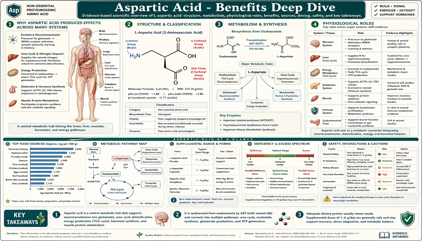
Aspartic acid is one of the most strategically placed metabolites in human biochemistry. The same simple four-carbon dicarboxylic amino acid activates N-methyl-D-aspartate (NMDA) receptors (whether nerves use it as a neurotransmitter is disputed), the only nitrogen donor for the argininosuccinate synthetase step of the urea cycle, an anaplerotic refill for the tricarboxylic acid (TCA) cycle, a gluconeogenic substrate, and the N1 atom of every purine nucleotide built de novo. Its chiral D-isomer takes a completely different path — concentrating in the pituitary, testes, and ovaries, where it modulates testosterone and the broader endocrine cascade. Four deep-dive pages below unpack the major biological roles, the supporting clinical evidence, and the practical implications for patients considering aspartate-containing supplements or facing diseases that disrupt aspartate metabolism.
Deep-Dive Articles
Neurotransmission & the N-Methyl-D-Aspartate Receptor
L-aspartate as a proposed second excitatory neurotransmitter alongside glutamate, a role researchers dispute; how the N-methyl-D-aspartate receptor functions as a coincidence-detection switch for long-term potentiation and memory formation, the distinction between L- and D-aspartate, the excitotoxicity cascade that drives neuronal death in stroke and traumatic brain injury, and the clinical role of N-methyl-D-aspartate antagonists like ketamine and memantine.
Read the deep dive →The Urea Cycle
Aspartate as the obligatory nitrogen donor at the argininosuccinate synthetase (ASS1) step of the urea cycle, the five urea-cycle enzyme defects (carbamoyl phosphate synthetase 1, ornithine transcarbamylase, argininosuccinate synthetase, argininosuccinate lyase and arginase 1) plus the cofactor and transporter disorders (N-acetylglutamate synthase deficiency, hyperornithinemia-hyperammonemia-homocitrullinuria syndrome and citrin deficiency), the malate-aspartate shuttle bridging cytosol and mitochondria, and the management of acute and chronic hyperammonemia including L-ornithine L-aspartate (LOLA) in hepatic encephalopathy (confusion caused by liver failure).
Read the deep dive →D-Aspartate & Testosterone
D-aspartate (the chiral isomer) and its accumulation in the anterior pituitary, testes, and ovaries; modulation of luteinizing hormone (LH), follicle-stimulating hormone (FSH), growth hormone, and testosterone release; Topo and D'Aniello's controversial 2009 Italian placebo-controlled trial showing a 42% testosterone increase (randomization not reported); Willoughby and Leutholtz's 2013 trial in resistance-trained men, which found no effect; the multi-million-dollar supplement market that persists despite mixed evidence; and what the data actually support.
Read the deep dive →Energy Production
Aspartate at the strategic crossroads of cellular energy: TCA-cycle anaplerosis via the aspartate-oxaloacetate equilibrium, the malate-aspartate shuttle moving cytosolic reduced nicotinamide adenine dinucleotide (NADH) into mitochondria for adenosine triphosphate (ATP) synthesis, the gluconeogenic pathway during fasting, the purine nucleotide cycle in exercising muscle, and the historical (and modern) story of potassium-magnesium aspartate as an ergogenic supplement.
Read the deep dive →Table of Contents
- Deep-Dive Articles
- Why Aspartic Acid Produces Effects Across Many Systems
- What Later D-Aspartate Trials Found
- Research Papers: Neurotransmission & N-Methyl-D-Aspartate
- Research Papers: Urea Cycle & Hyperammonemia
- Research Papers: D-Aspartate & Endocrine Function
- Research Papers: Energy Production & Mitochondrial Function
- Research Papers: Cross-Cutting (Mechanism, Status, Safety)
- External Authoritative Resources
- Connections
- Featured Videos
Why Aspartic Acid Produces Effects Across Many Systems
Most amino acids act through one principal mechanism — serving as protein building blocks plus, in some cases, one or two specialized metabolic roles. Aspartic acid is unusual because it operates through four fundamentally different mechanisms in two different molecular forms, and each maps to a distinct category of clinical effect.
- Excitatory neurotransmission via N-methyl-D-aspartate receptor activation (L-aspartate) — L-aspartate has been proposed as a neurotransmitter released at glutamatergic synapses in the central nervous system. It is a selective agonist of the N-methyl-D-aspartate receptor (a different job from that of the receptor's co-agonists such as D-serine), and activating this receptor brings in the calcium influx that drives long-term potentiation, memory formation, and (when over-activated) excitotoxic cell death. See Neurotransmission and the N-methyl-D-aspartate Receptor for the full mechanism, the long-term-potentiation-and-memory connection, the role in stroke and traumatic brain injury (TBI) excitotoxicity, and the clinically important N-methyl-D-aspartate-antagonist drug class.
- Urea cycle nitrogen donor and malate-aspartate shuttle carrier (L-aspartate) — the argininosuccinate synthetase (ASS1) step of the urea cycle is the only nitrogen-disposal pathway for one of the two nitrogens in each urea molecule, and aspartate is the obligatory donor. The malate-aspartate shuttle bridges cytosolic and mitochondrial reduced nicotinamide adenine dinucleotide pools, supporting oxidative adenosine triphosphate synthesis in heart, liver, kidney, and brain. See Aspartate in the Urea Cycle for the full urea cycle anatomy, the five enzyme defects, and the management of acute and chronic hyperammonemia.
- Endocrine modulator via D-aspartate accumulation in steroidogenic tissues — the chiral D-isomer concentrates in the pituitary, testes, ovaries, and adrenal cortex, where it modulates gonadotropin-releasing hormone, luteinizing hormone, follicle-stimulating hormone, growth hormone, testosterone, and progesterone. The enzyme that makes it is unsettled: an aspartate racemase was described in brain and neuroendocrine tissues in 2010 (Kim), while a 2016 report (Ito) found that serine racemase makes D-aspartate in some brain areas but not in the testes, and that the enzyme responsible for its making in mammals had not been identified. This role differs from L-aspartate's proposed neurotransmitter and metabolic functions, although D-aspartate also acts in the brain: mice given it long term showed N-methyl-D-aspartate-receptor effects on nerve-cell shape and synaptic plasticity (Errico 2014). See D-Aspartate and Testosterone for the tissue distribution, the pharmacology of the hypothalamic-pituitary-gonadal axis, and the controversial supplement market.
- TCA-cycle anaplerotic substrate and adenosine triphosphate-precursor donor (L-aspartate) — aspartate is in direct equilibrium with the TCA-cycle intermediate oxaloacetate via aspartate aminotransferase, supports gluconeogenesis during fasting, donates the N1 atom of every purine ring during de novo nucleotide synthesis (every adenosine triphosphate molecule), and participates in the purine nucleotide cycle that supports energy production in exercising muscle. See Aspartate and Energy Production for the metabolic networks and the history of aspartate as an ergogenic supplement.
Whether aspartate is a true neurotransmitter is disputed. It switches on N-methyl-D-aspartate receptors, and some evidence had revived the idea that nerves release it as a signal. A 2015 study (Herring, Journal of Neuroscience) used mice lacking the glutamate-selective vesicle transporter VGLUT1 and found that glutamate alone fully accounts for activation of these receptors at excitatory synapses in the hippocampus, which the authors say excludes a role for aspartate there. That study covers one brain region in mice.
The unifying picture is that aspartate sits at multiple strategic intersections in metabolism. It is both a proposed neurotransmitter and a metabolic node; both a building block (for proteins, for purines) and a regulator (of urea synthesis, of endocrine signaling); both an L-form workhorse (the three L-aspartate roles above) and a D-form specialist (best known in endocrine tissues, and also active in the developing brain). The same amino acid, in two stereoisomeric forms, threads through nearly every major physiological system.
The therapeutic complication is that this same versatility makes targeted aspartate supplementation difficult to translate into reliable clinical benefit. The endogenous synthesis system (from oxaloacetate via aspartate aminotransferase, AST) is robust under normal conditions, the blood-brain barrier restricts neural exposure, and feedback regulation in the hypothalamic-pituitary-gonadal axis dampens the endocrine effects of exogenous D-aspartate. The supplement market for aspartate-containing products has historically promised more than the controlled trial evidence supports — with the partial exception of L-ornithine L-aspartate for hepatic encephalopathy, where a 2018 Cochrane review rated the controlled-trial evidence as very low quality.
That Cochrane review (Goh) identified 36 randomized trials and could use outcome data from 29 with 1,891 participants. Compared with placebo or no treatment across all trials, L-ornithine L-aspartate was linked to less hepatic encephalopathy (risk ratio 0.70, 22 trials, 1,375 participants) and fewer deaths (risk ratio 0.42, 19 trials, 1,489 participants); a risk ratio below 1 favors the treatment. Those results did not hold in the better-run trials: in the one trial at low risk of bias, the encephalopathy result was risk ratio 0.96 (63 participants), and in the four low-risk trials for mortality it was risk ratio 0.47 with a confidence interval of 0.06 to 3.58 (244 participants). The authors wrote that they are "very uncertain about these findings".
For minimal hepatic encephalopathy, the mild form found only by testing, a 2024 meta-analysis (He) of 6 randomized trials with 292 patients found L-ornithine L-aspartate more effective than placebo or no treatment at reversing it (risk ratio 2.264) and at preventing progression to the overt form (risk ratio 0.220), with no significant difference in deaths or ammonia levels.
What Later D-Aspartate Trials Found
The published human trials differ in who took part, the dose and the length, and their results differ too:
- Topo 2009. 23 men took 3.12 g of sodium D-aspartate a day for 12 days and 20 took placebo. Testosterone rose from 4.5 to 6.4 ng/ml (42%) and luteinizing hormone 33%. The report does not describe randomization.
- Willoughby 2013. Resistance-trained men took 3 g a day or placebo for 28 days of heavy training. Total and free testosterone, luteinizing hormone, gonadotropin-releasing hormone and estradiol did not change, and strength and body composition improved about equally in both groups.
- Melville 2015. 24 resistance-trained men took 3 g, 6 g or plain flour for 14 days. Six grams lowered total and free testosterone; 3 g had no significant effect. The authors say the effect of this fall on strength and muscle gain is unknown.
- Melville 2017. 22 resistance-trained men took 6 g or placebo during 12 weeks of training. Testosterone did not change, estradiol fell 16%, and both groups gained strength and muscle size to a similar degree.
The authors of the 2017 trial contrast earlier increases in untrained men with no change or falls in resistance-trained men. A 2017 systematic review (Roshanzamir) found that D-aspartate raised testosterone in male animal studies, that human studies gave inconsistent results, and that the human evidence was still sparse.
Research Papers: Neurotransmission & N-Methyl-D-Aspartate
- Bliss TV, Lømo T (1973). Long-lasting potentiation of synaptic transmission in the dentate area of the anaesthetized rabbit — PubMed: Bliss-Lømo 1973 LTP
- Olney JW (1969). Brain lesions, obesity, and other disturbances in mice treated with monosodium glutamate — PubMed: Olney excitotoxicity 1969
- Tsien JZ et al. (1996). Hippocampal CA1 NMDA receptor and spatial memory — PubMed: Tsien 1996 CA1 knockout
- Choi DW (1988). Glutamate neurotoxicity and diseases of the nervous system — PubMed: Choi excitotoxicity review
- Bullock R et al. (1998). Excitatory amino acid release after severe human head injury — PubMed: Bullock 1998 TBI microdialysis
- Hardingham GE, Bading H (2010). Synaptic vs extrasynaptic NMDA receptor signalling — PubMed: Hardingham synaptic vs extrasynaptic
- Berman RM et al. (2000). Antidepressant effects of ketamine in depressed patients — PubMed: Berman 2000 ketamine antidepressant
- Reisberg B et al. (2003). Memantine in moderate-to-severe Alzheimer's disease — PubMed: Reisberg memantine Alzheimer's
- Rothstein JD et al. (2005). Beta-lactam antibiotics increase EAAT2 expression — PubMed: Rothstein 2005 EAAT2 ceftriaxone
- Hawkins RA (2009). The blood-brain barrier and glutamate. American Journal of Clinical Nutrition. 90(3):867S-874S. — PubMed PMID: 19571220
- Herring BE, Silm K, Edwards RH, Nicoll RA (2015). Is aspartate an excitatory neurotransmitter? Journal of Neuroscience. 35(28):10168-10171. — PubMed PMID: 26180193
- Coyle JT, Balu D, Wolosker H (2020). D-serine, the shape-shifting NMDA receptor co-agonist. Neurochemical Research. 45(6):1344-1353. — PubMed PMID: 32189130
Research Papers: Urea Cycle & Hyperammonemia
- Krebs HA, Henseleit K (1932). Original description of the urea cycle — PubMed: Krebs-Henseleit 1932
- Häberle J, Burlina A, Chakrapani A et al. (2019). Suggested guidelines for the diagnosis and management of urea cycle disorders: first revision. Journal of Inherited Metabolic Disease. 42(6):1192-1230. — PubMed PMID: 30982989
- Brusilow SW, Danney M, Waber LJ et al. (1984). Treatment of episodic hyperammonemia in children with inborn errors of urea synthesis (sodium benzoate and sodium phenylacetate). New England Journal of Medicine. 310(25):1630-1634. — PubMed PMID: 6427608
- Brusilow S, Tinker J, Batshaw ML (1980). Amino acid acylation: a mechanism of nitrogen excretion in inborn errors of urea synthesis. Science. 207(4431):659-661. — doi:10.1126/science.6243418
- Batshaw ML et al. (2014). Longitudinal study of urea cycle disorders — PubMed: Batshaw longitudinal UCD
- Saheki T, Kobayashi K (2002). Citrin deficiency (mitochondrial aspartate-glutamate carrier) — PubMed: Saheki citrin deficiency
- Kido J, Matsumoto S, Häberle J et al. (2021). Long-term outcome of urea cycle disorders: report from a nationwide study in Japan. Journal of Inherited Metabolic Disease. 44(4):826-837. — PubMed PMID: 33840128
- Goh ET, Stokes CS, Sidhu SS, Vilstrup H, Gluud LL, Morgan MY (2018). L-ornithine L-aspartate for prevention and treatment of hepatic encephalopathy in people with cirrhosis. Cochrane Database of Systematic Reviews. 5(5):CD012410. — PubMed PMID: 29762873
- Butterworth RF, McPhail MJW (2019). L-ornithine L-aspartate (LOLA) for hepatic encephalopathy in cirrhosis: results of randomized controlled trials and meta-analyses. Drugs. 79(Suppl 1):31-37. — PubMed PMID: 30706425
- He Q, Mao C, Chen Z, Zeng Y, Deng Y, Hu R (2024). Efficacy of L-ornithine L-aspartate for minimal hepatic encephalopathy in patients with cirrhosis: a meta-analysis of randomized controlled trials. Arab Journal of Gastroenterology. 25(2):84-92. — PubMed PMID: 38403493
- Vilstrup H, Amodio P, Bajaj J et al. (2014). Hepatic encephalopathy in chronic liver disease: 2014 practice guideline by the American Association for the Study of Liver Diseases and the European Association for the Study of the Liver. Hepatology. 60(2):715-735. — PubMed PMID: 25042402
- Bachmann C (2002). Mechanisms of hyperammonemia. Clinical Chemistry and Laboratory Medicine. 40(7):653-662. — PubMed PMID: 12241009
Research Papers: D-Aspartate & Endocrine Function
- Topo E, Soricelli A, D'Aniello A, Ronsini S, D'Aniello G (2009). The role and molecular mechanism of D-aspartic acid in the release and synthesis of LH and testosterone in humans and rats. Reproductive Biology and Endocrinology. 7:120. — PubMed PMID: 19860889
- Willoughby DS, Leutholtz B (2013). D-aspartic acid supplementation combined with 28 days of heavy resistance training has no effect on body composition, muscle strength, and serum hormones associated with the hypothalamo-pituitary-gonadal axis in resistance-trained men. Nutrition Research. 33(10):803-810. — PubMed PMID: 24074738
- Melville GW, Siegler JC, Marshall PW (2015). Three and six grams supplementation of d-aspartic acid in resistance trained men. Journal of the International Society of Sports Nutrition. 12:15. — PubMed PMID: 25844073
- Melville GW, Siegler JC, Marshall PWM (2017). The effects of d-aspartic acid supplementation in resistance-trained men over a three month training period: a randomised controlled trial. PLoS One. 12(8):e0182630. — PubMed PMID: 28841667
- Roshanzamir F, Safavi SM (2017). The putative effects of D-aspartic acid on blood testosterone levels: a systematic review (human results inconsistent). International Journal of Reproductive Biomedicine. 15(1):1-10. — PubMed PMID: 28280794
- D'Aniello A (2007). D-Aspartic acid: an endogenous amino acid with an important neuroendocrine role — PubMed: D'Aniello 2007 review
- D'Aniello A et al. (1996). D-aspartic acid in rat testosterone synthesis — PubMed: D'Aniello 1996 rat mechanism
- D'Aniello G, Ronsini S, Notari T et al. (2012). D-aspartate, a key element for the improvement of sperm quality. Advances in Sexual Medicine. 2(4):45-53. — doi:10.4236/asm.2012.24008
- Long Z, Lee JA, Okamoto T, Nimura N, Imai K, Homma H (2000). d-Aspartate in a prolactin-secreting clonal strain of rat pituitary tumor cells (GH3). Biochemical and Biophysical Research Communications. 276(3):1143-1147. — PubMed PMID: 11027602
- Lee JA, Long Z, Nimura N, Iwatsubo T, Imai K, Homma H (2001). Localization, transport, and uptake of D-aspartate in the rat adrenal and pituitary glands. Archives of Biochemistry and Biophysics. 385(2):242-249. — PubMed PMID: 11368004
- Furuchi T, Homma H (2005). Free D-aspartate in mammals. Biological & Pharmaceutical Bulletin. 28(9):1566-1570. — PubMed PMID: 16141517
- Kim PM, Duan X, Huang AS, Liu CY, Ming GL, Song H, Snyder SH (2010). Aspartate racemase, generating neuronal D-aspartate, regulates adult neurogenesis. Proceedings of the National Academy of Sciences of the USA. 107(7):3175-3179. — PubMed PMID: 20133766
- Ito T, Hayashida M, Kobayashi S, Muto N, Hayashi A, Yoshimura T, Mori H (2016). Serine racemase is involved in d-aspartate biosynthesis. Journal of Biochemistry. 160(6):345-353. — PubMed PMID: 27387750
Research Papers: Energy Production & Mitochondrial Function
- Krebs HA, Johnson WA (1937). Original description of the citric acid cycle — PubMed: Krebs-Johnson 1937 TCA
- Owen OE et al. (2002). Anaplerosis and cataplerosis in TCA cycle function — PubMed: Owen 2002 anaplerosis
- Birsoy K et al. (2015). Mitochondrial ETC supports aspartate biosynthesis in proliferating cells — PubMed: Birsoy 2015 Cell
- Sullivan LB et al. (2015). Aspartate biosynthesis as essential function of respiration — PubMed: Sullivan 2015 Cell
- Lu M, Zhou L, Stanley WC, Cabrera ME, Saidel GM, Yu X (2008). Role of the malate-aspartate shuttle on the metabolic response to myocardial ischemia. Journal of Theoretical Biology. 254(2):466-475. — PubMed PMID: 18603266
- Tullson PC, Terjung RL (1991). Adenine nucleotide metabolism in contracting skeletal muscle. Exercise and Sport Sciences Reviews. 19:507-537. — PubMed PMID: 1936094
- Brand MD (2005). Efficiency and plasticity of mitochondrial energy transduction — PubMed: Brand 2005 mitochondrial efficiency
- Rosen H, Blumenthal A, Agersborg HP (1962). Effects of the potassium and magnesium salts of aspartic acid on metabolic exhaustion. Journal of Pharmaceutical Sciences. 51:592-593. — PubMed PMID: 14493900
- Wesson M, McNaughton L, Davies P et al. (1988). Effects of oral administration of aspartic acid salts on the endurance capacity of trained athletes. Research Quarterly for Exercise and Sport. 59(3):234-239. — doi:10.1080/02701367.1988.10605509
- Cynober L (2002). Plasma amino acid levels and membrane transport — PubMed: Cynober plasma amino acids
Research Papers: Cross-Cutting (Mechanism, Status, Safety)
- Wolosker H, Dumin E, Balan L, Foltyn VN (2008). D-amino acids in the brain: D-serine in neurotransmission and neurodegeneration. FEBS Journal. 275(14):3514-3526. — doi:10.1111/j.1742-4658.2008.06515.x
- Errico F, Nisticò R, Di Giorgio A et al. (2014). Free D-aspartate regulates neuronal dendritic morphology, synaptic plasticity, gray matter volume and brain activity in mammals. Translational Psychiatry. 4(7):e417. — PubMed PMID: 25072322
- Topic search: aspartame metabolism safety reviews — PubMed: Aspartame safety reviews
- EFSA and FDA acceptable daily intake reviews for aspartame — PubMed: Aspartame ADI
- Vitamin B6 (pyridoxal-5-phosphate) and aminotransferase function — PubMed: B6 and aminotransferases
- Aspartate aminotransferase (AST) as clinical liver function marker — PubMed: AST as liver marker
- Plasma amino acid profiles in chronic disease — PubMed: Plasma amino acid profiles
- Branched-chain amino acid: aromatic amino acid ratio in cirrhosis — PubMed: Branched-chain to aromatic amino acid ratio
- Aspartame and phenylketonuria contraindication — PubMed: Aspartame and PKU
- Topic search: amino acid supplementation and dietary reference intakes — PubMed: Amino acid supplementation DRI
External Authoritative Resources
- Linus Pauling Institute — L-Carnitine — a page on L-carnitine, not on aspartic acid
- U.S. National Institutes of Health (NIH) Office of Dietary Supplements — Fact Sheets — comprehensive nutrient summaries
- UniProt — mitochondrial aspartate aminotransferase — protein database entry
- GeneReviews — Urea Cycle Disorders Overview — clinical genetics resource
- PubMed — All research on aspartic acid / aspartate (~200,000+ papers)
Connections
- All Amino Acids
- Aspartic Acid (Main Page)
- Aspartic Acid for Neurotransmission & N-Methyl-D-Aspartate
- Aspartic Acid in the Urea Cycle
- D-Aspartate & Testosterone
- Aspartic Acid and Energy Production
- Glutamic Acid
- Asparagine
- Arginine
- Glutamine
- Alanine
- Glycine
- Magnesium
- Zinc
- Potassium
- Vitamin B6
- Testosterone
- Liver Disease
- Stroke
- Fatigue
- Aspartame
- History
- Sources