⚠ Evidence warning: Clinical trials or other evidence do not support the claim that threonine supply is the most important single amino-acid bottleneck for antibody production, or that low threonine explains why gut barrier problems and recurrent sinus, ear, urinary and gut infections cluster in the same people. This page keeps it as a report of what people, websites or other media say.
Threonine for Immune Function
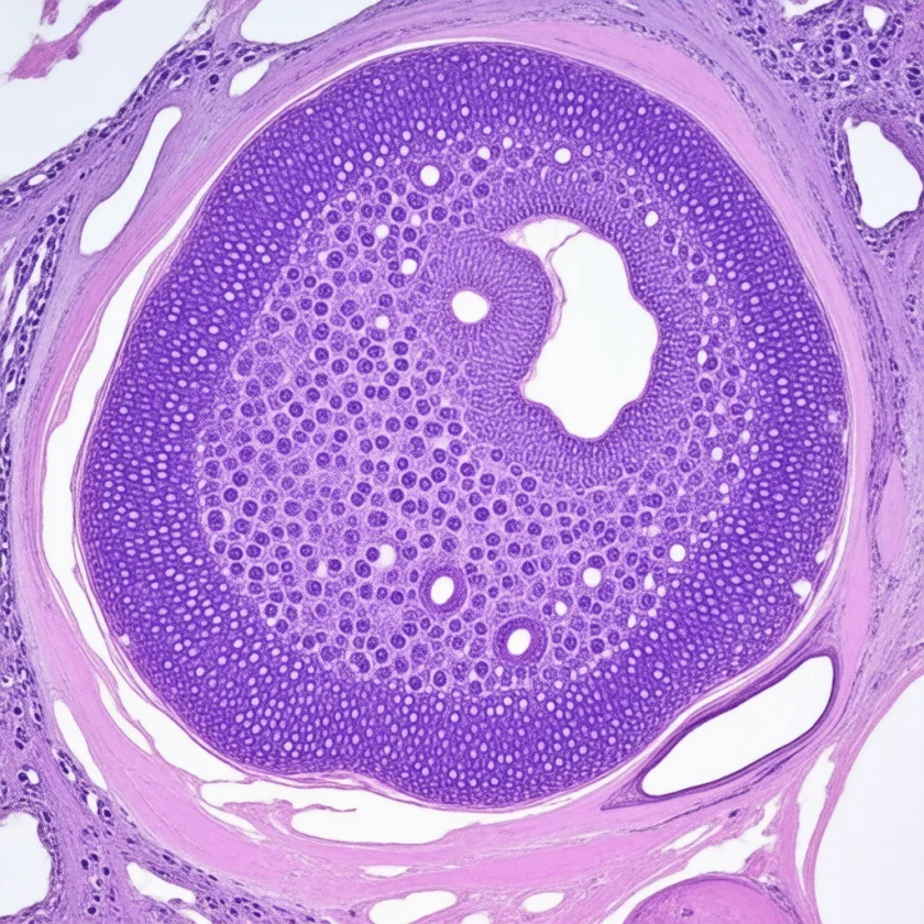
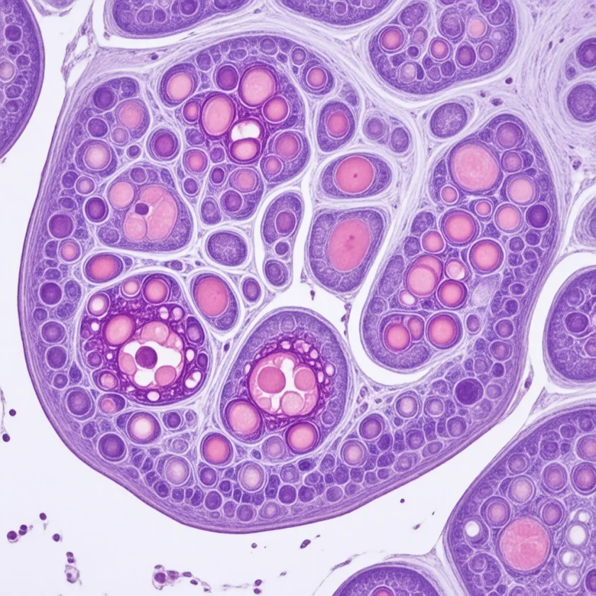
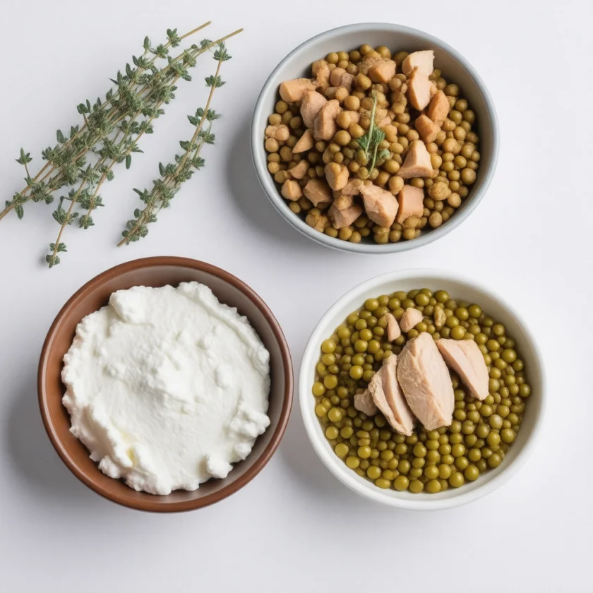
Threonine's role in immune function is one of those quiet biochemical facts that, once you see it, reframes the whole story of antibody biology. The hinge region of an antibody — the flexible linker between the antigen-binding Fab arms and the effector-function Fc tail — can carry sugar chains (O-glycans) attached to threonine and serine residues. In immunoglobulin G (IgG) this is sparse: in IgG3, each of three threonine sites in the hinge carries a sugar in only about 10% of molecules. The hinge of immunoglobulin A1 (IgA1) is the heavily decorated one: rich in threonine, serine and proline, it has nine potential sites for O-linked sugars that determine whether the antibody is functional, where it traffics, and how long it circulates. Without adequate threonine supply, immunoglobulin synthesis is constrained at the most quantitatively significant single amino acid bottleneck. This page walks through the antibody biology, the lymphocyte proliferation evidence, the experimental deficiency data, and the clinical contexts where threonine's immune role becomes practically relevant.
Table of Contents
- Antibody Biology — Why Threonine Matters
- The Immunoglobulin G Hinge Region
- The IgA1 Hinge — A Threonine-Rich Tandem Repeat
- Mucosal Immunoglobulin A and Secretory Defense
- Lymphocyte Proliferation
- Thymus Development and T-Cell Maturation
- Experimental Deficiency Studies
- GALT and the Gut-Centric Immune System
- Vaccine Response and Antibody Production
- Clinical Applications
- Cautions
- Key Research Papers
- Connections
- Featured Videos
Antibody Biology — Why Threonine Matters
The immune system depends on a continuous, massive production of antibody molecules. A healthy adult human B-cell compartment produces approximately 2-3 grams of immunoglobulin per day in the steady state, with substantial increases during active infection or after vaccination. Each antibody is a heterotetrameric protein composed of two heavy chains (about 450 amino acids each) and two light chains (about 220 amino acids each), held together by disulfide bonds.
The five major classes of human immunoglobulin — immunoglobulin G, immunoglobulin A (IgA), immunoglobulin M (IgM), IgD, and immunoglobulin E (IgE) — share the same basic architecture but differ in their heavy chain constant regions, which determine their effector functions and tissue distribution. All five classes share a distinctive structural feature called the hinge region, a flexible polypeptide linker that joins the antigen-binding Fab arms to the effector Fc tail, allowing the antibody to adopt different geometries when engaging antigen.
The hinge regions of the human immunoglobulins are unusually rich in proline (which provides backbone rigidity at specific positions), and equally unusually rich in threonine and serine. The functional reason for the threonine and serine enrichment is that these are the residues that serve as attachment sites for O-linked glycans, and the hinge is the most heavily O-glycosylated region of the antibody molecule. The O-glycan coat on the hinge protects this exposed, accessible region from proteolytic cleavage by bacterial proteases that would otherwise sever the molecule and abolish its function.
The composite consequence is that immunoglobulin synthesis at the B-cell level requires threonine in disproportionately large quantities relative to the threonine content of average tissue protein. When threonine is in short supply — whether from inadequate intake, malabsorption, or competing demand from rapidly turning over gut mucin — antibody production slows, and the immune response to infection becomes less robust.
The Immunoglobulin G Hinge Region
The immunoglobulin G hinge is the prototype hinge region and is divided into three sub-regions: an upper hinge between the Fab and the disulfide-bonded core, a middle hinge with the inter-heavy-chain disulfide bonds, and a lower hinge between the disulfides and the Fc. Across the four immunoglobulin G subclasses (IgG1, IgG2, IgG3, IgG4), the hinge length and composition vary, but all four are enriched in proline, cysteine (for disulfide bonding), threonine, and serine.
The most striking case is IgG3, which has the longest hinge of any human immunoglobulin — 62 amino acids in the most common allotype, compared to 15 in IgG1, 12 in IgG2, and 12 in IgG4. The IgG3 hinge contains three tandem repeats of a 15-residue motif rich in proline, cysteine, glutamate, and threonine. The threonine residues in this region carry O-linked glycans in some structural studies, although immunoglobulin G hinge O-glycosylation is generally less heavy than IgA1 hinge O-glycosylation. A 2015 mass-spectrometry study (Plomp and colleagues) found a sugar chain on the threonine of the repeat motif in the IgG3 hinge, but each of the three sites was occupied in only about 10% of IgG3 molecules from the blood of six donors (about 13% in two laboratory-made IgG3 antibodies) — partial decoration, far lighter than in IgA1.
The functional importance of the immunoglobulin G hinge for antibody function has been demonstrated through both natural variants and engineered modifications:
- Engineered immunoglobulin G molecules with deleted or shortened hinges show reduced flexibility and impaired complement activation
- The immunoglobulin G hinge is the site of the Fc-receptor and complement-binding interactions that determine effector function — subtle changes in hinge structure or glycosylation alter ADCC (antibody-dependent cellular cytotoxicity), complement-dependent cytotoxicity (CDC), and other downstream effector functions
The clinical implication is that the immunoglobulin G hinge is built from threonine, serine, and proline as its load-bearing residues, all of which need to be available at synthesis time in the B cell. While threonine alone is rarely the limiting factor in well-nourished individuals, it becomes potentially limiting in malnutrition, in active gut disease where threonine is competing with mucin demand, and during periods of intense antibody production after vaccination or infection.
The IgA1 Hinge — A Threonine-Rich Tandem Repeat
If the immunoglobulin G hinge is moderately threonine-rich, the IgA1 hinge is extreme. The IgA1 heavy chain contains a 26-amino-acid hinge region positioned between the CH1 and CH2 constant domains, and this hinge is built predominantly from threonine, serine, and proline. It carries nine potential O-glycosylation sites; threonine 225, threonine 228, serine 230, serine 232 and threonine 236 have been identified as the main places where the sugars attach (Takahashi and colleagues, 2012).
The composition of the IgA1 hinge sequence is approximately:
- Proline: ~38% of hinge residues
- Serine: ~23%
- Threonine: ~19%
- All other amino acids combined: ~20%
Of the nine potential O-glycosylation sites in the IgA1 hinge, typically three to five are occupied with O-linked GalNAc-based glycans; a 2004 analysis of more than 70 serum samples found a sixth sugar in about 5-10% of the IgA1 glycoforms (Tarelli and colleagues) (the same N-acetylgalactosamine that anchors mucin glycans, attached to the same kind of threonine and serine hydroxyl groups). These O-glycans are heavily decorated with galactose and sialic acid, creating a sugar coat that wraps the hinge like a thick rubber sleeve protecting an electrical cable.
The functional significance of IgA1 hinge O-glycosylation is dramatic:
- Protease resistance. Bacterial immunoglobulin A1 proteases (produced by Streptococci including Streptococcus pneumoniae, by Haemophilus influenzae and by Neisseria) cut specific bonds in the IgA1 hinge sequence as part of their mucosal-evasion strategy; they are specific to human IgA1, not immunoglobulin G. The O-glycan coat provides partial protection, and under-glycosylated IgA1 is cleaved more readily. Strains and individuals with sub-optimal hinge glycosylation are at increased risk for mucosal infection.
- Receptor binding. IgA1 binds to the polymeric Ig receptor (pIgR) for transport across mucosal epithelium, and to Fc-alpha receptors on neutrophils and macrophages. These interactions depend on the hinge geometry, which is maintained by the O-glycan coat.
- Immunoglobulin A nephropathy. The most common primary glomerulonephritis worldwide is immunoglobulin A nephropathy, in which under-galactosylated IgA1 (with truncated O-glycan chains that expose the underlying GalNAc) is deposited in glomeruli, triggering autoimmune attack and progressive kidney damage. The disease is a direct consequence of abnormal IgA1 hinge glycosylation.
The IgA1 hinge is therefore one of the most quantitatively important threonine sinks in the human body during active antibody production, and disturbances in hinge glycosylation underlie a major class of human disease.
Mucosal Immunoglobulin A and Secretory Defense
Immunoglobulin A is by far the most abundant immunoglobulin produced in the human body when measured in grams per day, exceeding immunoglobulin G and immunoglobulin M combined. Most of this immunoglobulin A is produced as dimeric immunoglobulin A in mucosa-associated lymphoid tissue (MALT) and secreted across the gut, airway, lacrimal, salivary, and mammary epithelium as secretory immunoglobulin A. This is the antibody class that lines the inner surface of every mucous membrane in the body and serves as the first specific-immunity defense against pathogens at those surfaces.
Secretory immunoglobulin A neutralizes pathogens by:
- Binding bacterial adhesins and preventing attachment to epithelial cells
- Aggregating pathogens for clearance by mucus flow
- Neutralizing toxins (such as cholera toxin and shiga toxin)
- Maintaining tolerance to commensal bacteria through low-affinity coating
- Trapping pathogens in the mucus layer for elimination
The synthesis of secretory immunoglobulin A requires not just the heavy chain (with its threonine-rich hinge) but also the J chain that links two immunoglobulin A monomers into a dimer, and the secretory component (a fragment of the polymeric Ig receptor) that wraps the dimer for transport. The secretory component itself is also a glycoprotein, and its glycans help protect the entire secretory immunoglobulin A complex from proteolytic degradation in harsh mucosal environments (such as the acidic stomach or protease-rich gut lumen).
The link between threonine and secretory immunoglobulin A comes from piglet studies, with mixed results. In piglets born small, adding threonine to the feed raised secretory immunoglobulin A in the gut (Zhang and colleagues, 2019). In weaned pigs infected with a diarrhoea-causing E. coli, slightly more threonine did not change total immunoglobulin A and only tended to raise gut immunoglobulin A against the bacterium (Trevisi and colleagues, 2015). Both are animal results; see Experimental Deficiency Studies below.
For more on the role of secretory immunoglobulin A at mucosal surfaces, see our pages on Vitamin A for Immune Function (where retinoic acid drives immunoglobulin A class switching) and the gut-immune axis discussed in Leaky Gut Syndrome.
Lymphocyte Proliferation
The adaptive immune response depends on rapid clonal expansion of antigen-specific T and B lymphocytes. When a B cell encounters its cognate antigen and receives appropriate T-cell help, it enters cell cycle and can undergo 8-10 rounds of division within 5-7 days, producing thousands of identical daughter cells from a single naive precursor. Each division requires duplication of the entire cellular protein content, the genome, organelles, and so on. The amino acid demand during clonal expansion is correspondingly large.
Threonine, along with the other essential amino acids, must be available in adequate quantity during clonal expansion. In vitro studies of lymphocyte proliferation in response to mitogens (phytohemagglutinin, concanavalin A, lipopolysaccharide) consistently show that threonine restriction reduces proliferation rate. Some studies have shown that lymphocytes are even more sensitive to threonine limitation than to limitation of other essential amino acids, possibly because of the disproportionate threonine demand of nascent antibody synthesis during the simultaneous differentiation that accompanies expansion.
In humans, the clinical correlate of impaired lymphocyte proliferation appears in the elderly, in protein-energy malnutrition, and in chronic illness with hypercatabolism. Delayed-type hypersensitivity skin test responses (a clinical measure of T-cell function) are reduced in protein-undernourished patients, and supplementation of total protein and specific amino acids can restore these responses. Whether targeted threonine supplementation has any independent clinical effect beyond optimization of overall protein intake remains underexplored.
Thymus Development and T-Cell Maturation
The thymus is the central lymphoid organ where T-cell progenitors from bone marrow mature into competent peripheral T cells. Thymic function is most active in early life — the thymus is largest in children and progressively involutes through adulthood, with the rate of new T-cell production declining sharply after age 60. The mature T-cell repertoire established during the active thymic years has to suffice for the rest of life, with peripheral T-cell expansion replacing thymic output as the primary source of T-cell replenishment.
Protein-energy malnutrition in childhood has long been known to produce dramatic thymic atrophy — the "nutritional thymectomy" phenomenon. Children with marasmus or kwashiorkor show thymic shrinkage on imaging and reduced thymic hormone production. The mechanism is multifactorial, but threonine specifically has been implicated in animal studies of protein-restricted weanling rats and pigs, where thymic weight, thymocyte count, and thymic hormone output drop with restricted threonine intake.
The clinical implication is that adequate childhood nutrition shapes the lifetime T-cell repertoire. The thymus that never grew properly in childhood cannot be made to grow in adulthood, and the T-cell diversity established in those years cannot be expanded later. This is part of why early childhood undernutrition correlates with elevated infectious disease risk decades later. For more on nutritional contributors to immune development, see our Pediatrics page, and the discussion in Vitamin A for Immune Function.
Experimental Deficiency Studies
The cleanest data on threonine and immune function comes from controlled animal-feeding studies in which threonine intake can be precisely manipulated. The dominant model has been the weanling piglet, both because of pig gastrointestinal similarity to human and because piglets have been the workhorse animal for swine-nutrition research with high-quality threonine-content data available.
Selected findings:
- Wang et al. (2007) — young pigs fed 0.37% (deficient) or 1.11% (excess) digestible threonine for 14 days made protein more slowly in the jejunal lining, gut mucins and muscle than pigs fed 0.74% (the requirement at the time); this study measured protein synthesis, not antibodies
- Wang et al. (2010) — in weanling piglets fed 0.37, 0.74, 0.89 or 1.11% digestible threonine for 14 days, the 0.37% and 1.11% diets reduced total duodenal mucins and MUC2 gene activity (mRNA) in the duodenum and jejunum compared with 0.89%
- Li et al. (2007) — a review, not a feeding experiment: it describes amino acids regulating the activation and multiplication of lymphocytes and the production of antibodies, and names arginine, glutamine and cysteine precursors as the best-studied examples of supplementation
- Faure et al. (2005) — rats fed 30% of their threonine requirement for 14 days made mucin more slowly in the duodenum, ileum and colon, while 60% did not differ from controls; mucin mRNA levels did not differ, so the authors placed the block at the translational level (the step where the protein is assembled)
- Trevisi et al. (2015) — 48 weaned pigs were fed 8.5 or 9.0 g threonine per kg of feed and then infected with a diarrhoea-causing E. coli. The extra threonine did not change days of diarrhoea or bacterial shedding after infection, did not change total immunoglobulin A or immunoglobulin M, and only tended to raise gut immunoglobulin A against the bacterium (p = 0.089). This is an animal study.
- Zhang et al. (2019) — in piglets born small (intrauterine growth restriction), adding 2 g L-threonine per kg of feed for 3 weeks raised gut mucin (Muc2) and secretory immunoglobulin A and lowered gut inflammatory signals. This is again an animal result.
Taken together, these animal studies show that threonine supply affects gut mucin production, while the effect on antibody output is mixed: higher threonine raised gut IgA in piglets born small, but made little difference in an E. coli challenge study. No study compares threonine with other individual amino acids as a driver of immune function.
Human studies are much more limited because controlled threonine restriction in humans is ethically constrained. The available data is largely correlative — reduced plasma threonine in inflammatory bowel disease (IBD), in severe burn injury, in critical illness, all of which are also associated with impaired antibody response. Causation cannot be cleanly inferred.
GALT and the Gut-Centric Immune System
Approximately 70% of the body's lymphoid tissue resides in the gut-associated lymphoid tissue (GALT), distributed across Peyer's patches, mesenteric lymph nodes, isolated lymphoid follicles, and the diffuse lamina propria lymphocyte population. The gut wall is the single largest interface between the body and the antigenic world (food antigens, commensal bacteria, occasional pathogens), and the immune system has invested heavily in gut-specific machinery.
Threonine sits at the intersection of two systems that protect the gut wall:
- The mucin barrier — threonine-rich MUC2 forms the physical mucus layer that keeps bacteria away from the epithelium (covered in detail in the Gut Mucin and Barrier deep-dive)
- The immunoglobulin A arm of the immune system — threonine-rich IgA1 and IgA2 are secreted across the epithelium into the mucus layer to neutralize any pathogens that get close to the cells
These two systems are functionally complementary — the mucus barrier provides the first physical line of defense, and secretory immunoglobulin A provides the second, antigen-specific layer within and just under the mucus. Both systems depend on threonine. A patient with marginal threonine status (gut disease, malnutrition, prolonged total parenteral nutrition (intravenous feeding), restrictive diet) experiences degradation of both layers simultaneously, compounding the loss of mucosal protection.
This is the deep biological explanation for why gut barrier dysfunction and recurrent mucosal infection (sinusitis, otitis media, urinary tract infection, gastroenteritis) often cluster in the same patients. They are not independent problems; they share a common nutritional substrate dependency.
Vaccine Response and Antibody Production
Vaccination relies on the host's ability to mount a specific antibody response to the vaccine antigen. The magnitude and durability of this response is one of the strongest predictors of vaccine effectiveness. Numerous studies have shown that protein-energy malnutrition reduces vaccine response — for example, undernourished children show reduced antibody titers to measles, polio, and hepatitis B vaccines compared to well-nourished children of the same age.
Within the broader category of nutritional contributors to vaccine response, individual amino acid status has received less attention than overall protein intake. However, the same biochemistry that drives antibody production for natural infection applies to vaccine response. Adequate threonine supply at the time of B-cell clonal expansion and antibody synthesis is presumably as important for vaccine response as for response to natural pathogens.
Nutrition points commonly described in connection with vaccine response include:
- Adequate overall protein intake (1.0-1.2 g/kg/day minimum) in the weeks leading up to and following vaccination
- Correcting any documented micronutrient deficiencies (vitamin D, vitamin A, zinc, iron) before vaccination
- No extreme calorie restriction or prolonged fasting in the immediate post-vaccination window
- For older people (who already have reduced vaccine response), a total protein intake of 1.2-1.5 g/kg/day, which may help
Direct evidence for threonine supplementation specifically improving human vaccine response is lacking. What has been studied is total nutritional status, not individual nutrient targets.
Clinical Applications
The clinical contexts where threonine supply meaningfully affects immune function include:
- Inflammatory bowel disease — reduced plasma threonine, increased demand at gut wall for mucin synthesis, and impaired antibody response combine to produce a measurable threonine inadequacy. Protein intakes of 1.2-1.5 g/kg/day from high-biological-value sources are the figures commonly given. Whey protein is particularly threonine-dense and often well-tolerated.
- Severe burn injury — hypercatabolism, dramatically increased wound-healing demand, and impaired immune function during the acute period. Specialized burn nutrition formulas provide elevated total protein and meaningful threonine; standalone L-threonine is not typically added beyond what formulas provide.
- Recurrent mucosal infection in adults — the factors described are total protein intake, secretory immunoglobulin A levels if measurable, and gut barrier integrity. Overall nutrition is usually described as producing more benefit than isolated amino acid supplementation.
- Elderly patients — protein intake commonly drops below 0.8 g/kg/day with corresponding immune function decline. Intakes toward 1.2 g/kg/day from high-biological-value sources are the figure commonly given for this group.
- Cystic fibrosis — chronic respiratory infection, malabsorption, and elevated metabolic demand combine. Pancreatic enzyme replacement, fat-soluble vitamin supplementation, and aggressive nutritional support are standard care.
- Post-bariatric surgery — reduced food volume can drop total protein and amino acid intake. The figure commonly given is a lifelong protein intake of at least 60-80 g/day, with periodic checks of nutritional status.
Standalone L-threonine for immune support is rarely the first-line intervention. Total protein optimization, addressing co-existing micronutrient deficiencies (zinc, vitamin A, vitamin D), and treating any underlying gut barrier dysfunction usually delivers the meaningful clinical benefit.
Cautions
- Whole food protein. Single amino acid supplementation rarely outperforms optimized whole-food protein intake. Threonine in particular is well supplied by ordinary mixed diets containing eggs, dairy, fish, poultry, legumes, or soy.
- Immunoglobulin A nephropathy — there is no evidence that boosting threonine intake helps; the disease pathology is in IgA1 hinge glycosylation chemistry (galactose attachment to the GalNAc), not in threonine supply per se. The disease can be triggered or worsened by mucosal infection (which drives IgA1 production), so anything that boosts antibody production without correcting the glycosylation defect could theoretically worsen disease.
- Active autoimmune disease — the goal of nutritional support is balanced immune function, not maximalism. Pushing total protein or single amino acids to levels above what the body actually uses does not appear to help autoimmune conditions and may complicate management.
- Renal impairment — diets in advanced chronic kidney disease (CKD) restrict total protein, and isolated amino acid supplements add to the nitrogen load the kidneys have to clear.
- The Vitamin A / Vitamin D / Zinc trio drives much more of the antibody-response biology than threonine does in well-nourished adults. In recurrent infection, gaps in those three nutrients are the ones usually looked at first.
Key Research Papers
- Wang X, Qiao S, Yin Y, Yue L, Wang Z, Wu G (2007). A deficiency or excess of dietary threonine reduces protein synthesis in jejunum and skeletal muscle of young pigs. Journal of Nutrition. — PubMed PMID: 17513404
- Wang W, Zeng X, Mao X, Wu G, Qiao S (2010). Optimal dietary true ileal digestible threonine for supporting the mucosal barrier in small intestine of weanling pigs. Journal of Nutrition. — PubMed PMID: 20335627
- Li P, Yin YL, Li D, Kim SW, Wu G (2007). Amino acids and immune function. British Journal of Nutrition. — PubMed PMID: 17403271
- Mestecky J, Russell MW (1986). IgA subclasses. Monographs in Allergy. (Foundational on IgA1 hinge composition.) — PubMed PMID: 3093850
- Kerr MA (1990). The structure and function of human IgA. Biochemical Journal. — PubMed PMID: 2241915
- Suzuki H et al. (2009). Aberrantly glycosylated IgA1 in IgA nephropathy patients is recognized by IgG antibodies with restricted heterogeneity. Journal of Clinical Investigation. — PubMed PMID: 19478457
- Field CJ, Johnson IR, Schley PD (2002). Nutrients and their role in host resistance to infection. Journal of Leukocyte Biology. — PubMed PMID: 11781377
- Calder PC (2006). Branched-chain amino acids and immunity. Journal of Nutrition. (Context for comparing different amino acids' immune effects.) — PubMed PMID: 16365100
- Faure M, Moënnoz D, Montigon F, Mettraux C, Breuillé D, Ballèvre O (2005). Dietary threonine restriction specifically reduces intestinal mucin synthesis in rats. Journal of Nutrition. — PubMed PMID: 15735082
- Faure M et al. (2007). Threonine utilization for synthesis of acute-phase proteins, intestinal proteins, and mucins is increased during sepsis in rats. Journal of Nutrition. — PubMed PMID: 17585034
- Cuthbertson D et al. (2005). Anabolic signaling deficits underlie amino acid resistance of wasting, aging muscle. FASEB Journal. — PubMed PMID: 15596483
- Hosomi K, Kunisawa J (2017). The specific roles of vitamins in the regulation of immunosurveillance and maintenance of immunologic homeostasis in the gut. Immune Network. — PubMed PMID: 28261016
- Trevisi P, Corrent E, Mazzoni M, Messori S, Priori D, Gherpelli Y, Simongiovanni A, Bosi P (2015). Effect of added dietary threonine on growth performance, health, immunity and gastrointestinal function of weaning pigs with differing genetic susceptibility to Escherichia coli infection and challenged with E. coli K88ac. Journal of Animal Physiology and Animal Nutrition. — PubMed PMID: 24965751
- Zhang H, Chen Y, Li Y, Zhang T, Ying Z, Su W, Zhang L, Wang T (2019). l-Threonine improves intestinal mucin synthesis and immune function of intrauterine growth-retarded weanling piglets. Nutrition. — PubMed PMID: 30504005
- Plomp R, Dekkers G, Rombouts Y, Visser R, Koeleman CA, Kammeijer GS, Jansen BC, Rispens T, Hensbergen PJ, Vidarsson G, Wuhrer M (2015). Hinge-Region O-Glycosylation of Human Immunoglobulin G3 (IgG3). Molecular & Cellular Proteomics. — PubMed PMID: 25759508
- Takahashi K, Smith AD, Poulsen K, Kilian M, Julian BA, Mestecky J, Novak J, Renfrow MB (2012). Naturally occurring structural isomers in serum IgA1 o-glycosylation. Journal of Proteome Research. — PubMed PMID: 22067045
- Tarelli E, Smith AC, Hendry BM, Challacombe SJ, Pouria S (2004). Human serum IgA1 is substituted with up to six O-glycans as shown by matrix assisted laser desorption ionisation time-of-flight mass spectrometry. Carbohydrate Research. — PubMed PMID: 15337464
- Mistry D, Stockley RA (2006). IgA1 protease. International Journal of Biochemistry & Cell Biology. — PubMed PMID: 16293440
- Allen LH (2009). How common is vitamin B-12 deficiency? American Journal of Clinical Nutrition. (Context for micronutrient gaps that compound amino acid limitations.) — PubMed PMID: 19116323
PubMed Topic Searches
Connections
- All Amino Acids
- Threonine Overview
- Threonine Benefits Hub
- Threonine for Gut Mucin
- Threonine for Collagen
- Threonine for Liver Health
- Glutamine (Lymphocyte Fuel)
- Arginine (Immune Modulation)
- Serine (Co-glycosylation Residue)
- Vitamin A for Immune Function
- Vitamin D3
- Vitamin C
- Zinc
- Immune Boosting
- Inflammatory Bowel Disease
- Nephrology (Immunoglobulin A Nephropathy)
- Bone Broth
- Eggs
- History
- Sources