⚠ Evidence warning: Clinical trials or other evidence do not support the claims that carnosine and beta-alanine slow diabetes complications, that muscle carnosine falls about 60% between ages 25 and 70 with a body-wide effect, and that one CNDP1 gene variant is among the strongest genetic protections against diabetic kidney disease. This page keeps it as a report of what people, websites or other media say.
Histidine, Carnosine, and Antioxidant Defense
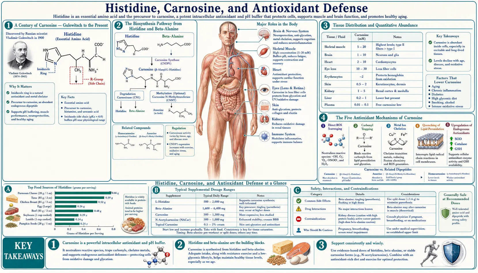
Carnosine is the second-most-abundant nitrogen compound in muscle tissue (after creatine), and the body manufactures essentially all of it from histidine and beta-alanine using a single adenosine triphosphate-dependent ligase. Russian biochemist V. S. Gulewitsch first isolated carnosine from a Liebig's meat extract in 1900 and named it after the Latin carnis (flesh) — the very word "carnivore" shares the same root. Over the next century, carnosine emerged as one of the most multi-functional small molecules in biology: a pH buffer (the share of muscle's lactate tolerance that comes from carnosine is still debated: a 2015 position stand reports early estimates of as much as 40% of muscle's buffering capacity in animals, and more recent human research as low as 7%), a direct free-radical scavenger, a specific quencher of the lipid peroxidation product 4-hydroxynonenal, a chelator of pro-oxidant copper and iron, and the most efficient endogenous inhibitor of advanced glycation end-product (AGE) formation. Muscle carnosine concentration is reported to decline roughly 60% between ages 25 and 70, mirroring the broader age-related rise in oxidative stress and protein damage. The dipeptide also exists as a related compound anserine (beta-alanyl-N-methylhistidine) in poultry and fish muscle — chicken breast and tuna are nature's richest carnosine/anserine sources. This deep-dive walks through the carnosine biosynthesis pathway from histidine and beta-alanine, the five distinct antioxidant mechanisms, the AGE-quenching role in diabetes complications, the age-related decline, and how histidine, beta-alanine (the rate-limiting precursor), and carnosine itself compare as supplements.
Table of Contents
- A Century of Carnosine — Gulewitsch to the Present
- The Biosynthesis Pathway from Histidine and Beta-Alanine
- Tissue Distribution and Quantitative Abundance
- The Five Antioxidant Mechanisms of Carnosine
- Advanced Glycation End-Product (AGE) Quenching
- Carnosine in Diabetic Complications
- Age-Related Decline of Muscle Carnosine
- Anserine in Poultry and Fish, Balenine in Whale Muscle
- Brain Carnosine and Polaprezinc (Zinc-Carnosine)
- How Histidine, Beta-Alanine and Carnosine Compare as Supplements
- Key Research Papers
- Connections
- Featured Videos
A Century of Carnosine — Gulewitsch to the Present
Carnosine was first isolated in 1900 by Vladimir Sergeyevich Gulewitsch at the University of Moscow from a Liebig's meat extract — a 19th-century beef bouillon concentrate that was a staple convenience food and laboratory reagent of the era. Gulewitsch named the compound carnosine, from the Latin carnis (flesh), reflecting both its source and its abundance in muscle. The structure as a beta-alanyl-histidine dipeptide was determined a decade later. The closely related compound anserine (beta-alanyl-N-methylhistidine) was isolated from goose muscle in 1929 — the name comes from anser, Latin for goose. A third relative, balenine (beta-alanyl-N-tau-methylhistidine), was identified in whale muscle in 1962.
For most of the 20th century, carnosine was treated as a curiosity — abundant in muscle, but with no clear function beyond a vague role in pH buffering. The renaissance came in the 1980s and 1990s through the work of Russian biochemist Alexander A. Boldyrev and his colleagues at Moscow State University, who systematically demonstrated carnosine's direct antioxidant activity, its quenching of advanced glycation end-products, its protective effects in cardiac ischemia, and its specific role in scavenging the lipid peroxidation product 4-hydroxynonenal. By the early 2000s, carnosine had been recognized as one of the most multi-functional small molecules in muscle biology, and the broader implications — for diabetes complications, neurodegenerative disease, age-related sarcopenia, and exercise performance — began to emerge in the Western literature.
The exercise-physiology angle drove the commercial supplement industry. Roger Harris and Craig Sale (UK) and a number of Asian groups demonstrated in the early 2000s that beta-alanine supplementation (the rate-limiting precursor) dose-dependently raises muscle carnosine, and that the elevated muscle carnosine measurably improves high-intensity exercise performance by increasing intramuscular buffering capacity. Beta-alanine became one of the few sports supplements with strong randomized controlled trial (RCT) evidence for ergogenic effect, and the "tingling" sensation from beta-alanine ingestion (paresthesia from MRGPRD receptor activation) became a recognizable feature of pre-workout supplement formulations.
The Biosynthesis Pathway from Histidine and Beta-Alanine
Carnosine biosynthesis is remarkably simple compared to most peptide synthesis pathways. The body does not assemble carnosine on the ribosome (which would require a tRNA for beta-alanine, which does not exist). Instead, a dedicated soluble enzyme, carnosine synthase (also called ATP-grasp domain-containing protein 1, or ATPGD1, encoded by the CARNS1 gene on chromosome 11q13.2), directly ligates beta-alanine to L-histidine, using adenosine triphosphate (ATP) hydrolysis to drive the reaction:
beta-alanine + L-histidine + adenosine triphosphate → carnosine + adenosine diphosphate + inorganic phosphate
The enzyme was long thought to produce adenosine monophosphate and pyrophosphate. The 2010 study that identified it (Drozak and colleagues) measured adenosine diphosphate instead, formed in step with the carnosine, which places the enzyme in the "ATP-grasp" family of ligases.
The enzyme has a Km for histidine of approximately 16 micromolar (well below muscle histidine concentration of around 300 micromolar) and a Km for beta-alanine of approximately 2 millimolar (around the muscle beta-alanine concentration). This kinetic asymmetry is critical — histidine is essentially always saturating, while beta-alanine is essentially always rate-limiting for carnosine synthesis. The implication: you cannot raise muscle carnosine by supplementing histidine alone (you are already saturating the enzyme on the histidine side), but you can substantially raise it by supplementing beta-alanine (which is below the Km and therefore directly controls the synthesis rate).
Carnosine synthase is expressed at highest levels in skeletal muscle, with substantial expression also in cardiac muscle, kidney, and certain brain regions (olfactory bulb, ventral mesencephalon). The enzyme is essentially absent from liver and from erythrocytes, which is why circulating plasma carnosine is very low (most of it is rapidly degraded by serum carnosinase) and almost all of the body's carnosine is located inside the cells where it was synthesized.
The serum carnosinase enzyme (CN1, encoded by CNDP1 on chromosome 18q22) is a critical regulator of carnosine pharmacokinetics. CN1 degrades free carnosine in plasma back to histidine and beta-alanine within minutes. This is why little intact oral carnosine reaches muscle in adults — the carnosine is degraded in the gut and bloodstream before reaching muscle. Its beta-alanine still counts, though: a 2012 review (Harris and colleagues) reports the same rise in muscle carnosine from an equal molar quantity of carnosine, acting as another source of beta-alanine. Beta-alanine supplementation works because beta-alanine is resistant to CN1 and reaches muscle in intact form for in-situ synthesis.
CNDP1 genetic variants have clinical relevance. A trinucleotide repeat polymorphism in CNDP1 (the "Mannheim allele") produces a less-active CN1 enzyme. Carriers have higher plasma carnosine and reduced risk of diabetic nephropathy in type 2 diabetes (a link later studies found to be sex specific; see the diabetes section below) — one of the strongest genetic protective signals against diabetic kidney disease ever identified.
Tissue Distribution and Quantitative Abundance
Carnosine is one of the most abundant nitrogen compounds in skeletal muscle, second only to creatine and creatine phosphate. Type II (fast-twitch, glycolytic) muscle fibers contain approximately twice as much carnosine as type I (slow-twitch, oxidative) fibers, reflecting the greater need for pH buffering during anaerobic glycolysis.
Typical concentrations:
- Human vastus lateralis (mixed-fiber thigh muscle): 15-30 mmol/kg dry muscle
- Type II human muscle fibers (after biopsy fiber typing): 30-40 mmol/kg dry
- Type I human muscle fibers: 12-18 mmol/kg dry
- Sprinter / power athlete muscle: can reach 35-45 mmol/kg dry, reflecting both fiber-type composition and training adaptation
- Cardiac muscle: 5-10 mmol/kg dry
- Olfactory bulb (the highest-carnosine brain region): 1-3 mmol/kg wet
- Most other brain regions: below 0.5 mmol/kg wet
For comparison, adenosine triphosphate in muscle is around 5-8 mmol/kg dry, creatine plus phosphocreatine is around 50-80 mmol/kg dry. Carnosine is therefore in the same molar range as adenosine triphosphate and accounts for a sizable fraction of total muscle nitrogen.
The sex difference is real: in a study of 149 healthy people (Everaert 2011), men had 36%, 28% and 82% higher carnosine than women in the soleus, gastrocnemius and tibialis anterior muscles of the lower leg. The higher type II fiber proportion in male muscle is one proposed reason. Androgen-driven effects on beta-alanine handling have also been proposed, although in that study circulating testosterone was unrelated to muscle carnosine in healthy men.
The dietary contribution to muscle carnosine is substantial in mixed omnivorous diets. Each 100g of beef contains approximately 365 mg of carnosine; chicken breast approximately 750 mg of carnosine and anserine combined; pork around 425 mg. A typical Western omnivore intake of 100-300 mg of carnosine per day is rapidly degraded by serum carnosinase to beta-alanine and histidine, which then re-enter the muscle for in-situ resynthesis. Strict vegetarians and vegans have measurably lower muscle carnosine than omnivores, reflecting the absence of dietary beta-alanine and dietary carnosine, though the size of the gap differs by report: vegetarians were 26% lower in the gastrocnemius in the Everaert study, while the 2012 Harris review describes omnivore levels as two or more times higher. In the Everaert study, omnivores with high and low beta-alanine intakes did not differ, and CNDP1 genotype did not affect muscle carnosine. This is one of the few clearly-demonstrated nutrient deficits of plant-based eating that is not easily replaced by plant food sources (beta-alanine is almost absent from plant foods).
Chicken broth supplies beta-alanine too. The 2012 Harris review reports that the dipeptides in chicken broth delivered close to the theoretical amount of beta-alanine to the blood, and that a dose of 40 mg per kilogram of body weight, which caused intense tingling in everyone tested as pure beta-alanine, caused none as broth. An 800 mg dose of beta-alanine equals what about 150 g of chicken breast supplies, assuming its dipeptides are fully broken down.
The Five Antioxidant Mechanisms of Carnosine
Carnosine's antioxidant activity is multifactorial, with five distinct mechanisms that together make it more than just another general-purpose radical scavenger:
1. Direct hydroxyl radical scavenging. The imidazole ring (contributed by the histidine moiety) reacts with hydroxyl radicals at near-diffusion-limited rate constants. This is the same chemistry that makes free histidine an antioxidant, but the dipeptide is more stable and has a longer intracellular half-life than free histidine, providing more sustained protection.
2. Singlet oxygen quenching. Carnosine quenches singlet oxygen (a high-energy excited form of O2 generated by photochemistry and certain enzyme reactions) with a rate constant approximately 10-fold higher than histidine and 100-fold higher than glutathione. This is particularly relevant in tissues exposed to light (eye, skin) and in tissues with high peroxidase activity.
3. Specific quenching of 4-hydroxynonenal (4-HNE). The most distinctive carnosine activity. 4-HNE is a particularly toxic lipid peroxidation product — an alpha-beta unsaturated aldehyde that forms when polyunsaturated fatty acids undergo radical-mediated peroxidation. 4-HNE forms covalent adducts with lysine, cysteine, and histidine residues in proteins, damaging enzymes and triggering pro-inflammatory cell signaling. Carnosine reacts with 4-HNE via a Michael addition between the imidazole ring of the histidine moiety and the alpha-beta unsaturated carbonyl of 4-HNE, forming a stable carnosine-4-HNE adduct that is then excreted. This effectively detoxifies 4-HNE before it can damage essential proteins. In a test-tube study (Aldini 2002), carnosine was 10 times more active at quenching 4-HNE than free histidine, reflecting kinetic catalysis from the beta-alanine moiety and from the imidazole orientation in the dipeptide.
4. Transition metal chelation. Carnosine chelates copper (II) and zinc with moderate affinity, and iron (II) more weakly. This is biologically significant because both free copper and free iron catalyze the Fenton reaction (Fe2+ or Cu+ + H2O2 → OH· + OH- + Fe3+/Cu2+), which generates hydroxyl radicals from cellular hydrogen peroxide. By sequestering catalytic metals, carnosine reduces the rate of hydroxyl radical formation in tissues with metal overload or under oxidative stress.
5. Inhibition of advanced glycation end-product (AGE) formation. The largest and most clinically important carnosine activity. Discussed in detail in the next section.
These five mechanisms together explain why carnosine activity is hard to reproduce with simpler general antioxidants — vitamin C, vitamin E, and glutathione each cover a subset of these activities, but no single small molecule covers all five. Carnosine is unique in this respect, which is why its decline with age has such a noticeable systemic effect.
Advanced Glycation End-Product (AGE) Quenching
Advanced glycation end-products (AGEs) are formed when reducing sugars (primarily glucose and fructose, but also methylglyoxal and other reactive carbonyl intermediates) react non-enzymatically with the free amino groups of lysine, arginine, and other amino acid residues on proteins. The reaction proceeds in stages: initial Schiff base formation, rearrangement to a stable Amadori product, and then further reactions over weeks to months to form the irreversible cross-linked end-products that we call AGEs.
AGEs accumulate progressively in long-lived proteins — collagen in skin and tendons, crystallins in the lens of the eye, vascular basement membranes, neuronal proteins. They cross-link extracellular matrix proteins, stiffening arteries (contributing to systolic hypertension and pulse-wave reflection), thickening basement membranes (contributing to diabetic nephropathy), causing lens opacification (contributing to senile cataracts), and triggering pro-inflammatory signaling through the RAGE (receptor for AGE) on macrophages and endothelial cells.
Carnosine inhibits AGE formation at multiple steps:
- Direct competition with protein lysine — the beta-alanine N-terminus of carnosine has a primary amino group that competes with protein lysine residues for the initial glycation reaction. Sugars react with carnosine rather than with protein, and the carnosine-sugar adduct is excreted.
- Methylglyoxal scavenging — methylglyoxal is a highly reactive intermediate in glycolysis that forms AGEs much faster than glucose itself. Carnosine reacts with methylglyoxal at high rate constants, intercepting it before it can damage proteins.
- Imidazole-aldehyde Michael addition — similar to the 4-HNE mechanism, the imidazole ring of carnosine forms stable adducts with reactive aldehyde intermediates in the AGE pathway.
- Carbonyl scavenging — carnosine is the most efficient endogenous "carbonyl scavenger" identified, with broad activity against the reactive carbonyl species that drive both glycation and lipid peroxidation.
The net effect is that carnosine acts as a sacrificial substrate for glycation, taking the chemical damage that would otherwise accumulate on long-lived structural proteins. This is the mechanistic basis for the strong epidemiological signal linking low plasma carnosine to accelerated tissue aging, and for the protective effect of CNDP1 genetic variants that raise plasma carnosine in diabetic patients.
Carnosine in Diabetic Complications
Type 2 diabetes is essentially a disease of accelerated glycation. Chronic hyperglycemia drives a several-fold increase in the rate of AGE formation throughout the body, producing the long-term complications that account for most diabetes-associated morbidity: diabetic nephropathy, diabetic retinopathy, diabetic neuropathy, accelerated atherosclerosis, and impaired wound healing.
Carnosine's anti-glycation activity makes it a logical therapeutic candidate, and the evidence base has been growing for two decades:
- Diabetic nephropathy — the most-studied indication. The CNDP1 (carnosinase) genetic polymorphism epidemiology was the original signal: patients homozygous for the "Mannheim allele" (5L/5L) of CNDP1 have low-activity serum carnosinase and elevated plasma carnosine. The first study (Janssen 2005) found the variant more common in people with diabetes who had no nephropathy (odds ratio 2.56). A later study (Mooyaart 2010) found the link sex specific: the estimated genotype risk was 0.5, about half, in women and 1.2 (confidence interval 0.77-1.69) in men with type 2 diabetes. A 2017 study (Albrecht) again found it likely gender-specific, and a 2026 review describes protective associations "in selected populations". These studies compare who has nephropathy, not how fast it progresses.
- Diabetic retinopathy — carnosine reduces the formation of advanced glycation end-products in lens crystallins and retinal extracellular matrix. N-acetylcarnosine eye drops (the "carnosine cataract" product) have been studied in age-related cataract rather than diabetic eye disease, with positive signals reported in some Russian and Asian trials and less consistent results in Western trials. A 2017 Cochrane review found only two potentially eligible studies (Russia and the United States, 114 people), could not classify either, and concluded there is "currently no convincing evidence" that the drops reverse cataract or prevent its progression.
- Glycemic control — small randomized controlled trials (RCTs) of oral carnosine (1-2 g/day) or beta-alanine (3-6 g/day) in pre-diabetic and type 2 diabetic patients have reported improvements in hemoglobin A1c (HbA1c), insulin sensitivity, and inflammatory markers. A 2021 meta-analysis (Matthews; 4 human studies, moderate certainty) estimated a change in HbA1c of -0.91% (90% credible interval -1.46 to -0.39) and in fasting glucose of -0.95 mmol/L (90% credible interval -2.1 to 0.08, which just crosses zero). Effects are smaller than diet, exercise, or metformin, but the safety profile is excellent.
- Diabetic vascular disease — carnosine protects endothelial function in animal models and in some small human trials. The mechanism involves both AGE-reduction and direct preservation of nitric oxide signaling.
Carnosine and beta-alanine have been described as possible adjuncts to standard diabetes management, particularly for patients with established complications or with the high-activity CNDP1 genotype that puts them at elevated nephropathy risk. They are not a replacement for insulin, metformin, glucagon-like peptide-1 (GLP-1) agonists, sodium-glucose cotransporter-2 (SGLT2) inhibitors, or other primary diabetes therapies; the claim made for them is that they may meaningfully slow the rate of complication progression.
Age-Related Decline of Muscle Carnosine
Muscle carnosine concentration is reported to decline with age in adults; a roughly 60% reduction between ages 25 and 70 is a figure the human studies listed on this page do not give. What they do report: carnosine in the soleus muscle was lower in older people among 149 healthy subjects (Everaert 2011); age predicted lower carnosine in patients with neuromuscular disease (R=-0.40; Stuerenburg 1999); and older people with knee osteoarthritis had lower carnosine in type II fibers but unchanged levels in type I fibers (described in the 2012 Harris review, which adds that human studies "lack sufficient control of the effects of diet and changes in muscle fibre composition"). The decline is parallel to (but distinct from) the better-known age-related declines in muscle mass, mitochondrial function, and creatine concentration.
Several factors contribute:
- Reduced beta-alanine supply — reduced meat intake with age, reduced gastric acid for protein digestion (atrophic gastritis is common after age 60), and reduced enterocyte transport capacity all reduce beta-alanine absorption.
- Reduced fast-twitch fiber mass — sarcopenia preferentially affects type II (high-carnosine) fibers, so total muscle carnosine declines partly as a function of overall fiber-type composition shift.
- Reduced carnosine synthase activity — aged muscle has reduced expression of carnosine synthase, possibly secondary to anabolic signaling decline.
- Increased oxidative carnosine consumption — aged tissue has higher baseline oxidative stress, which is "buffered" by carnosine via the mechanisms above, and this turnover depletes the pool faster than synthesis can replace it.
The functional consequence is reduced muscle buffering capacity, reduced peak power output, slower exercise recovery, and greater susceptibility to oxidative protein damage. Beta-alanine supplementation has been shown to reverse the carnosine decline in older adults to a substantial degree — typical regimens of 3-6 g/day for 12 weeks restore muscle carnosine to levels seen in much younger adults, with measurable improvements in functional capacity, exercise performance, and quality-of-life metrics in some trials. In one randomized trial of 18 healthy people aged 60-80 (del Favero 2012), 3.2 g/day for 12 weeks raised carnosine in the gastrocnemius muscle by 85.4% against 7.2% on placebo, and time to exhaustion in a constant-load test by 36.5% against 8.6%; the abstract makes no comparison with younger adults.
For more on age-related sarcopenia, see our page on Sarcopenia, or our general Amino Acids landing page.
Anserine in Poultry and Fish, Balenine in Whale Muscle
Carnosine has two close relatives in the histidine-dipeptide family:
Anserine (beta-alanyl-N-pi-methylhistidine) is identical to carnosine except for a methyl group on the imidazole ring of the histidine moiety. It is the dominant histidine-dipeptide in poultry muscle (chicken breast, turkey breast), in salmonid fish (salmon, trout), and in tuna. Anserine has antioxidant and AGE-quenching activities similar to carnosine, but with somewhat different metal-chelation properties (the methyl group changes the imidazole pKa and electron density). Anserine is rapidly metabolized in human tissue to carnosine's constituents (beta-alanine and the methylated histidine), and the methylated histidine cannot be reincorporated into protein but is excreted in urine.
Anserine is the basis for the high carnosine-equivalent content of chicken breast, which delivers approximately 750 mg of combined carnosine + anserine per 100 g serving — far more than beef. This is one reason why poultry is consistently associated with skeletal-muscle benefits in older adult nutrition studies, beyond the simple high-quality-protein effect.
Balenine (beta-alanyl-N-tau-methylhistidine) is found in cetacean muscle (whales, dolphins) and certain reptile muscle. It is essentially absent from human dietary sources (whale meat consumption is now negligible in developed countries) but represents an interesting evolutionary variant — the muscle of large diving mammals contains extraordinary concentrations of these histidine dipeptides, contributing to dive-tolerance through enhanced pH buffering during the prolonged anaerobic exercise of breath-holding dives.
The pattern across species is consistent: muscle that needs to operate at high intensity under acidotic conditions (sprint muscle of mammals, fish on rapid escape responses, diving muscle of whales) is rich in histidine dipeptides. This has been one of the more elegant evolutionary signals for the importance of these compounds in real-world athletic performance.
Brain Carnosine and Polaprezinc (Zinc-Carnosine)
Beyond muscle, carnosine has clinically relevant activities in two other compartments:
Brain (particularly olfactory bulb). Olfactory bulb has the highest carnosine concentration of any brain region (1-3 mmol/kg wet), where it acts as a co-released signaling molecule from olfactory sensory neurons and provides antioxidant protection in the high-turnover olfactory epithelium. Cerebellar Purkinje cells and certain hypothalamic nuclei also express moderate carnosine. The role in cognition and neurodegeneration is an active research area — carnosine and its zinc complex have been studied as adjuncts in autism spectrum disorder (some signals in small randomized controlled trials), in Alzheimer's disease (mechanistic plausibility for amyloid-beta interaction), and in Parkinson's disease (animal-model evidence for protection of dopaminergic neurons).
Polaprezinc (zinc-L-carnosine). A chelate compound of zinc and carnosine, marketed in Japan since 1994 as a treatment for gastric ulcer disease, and now widely used internationally as an over-the-counter supplement for gut health. The chelate has dual mechanisms: the carnosine moiety provides direct mucosal antioxidant protection and quenches the lipid peroxidation that contributes to gastric mucosal damage, while the zinc provides additional epithelial barrier support and antimicrobial activity against Helicobacter pylori (the dominant cause of peptic ulcer disease).
In randomized trials polaprezinc has been tested mainly as an add-on: to antibiotic regimens for H. pylori (results below), for painkiller-related injury of the small bowel, and for chemotherapy-related mouth sores. In a 2013 pilot trial in 20 long-term low-dose aspirin users, 150 mg a day for 4 weeks reduced the number of small-bowel reddened lesions and erosions, while the untreated group did not change; the trial was small and open-label. For chemotherapy-induced oral mucositis the evidence is low certainty: a 2026 meta-analysis of 8 studies (544 patients) found fewer cases of severe mucositis under one statistical model (odds ratio 0.48), but the result was no longer significant under the random-effects model (odds ratio 0.44, 95% confidence interval 0.18-1.06), and in the 2023 ToPaZ trial in 108 stem-cell-transplant patients a polaprezinc mouthwash had no effect (severe mucositis 35% against 36% with sodium bicarbonate). Polaprezinc is a zinc medicine, and long-term zinc can cause copper deficiency: a 2020 case report describes a 75-year-old man on hemodialysis whose blood counts fell across all cell types, with blood copper below 2 µg/dL, three months after starting polaprezinc. The site's zinc carnosine (polaprezinc) page covers these trials and the safety data in detail.
A 2022 meta-analysis of 3 randomized trials (Mahmoud; 396 people) found that adding polaprezinc to antibiotic triple therapy raised Helicobacter pylori eradication (odds ratio 2.01 by intention to treat, 95% confidence interval 1.27-3.21) with no difference in side effects; the authors call the evidence "still scarce". In one manufacturer-sponsored trial in China (Tan 2017), eradication by intention to treat was 77.0% with 75 mg twice daily added, against 58.6% with triple therapy alone.
How Histidine, Beta-Alanine and Carnosine Compare as Supplements
The three candidates for raising muscle carnosine concentration differ in what the research shows.
Direct carnosine supplementation delivers little intact carnosine to muscle in healthy adults. Oral carnosine is rapidly hydrolyzed by intestinal and serum carnosinase to its constituent beta-alanine and histidine within minutes. The intact carnosine that reaches the systemic circulation is negligible. Its beta-alanine still counts: the 2012 Harris review reports that an equal molar quantity of carnosine produces the same rise in muscle carnosine as beta-alanine. There is some evidence for direct carnosine effects on gut epithelium and on certain mucosal sites (polaprezinc above is a special case), but for muscle, oral carnosine acts mainly as a source of beta-alanine.
Histidine supplementation does not raise muscle carnosine in healthy adults, because histidine is essentially never rate-limiting in carnosine synthesis (see the kinetic discussion above). The exception is in chronic kidney disease, where dialysis depletes plasma histidine; in that setting, histidine supplementation does support both hemoglobin synthesis and muscle carnosine, but the indication is the underlying histidine deficiency, not the carnosine elevation per se.
Beta-alanine supplementation is the established protocol for raising muscle carnosine. Typical regimen: 3-6 g/day for 8-12 weeks. The dose is split into multiple smaller doses (e.g., 800 mg four times daily) to minimize the "tingling" sensation (paresthesia from MRGPRD activation) that is unpleasant but harmless. Sustained-release beta-alanine formulations reduce the paresthesia effectively.
Expected results from a 12-week beta-alanine loading:
- Muscle carnosine increases 40-80% from baseline
- High-intensity exercise capacity (1-4 minute duration) improves 2-3% on average
- Buffering capacity in working muscle increases meaningfully
- Lean body mass changes are variable and typically modest
- Reduced markers of exercise-induced oxidative stress in some studies
A 2017 meta-analysis (Saunders; 40 trials, 1,461 people) found an overall effect size of 0.18 (95% confidence interval 0.08-0.28). For exercise lasting half a minute to 10 minutes, the effect was larger on capacity tests (0.50) than on performance tests (0.11).
Beta-alanine is one of the few sports-nutrition supplements with sufficient randomized controlled trial evidence to be on the International Olympic Committee's "Strong" evidence list for ergogenic effect (alongside creatine, caffeine, sodium bicarbonate, and nitrate).
Combined beta-alanine + histidine is sometimes promoted as superior to beta-alanine alone, but the kinetic argument suggests no additional benefit unless the individual is in a histidine-deficient state.
Beta-alanine may lower histidine, and this question is still open. In one 23-day trial of 30 people (Blancquaert 2017), 6 g/day of beta-alanine lowered histidine by 30.6% in plasma and 31.6% in muscle, which did not happen when 3.5 g/day of histidine was taken with it. Histidine alone left carnosine unchanged, and the two together raised carnosine no more than beta-alanine alone. A 2019 meta-analysis of human data (Dolan) found no main effect of beta-alanine on muscle histidine.
That 2019 safety review covered 101 human and 50 animal studies. Tingling was the only reported side effect (odds ratio 8.9), dropout rates matched placebo, and the liver enzyme alanine aminotransferase rose slightly while staying within reference ranges. After supplementation stops, muscle carnosine drifts back to its earlier level with an estimated half-life of 5-9 weeks (Harris 2012).
Key Research Papers
- Gulewitsch W, Amiradžibi S (1900). Ueber das Carnosin, eine neue organische Base des Fleischextractes. Berichte der deutschen chemischen Gesellschaft. 33:1902-1903. The original isolation paper. — doi:10.1002/cber.19000330275
- Boldyrev AA et al. (2013). Physiology and pathophysiology of carnosine. Physiological Reviews. The definitive modern review. — PubMed PMID: 24137022
- Harris RC et al. (2006). The absorption of orally supplied beta-alanine and its effect on muscle carnosine synthesis in human vastus lateralis. Amino Acids. — PubMed PMID: 16554972
- Hill CA et al. (2007). Influence of beta-alanine supplementation on skeletal muscle carnosine concentrations and high intensity cycling capacity. Amino Acids. — PubMed PMID: 16868650
- Hipkiss AR (2009). Carnosine and its possible roles in nutrition and health. Advances in Food and Nutrition Research. — PubMed PMID: 19595386
- Aldini G et al. (2002). Carnosine is a quencher of 4-hydroxy-nonenal: through what mechanism of reaction? Biochemical and Biophysical Research Communications. — PubMed PMID: 12419310
- Janssen B et al. (2005). Carnosine as a protective factor in diabetic nephropathy: association with a leucine repeat of the carnosinase gene CNDP1. Diabetes. The Mannheim allele paper. — PubMed PMID: 16046297
- Sale C et al. (2010). Effect of beta-alanine supplementation on muscle carnosine concentrations and exercise performance. Amino Acids. — PubMed PMID: 20091069
- Stegen S et al. (2015). Plasma carnosine, but not muscle carnosine, attenuates high-fat diet-induced metabolic stress. Applied Physiology, Nutrition, and Metabolism. — PubMed PMID: 26307517
- Tan B et al. (2017). Polaprezinc combined with clarithromycin-based triple therapy for Helicobacter pylori-associated gastritis: A prospective, multicenter, randomized clinical trial. PLoS One. — PubMed PMID: 28407007
- Trexler ET et al. (2015). International society of sports nutrition position stand: beta-alanine. Journal of the International Society of Sports Nutrition. — PubMed PMID: 26175657
- Hipkiss AR (2015). Possible Benefit of Dietary Carnosine towards Depressive Disorders. Aging and Disease. — PubMed PMID: 26425385
- Drozak J et al. (2010). Molecular identification of carnosine synthase as ATP-grasp domain-containing protein 1 (ATPGD1). Journal of Biological Chemistry. — PubMed PMID: 20097752
- Harris RC et al. (2012). Determinants of muscle carnosine content. Amino Acids. — PubMed PMID: 22327512
- Everaert I et al. (2011). Vegetarianism, female gender and increasing age, but not CNDP1 genotype, are associated with reduced muscle carnosine levels in humans. Amino Acids. — PubMed PMID: 20865290
- Stuerenburg HJ, Kunze K (1999). Concentrations of free carnosine (a putative membrane-protective antioxidant) in human muscle biopsies and rat muscles. Archives of Gerontology and Geriatrics. — PubMed PMID: 15374064
- Mooyaart AL et al. (2010). Association between CNDP1 genotype and diabetic nephropathy is sex specific. Diabetes. — PubMed PMID: 20332346
- Albrecht T et al. (2017). The CNDP1 (CTG)(5) Polymorphism Is Associated with Biopsy-Proven Diabetic Nephropathy, Time on Hemodialysis, and Diabetes Duration. Journal of Diabetes Research. — PubMed PMID: 28553654
- Delibasi BT et al. (2026). CNDP1 and Diabetic Kidney Disease: From Genetic Susceptibility to Therapeutic Targeting. Genes. — PubMed PMID: 42074485
- Matthews JJ et al. (2021). Effect of Carnosine or β-Alanine Supplementation on Markers of Glycemic Control and Insulin Resistance in Humans and Animals: A Systematic Review and Meta-analysis. Advances in Nutrition. — PubMed PMID: 34333586
- Dubois VD, Bastawrous A (2017). N-acetylcarnosine (NAC) drops for age-related cataract. Cochrane Database of Systematic Reviews. — PubMed PMID: 28245346
- del Favero S et al. (2012). Beta-alanine (Carnosyn™) supplementation in elderly subjects (60-80 years): effects on muscle carnosine content and physical capacity. Amino Acids. — PubMed PMID: 22143432
- Blancquaert L et al. (2017). Effects of Histidine and β-alanine Supplementation on Human Muscle Carnosine Storage. Medicine and Science in Sports and Exercise. — PubMed PMID: 28106620
- Dolan E et al. (2019). A Systematic Risk Assessment and Meta-Analysis on the Use of Oral β-Alanine Supplementation. Advances in Nutrition. — PubMed PMID: 30980076
- Saunders B et al. (2017). β-alanine supplementation to improve exercise capacity and performance: a systematic review and meta-analysis. British Journal of Sports Medicine. — PubMed PMID: 27797728
- Mahmoud A et al. (2022). Efficacy and Safety of Polaprezinc-Based Therapy versus the Standard Triple Therapy for Helicobacter pylori Eradication: A Systematic Review and Meta-Analysis of Randomized Controlled Trials. Nutrients. — PubMed PMID: 36235778
- Watari I et al. (2013). Effectiveness of polaprezinc for low-dose aspirin-induced small-bowel mucosal injuries as evaluated by capsule endoscopy: a pilot randomized controlled study. BMC Gastroenterology. — PubMed PMID: 23826914
- De Francesco P et al. (2026). Protective and Healing Effects of Zinc L-Carnosine on the Oral Mucosa: A Systematic Review and Meta-Analysis. Dental Journal (Basel). — PubMed PMID: 42505716
- Nakagaki M et al. (2023). A randomised trial of topical polaprezinc to prevent oral mucositis in patients undergoing haematopoietic stem cell transplantation (ToPaZ study). Supportive Care in Cancer. — PubMed PMID: 38112867
- Koi S et al. (2020). Impaired hematopoiesis due to copper deficiency in a hemodialysis patient supplemented with zinc [Article in Japanese]. Rinsho Ketsueki. — PubMed PMID: 33162445
PubMed Topic Searches
Connections
- All Amino Acids
- Histidine Overview
- Histidine Benefits Hub
- Histidine, Histamine, and Allergy
- Histidine for Hemoglobin
- Histidine for Wound and Joint Health
- Beta-Alanine
- Zinc
- Copper
- Oxidative Stress
- Type 2 Diabetes
- Diabetic Nephropathy
- Cataracts
- Chicken (Anserine Source)
- Tuna (Anserine Source)
- History
- Sources