Arginine for Wound Healing
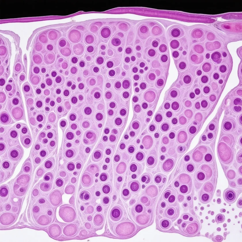
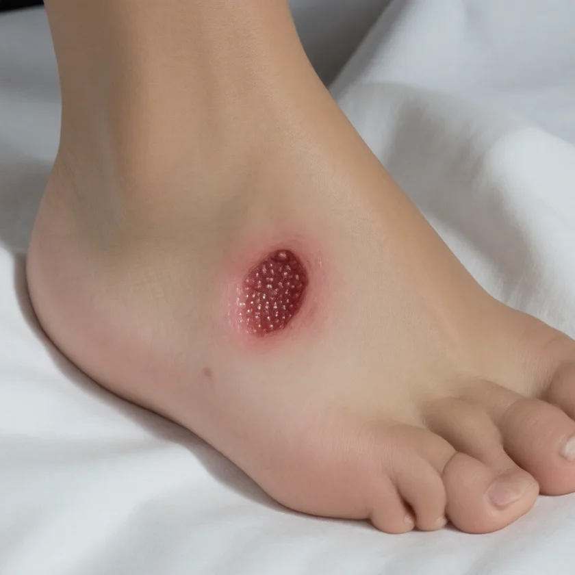
Arginine is the substrate of two competing enzymes: nitric oxide synthase, which turns it into nitric oxide (NO), a vasodilator and antimicrobial molecule, and arginase, which turns it into ornithine, the precursor of proline (for collagen) and of the polyamines (for cell proliferation). Reviews describe both arms as taking part in wound repair (Witte 2003), and in a rat wound model the NO pathway was most active in the first days of healing and the arginase pathway afterwards (Albina 1990). The human evidence comes from small experimental wounds in healthy volunteers, from trials in surgical patients of arginine-enriched immunonutrition formulas that also contain omega-3 fatty acids and nucleotides (so the effect of arginine alone cannot be separated), from pressure ulcer trials, and from diabetic foot ulcer trials. This deep-dive covers the biochemistry of the NOS/arginase fork, the wound-healing phases that involve each branch, the trial evidence in each population, the immunonutrition formulas, and the safety findings in critical illness.
Table of Contents
- Arginine's Dual-Substrate Position
- The Four Phases of Wound Healing
- The NO Arm: Inflammatory Phase
- The Ornithine Arm: Proline and Collagen
- Polyamines and Cell Proliferation
- Experimental-Wound and Surgical Trials
- Pressure Ulcer Trials
- Diabetic Foot Ulcers
- Immunonutrition Formulations
- Doses Used in Trials
- Cautions in Critical Illness
- Key Research Papers
- Connections
- Featured Videos
Arginine's Dual-Substrate Position
Arginine has more metabolic fates than most amino acids. Reviews of wound healing list protein synthesis, nitric oxide production, and metabolism to ornithine and the polyamines (Witte 2003; Stechmiller 2005). Its main fates are:
- Protein synthesis — like all amino acids, arginine is incorporated into newly synthesized proteins, including the collagen, elastin, fibronectin, and other extracellular matrix components that scaffold healing wounds
- Nitric oxide synthesis — the NOS pathway, discussed on the Cardiovascular and NO page. In wounds it is most active in the early, inflammatory phase.
- Urea cycle / ornithine production — arginase converts arginine to ornithine and urea. Ornithine is the precursor to proline (via ornithine aminotransferase and pyrroline-5-carboxylate reductase) and to polyamines (via ornithine decarboxylase). Both proline and polyamines are used in the proliferative phase of healing.
- Creatine synthesis — arginine and glycine form guanidinoacetate (arginine:glycine amidinotransferase), which guanidinoacetate methyltransferase then methylates, using a methyl group derived from methionine, to make creatine. This is not a direct part of wound healing.
- Agmatine synthesis — arginine decarboxylase produces agmatine, a biogenic amine studied for effects on imidazoline and NMDA receptors.
For wound healing, the relevant fork is between NOS (producing NO for inflammatory-phase signaling and antimicrobial defense) and arginase (producing ornithine for proline and polyamine synthesis). The two enzymes compete for the same substrate. In a rat wound model (polyvinyl alcohol sponges implanted under the skin, sampled from 6 hours to 15 days), wound fluid contained the most citrulline and nitrite, products of the NO pathway, during the first three days, the phase dominated by polymorphonuclear leukocytes; after day 3, ornithine accumulated and arginase activity was high (Albina 1990). An earlier study by the same group found low arginine, raised ornithine and high arginase activity in three different rat wound models (Albina 1988).
Because the wound uses arginine for different purposes at different times, “more arginine is better” does not capture the biology. The human trials below gave supplements throughout the period they studied rather than in a single phase.
The Four Phases of Wound Healing
Wound healing is described as four overlapping phases, each with distinct cellular and molecular events:
- Hemostasis (minutes) — platelet aggregation, fibrin clot formation, vasoconstriction. Arginine is not known to be limiting here; NO inhibits platelet aggregation, and the Albina study lists inhibition and reversal of platelet aggregation among the possible actions of the early NO pathway in wounds (Albina 1990).
- Inflammation (first days) — neutrophils first, then macrophages migrate to the wound, releasing inflammatory cytokines, phagocytosing debris and bacteria, and producing antimicrobial NO via iNOS. In the rat wound model, arginine in wound fluid fell well below plasma levels in this period while NO-pathway products peaked (Albina 1990).
- Proliferation (days to weeks) — fibroblasts migrate in and lay down new extracellular matrix (collagen, fibronectin, elastin), new capillaries form (angiogenesis), and keratinocytes migrate across the wound edge to re-epithelialize. In the rat model, arginine stayed low and ornithine accumulated after day 3, with high arginase activity (Albina 1990); ornithine feeds proline (collagen) and polyamine synthesis.
- Remodeling (weeks to years) — collagen is continuously degraded and resynthesized, changing from disorganized type III collagen in early scar to more organized type I collagen in mature scar.
Arginine is classed as conditionally essential: a 2005 review describes it as a nonessential amino acid that becomes a conditionally essential substrate in stressed adults (Stechmiller 2005). The arginine requirement of a healing wound in grams per day has not been established; the same review states that how much arginine meets metabolic needs during the phases of wound healing is still to be determined.
The NO Arm: Inflammatory Phase
The inflammatory phase of wound healing involves nitric oxide produced by inducible NOS (iNOS, NOS2) in activated inflammatory cells. Unlike the constitutive endothelial NOS, which produces small amounts of NO for vascular signaling, iNOS produces much larger amounts over sustained periods, enough to be toxic to microbes. The NO acts via several mechanisms:
- Direct antimicrobial activity — NO inhibits bacterial respiratory chain enzymes, DNA replication, and iron-containing enzymes essential for microbial metabolism
- Reactive nitrogen species generation — NO reacts with superoxide (also produced by phagocyte NADPH oxidase) to form peroxynitrite, a far more potent oxidant that nitrates protein tyrosine residues, oxidizes lipids, and damages microbial DNA
- Macrophage activation signaling — NO functions as both effector and autocrine signaling molecule, helping coordinate macrophage polarization and cytokine production
- Vasodilation and vascular permeability — NO from endothelial cells dilates wound-bed capillaries, increasing delivery of immune cells, oxygen, and nutrients
In the rat wound model, arginine concentrations in wound fluid fell markedly below plasma levels in two separate periods of healing (Albina 1990). Reviews propose that a limited arginine supply could reduce NO production and so the wound's antimicrobial defense (Witte 2003). The sources cited on this page do not measure wound infection rates in people according to their arginine status.
Animal studies of NOS inhibition give a mixed picture. In mice with implanted wound sponges, NOS inhibitors given in drinking water or by daily injection, including L-NAME, had no effect on wound NO products or on collagen deposition; when two inhibitors (aminoguanidine and S-methyl isothiouronium) were given continuously by an implanted pump, wound NO products and collagen accumulation both fell, and the authors concluded that NO is beneficial in wound healing (Schäffer 1999). In a mouse model of trauma and severe blood loss, L-arginine given during resuscitation restored wound hydroxyproline and collagen I synthesis and increased wound breaking strength (Wittmann 2005). These are animal studies, not human results.
The Ornithine Arm: Proline and Collagen
The arginase → ornithine pathway diverts arginine away from NO synthesis toward biosynthetic precursors. Ornithine has two principal fates in wound healing:
- Ornithine → pyrroline-5-carboxylate → proline — via ornithine aminotransferase (OAT) and pyrroline-5-carboxylate reductase. This is one of the body's routes to proline.
- Ornithine → putrescine → spermidine → spermine — via ornithine decarboxylase (ODC) and the subsequent polyamine biosynthetic enzymes. These polycationic small molecules are required for cell proliferation.
The proline pathway matters because glycine occupies every third position in the collagen chain and proline and hydroxyproline are abundant in it. Hydroxyproline is generated after translation by prolyl 4-hydroxylase, which converts proline residues already incorporated into the collagen polypeptide and stabilizes the collagen triple helix. The human trials below measured hydroxyproline as an index of new collagen synthesis and deposition (Barbul 1990).
Reviews propose that a shortfall in arginine supply to this pathway could limit collagen synthesis (Witte 2003). The human trials below measured collagen deposition in small experimental wounds; they did not measure wound breakdown after surgery.
Collagen synthesis also depends on other nutrients: vitamin C as cofactor for prolyl and lysyl hydroxylases; iron as cofactor for the same hydroxylases; zinc for matrix metalloproteinase function during remodeling; copper for lysyl oxidase, which catalyzes the cross-linking of collagen and elastin; and glycine. Collagen is covered on the Collagen page.
Polyamines and Cell Proliferation
The polyamines — putrescine, spermidine, and spermine — are required for cell proliferation. They are present in all mammalian cells and are involved in:
- DNA stabilization (polycationic binding to anionic phosphate backbone)
- Ribosome assembly and translation
- Membrane channel modulation
- Hypusine biosynthesis (post-translational modification of eIF5A, an essential translation factor)
- Eukaryotic gene expression regulation
Ornithine decarboxylase (ODC), the rate-limiting enzyme of polyamine biosynthesis, is rapidly induced in proliferating tissue and rapidly degraded, so polyamine production can change quickly. Reviews link the cell division of the proliferative phase to demand on the ornithine → polyamine pathway (Witte 2003).
Reviews propose the polyamine pathway as a second route by which arginine could aid repair, alongside collagen deposition. In the human experimental-wound trial in older volunteers, however, arginine increased collagen deposition but did not change the DNA content of the implanted wound tubing (an index of cell infiltration) or the rate at which a skin wound re-epithelialized (Kirk 1993).
Difluoromethylornithine (DFMO, eflornithine), an inhibitor of ornithine decarboxylase, is used clinically against African trypanosomiasis and has been studied in cancer. Its effect on human wound healing is not covered by the sources on this page.
Experimental-Wound and Surgical Trials
The first human trials measured collagen deposition in small experimental wounds in healthy volunteers, not in surgical patients. Barbul et al. (1990), at Sinai Hospital of Baltimore, studied 36 healthy, non-smoking volunteers. Under local anesthesia, a 5 cm segment of expanded polytetrafluoroethylene tubing was inserted under the skin of the upper arm (deltoid region), and the volunteers were randomized to 30 g/day of arginine hydrochloride (24.8 g free arginine), 30 g/day of arginine aspartate (17 g free arginine), or placebo, taken by mouth for 2 weeks; diet was not controlled. At 2 weeks the tubing was removed and its hydroxyproline content measured as an index of new collagen: 10.1 nmol/cm in the control group, 17.57 nmol/cm with arginine aspartate (p = 0.028) and 23.85 nmol/cm with arginine hydrochloride (p < 0.001). Both arginine groups also had greater lymphocyte responses to mitogens (Barbul 1990).
Kirk et al. (1993), from the same group, used the same tubing model in healthy volunteers over 65 years of age, again not surgical patients. In a randomized double-blind trial, 30 volunteers took 30 g/day of arginine aspartate (17 g free arginine) and 15 took a placebo syrup for 2 weeks. Arginine increased hydroxyproline accumulation in the tubing (26.49 vs 17.41 nmol/cm) and its total protein content, and raised serum IGF-1 and lymphocyte responses; it did not change the DNA content of the tubing or the rate of re-epithelialization of a 2 × 2 cm split-thickness skin wound on the thigh (Kirk 1993).
Trials in surgical patients tested arginine as part of a formula. Daly et al., at the University of Pennsylvania, randomized 85 patients having surgery for upper gastrointestinal cancers to an enteral formula supplemented with arginine, RNA and omega-3 fatty acids or to a standard enteral diet after surgery; among 77 eligible patients, infectious and wound complications occurred in 11% vs 37% (p = 0.02), and mean hospital stay was 15.8 vs 20.2 days (Daly 1992). A 1995 trial by the same group in 60 patients with esophageal, gastric or pancreatic cancer, fed by jejunostomy, reported infectious/wound complications in 10% of the supplemented group vs 43% of the standard group and a mean stay of 16 vs 22 days (Daly 1995). Because these formulas combined several nutrients, the trials do not show the effect of arginine alone.
Marimuthu et al. (2012) pooled 26 randomized trials (2,496 patients) comparing immune-modulating enteral formulas with isocaloric, isonitrogenous standard diets in major open gastrointestinal surgery. Infectious complications had a risk ratio of 0.64 (95% CI 0.55–0.74) and hospital stay had a mean difference of −1.88 days (95% CI −2.91 to −0.84), with no significant effect on mortality (RR 0.83, 95% CI 0.49–1.41); the authors noted that strong evidence for routine use had been lacking (Marimuthu 2012). Drover et al. (2011) systematically reviewed perioperative arginine-supplemented diets; its PubMed record carries no abstract, so no figures from it are restated here. In head and neck cancer surgery, a 2018 Cochrane review of 19 trials (1,099 participants; 16 of the 19 used formulas containing arginine) found no evidence of a difference in hospital stay or wound infection, a possible reduction in fistula formation (11.3% with standard care vs 5.4% with immunonutrition; RR 0.48), and rated the evidence low or very low quality (Howes 2018).
An earlier systematic review of 22 trials (2,419 surgical and critically ill patients) found immunonutrition associated with fewer infectious complications (RR 0.66, 95% CI 0.54–0.80) but no overall mortality difference (RR 1.10, 95% CI 0.93–1.31); the reduction in infections was larger in studies of surgical patients than in studies of critically ill patients (Heyland 2001).
Pressure Ulcer Trials
Pressure ulcers (also called pressure injuries, decubitus ulcers or bedsores) are chronic wounds resulting from sustained tissue pressure over bony prominences in immobile patients. They are common in nursing home residents, ICU patients, and people with spinal cord injury, and they are often slow to heal because the conditions that caused them (immobility, poor perfusion, often malnutrition) persist into the healing period.
Cereda et al. (2009) randomized 28 older residents of four long-term care facilities in Italy with recent stage II to IV pressure ulcers. All received 30 kcal/kg per day; 13 also received a 400 mL oral supplement or enteral formula enriched with protein, arginine, zinc and vitamin C. After 12 weeks the PUSH healing score had improved more in the enriched-formula group (−6.1 vs −3.3), and ulcer area had fallen more by week 8 (about 57% vs about 33%); the authors wrote that the results needed confirmation in larger trials (Cereda 2009).
The larger trial followed in 2015 (the OligoElement Sore Trial): 200 malnourished adults with stage II, III or IV pressure ulcers in long-term care or home care received 400 mL/day of an energy-dense, protein-rich formula enriched with arginine, zinc and antioxidants, or an equal volume of an isocaloric, isonitrogenous control formula, for 8 weeks, in a blinded multicenter trial. Ulcer area fell by a mean 60.9% with the enriched formula and 45.2% with the control (adjusted mean difference 18.7%, 95% CI 5.7%–31.8%); a reduction in area of 40% or more was more frequent (odds ratio 1.98), but complete healing, wound infections and the number of dressings did not differ (Cereda 2015).
Among smaller trials, Desneves et al. (2005) randomized 16 inpatients with stage 2 to 4 ulcers to a standard hospital diet, the diet plus two high-protein/energy supplements, or the same supplements with added arginine (9 g), vitamin C (500 mg) and zinc (30 mg) daily; over 3 weeks, only the arginine, vitamin C and zinc group showed a clinically significant improvement in PUSH score, and the authors called for a larger study (Desneves 2005). The 2019 international pressure injury guideline from the European Pressure Ulcer Advisory Panel, the National Pressure Injury Advisory Panel and the Pan Pacific Pressure Injury Alliance includes nutrition recommendations for pressure injury management, reviewed by Munoz et al. (2020).
Diabetic Foot Ulcers
Diabetic foot ulcers are chronic wounds in people with diabetes, arising from the combination of peripheral neuropathy (loss of protective sensation), peripheral arterial disease (impaired perfusion), and impaired wound healing. Among people with diabetes, 10–25% will experience a foot ulcer (Armstrong 2014).
Reviews propose several reasons the arginine–NO pathway may be impaired in diabetic wounds: raised ADMA (an endogenous NOS inhibitor), endothelial dysfunction that limits NO production, and arginase activity in wound macrophages that diverts arginine away from NO synthesis. These are proposed mechanisms, not measured trial outcomes.
Armstrong et al. (2014) gave 270 people with diabetic foot ulcers, alongside standard care, either a drink of arginine, glutamine and β-hydroxy-β-methylbutyrate (HMB) or a control drink, twice a day for 16 weeks, double-blind. Overall there was no difference in wound closure or time to healing at 16 weeks. In a post-hoc analysis, among people with serum albumin of 40 g/L or less, or an ankle-brachial index below 1.0, a greater proportion healed in the supplement group (1.70 and 1.66 times as likely to heal, respectively) (Armstrong 2014).
Eneroth et al. (2004) randomized people over 60 with Wagner grade I–II foot ulcers to 400 mL/day of a general oral nutritional supplement (26 people) or placebo (27 people) for six months, double-blind. Among those who completed the study, ulcers had healed in 7 of 17 on the supplement and 8 of 23 on placebo, a non-significant difference; the authors reported several methodological problems and could not demonstrate improved healing (Eneroth 2004).
The diabetic foot ulcer evidence is therefore one trial with no overall effect and a positive post-hoc subgroup, and one small trial that found no difference. In both, the supplements were given alongside standard care rather than in place of it.
Immunonutrition Formulations
Immunonutrition refers to nutritional formulas designed to modulate immune function and recovery in surgical and critically ill patients. The trials pooled by Heyland and colleagues used enteral nutrition supplemented with some combination of arginine, glutamine, nucleotides and omega-3 fatty acids (Heyland 2001); the early surgical trials used arginine, RNA and omega-3 fatty acids (Daly 1992). The components and their proposed roles:
- L-arginine — substrate for NOS-driven antimicrobial NO, ornithine-driven proline/polyamine biosynthesis, and protein synthesis
- L-glutamine — a major fuel for enterocytes and lymphocytes, studied for gut mucosal integrity and immune function; see Glutamine
- Omega-3 fatty acids (EPA + DHA) — shift eicosanoid production toward less inflammatory mediators; in the 1995 Daly trial, the arginine and omega-3 formula raised the omega-3/omega-6 ratio in plasma and white blood cells and lowered white-cell PGE2 production (Daly 1995)
- Nucleotides (RNA, often from yeast) — providing purine and pyrimidine bases for DNA/RNA synthesis in rapidly proliferating cells (gut mucosa, immune cells, healing wound)
Arginine-enriched immunonutrition formulas are made by several manufacturers and differ in arginine content and in the other nutrients they contain. In the Heyland review, studies using commercial formulas with high arginine content were associated with a significant reduction in infectious complications and a trend toward lower mortality compared with other immune-enhancing diets (Heyland 2001).
The surgical evidence is strongest for major open gastrointestinal surgery, where the Marimuthu meta-analysis found fewer infectious complications and a shorter hospital stay. In head and neck cancer surgery, the 2018 Cochrane review found only a possible reduction in fistulas and no evidence of an effect on wound infection or hospital stay (Howes 2018).
Doses Used in Trials
The amounts below are what the trials cited on this page used; they describe the research, not a dosing schedule.
- Experimental wounds in healthy volunteers — 30 g/day of arginine hydrochloride (24.8 g free arginine) or arginine aspartate (17 g free arginine) by mouth for 2 weeks (Barbul 1990; Kirk 1993).
- Surgery for upper gastrointestinal cancer — an enteral formula supplemented with arginine, RNA and omega-3 fatty acids, started after surgery (Daly 1992), given by jejunostomy in the 1995 trial (Daly 1995).
- Pressure ulcers — 400 mL/day of an energy-dense, protein-rich formula enriched with arginine, zinc and antioxidants for 8 weeks (Cereda 2015); two high-protein supplements with 9 g arginine, 500 mg vitamin C and 30 mg zinc a day for 3 weeks (Desneves 2005).
- Diabetic foot ulcers — a drink of arginine, glutamine and HMB twice a day for 16 weeks, alongside standard care (Armstrong 2014).
In these trials arginine was one part of overall nutrition. Vitamin C is the cofactor for the prolyl hydroxylase step, so collagen hydroxylation depends on it whatever the arginine supply. Decisions about supplements and medical nutrition belong with a clinician.
Cautions in Critical Illness
The arginine findings in critical illness differ from those in elective surgery. Studies in septic ICU patients recorded potential harm:
- The Bertolini sepsis trial (2003) — a randomized, unblinded multicentre trial in 33 Italian intensive care units compared enteral nutrition with an immune-enhancing formula (extra L-arginine, omega-3 fatty acids, vitamin E, beta-carotene, zinc and selenium) against parenteral nutrition. Of 237 recruited patients, 39 had severe sepsis or septic shock; in that subgroup, a planned interim analysis found ICU mortality of 44.4% with the enteral formula and 14.3% with parenteral nutrition (p = 0.039), and recruitment of patients with severe sepsis was stopped (Bertolini 2003). One proposed explanation is that extra arginine in sepsis, when iNOS is already upregulated, could add to NO production and hypotension; the trial did not test this.
- The Heyland meta-analysis (2001) — across 22 trials there was no overall mortality difference (RR 1.10, 95% CI 0.93–1.31); among studies of critically ill patients, those with high methodological quality scores were associated with increased mortality (and fewer infections) compared with lower-quality studies (Heyland 2001).
- Surgical versus septic patients — Heyland found the reduction in infections concentrated in surgical studies, and the Bertolini authors concluded that enteral immunonutrition, compared with parenteral nutrition, may be associated with excess mortality in severe sepsis. The evidence for benefit therefore comes from elective surgical patients, and the signal of harm from patients with severe sepsis.
Other cautions:
- Herpes simplex — herpes simplex virus did not grow in cultured human cells when arginine was withheld (Tankersley 1964); whether arginine supplements make outbreaks more frequent has not been tested in a trial found for this page. The Cardiovascular page covers this in more detail.
- Recent myocardial infarction — in the VINTAGE MI trial, in people after a first heart attack, 6 people taking arginine died during the 6-month study and none on placebo, and enrolment was closed for safety (Schulman 2006).
- Kidney and liver disease — two patients with severe liver disease and moderate kidney insufficiency developed marked hyperkalemia during and after an intravenous infusion of arginine hydrochloride, with a fatal cardiac arrhythmia in one; the authors attribute it to an inability to metabolize the arginine and excrete the potassium it shifts out of cells, and advise caution with arginine in renal or hepatic insufficiency (Bushinsky 1978). That report concerns intravenous infusion.
ICU care is also covered on the ARDS page.
Key Research Papers
- Barbul A, Lazarou SA, Efron DT, Wasserkrug HL, Efron G (1990). Arginine enhances wound healing and lymphocyte immune responses in humans. Surgery 108(2):331-6; discussion 336-7. — PubMed PMID: 2382229 — healthy volunteers, experimental wound.
- Kirk SJ, Hurson M, Regan MC, Holt DR, Wasserkrug HL, Barbul A (1993). Arginine stimulates wound healing and immune function in elderly human beings. Surgery 114(2):155-9; discussion 160. — PubMed PMID: 8342121 — healthy volunteers over 65, experimental wound.
- Daly JM, Weintraub FN, Shou J, Rosato EF, Lucia M (1995). Enteral nutrition during multimodality therapy in upper gastrointestinal cancer patients. Ann Surg 221(4):327-38. — PubMed PMID: 7726669
- Cereda E, Klersy C, Serioli M, Crespi A, D'Andrea F; OligoElement Sore Trial Study Group (2015). A nutritional formula enriched with arginine, zinc, and antioxidants for the healing of pressure ulcers: a randomized trial. Ann Intern Med 162(3):167-74. — PubMed PMID: 25643304
- Marimuthu K, Varadhan KK, Ljungqvist O, Lobo DN (2012). A meta-analysis of the effect of combinations of immune modulating nutrients on outcome in patients undergoing major open gastrointestinal surgery. Ann Surg 255(6):1060-8. — PubMed PMID: 22549749
- Drover JW, Dhaliwal R, Weitzel L, Wischmeyer PE, Ochoa JB, Heyland DK (2011). Perioperative use of arginine-supplemented diets: a systematic review of the evidence. J Am Coll Surg 212(3):385-99, 399.e1. — PubMed PMID: 21247782
- Witte MB, Barbul A (2003). Arginine physiology and its implication for wound healing. Wound Repair Regen 11(6):419-23. — PubMed PMID: 14617280
- Stechmiller JK, Childress B, Cowan L (2005). Arginine supplementation and wound healing. Nutr Clin Pract 20(1):52-61. — PubMed PMID: 16207646
- Armstrong DG, Hanft JR, Driver VR, Smith AP, Lazaro-Martinez JL, Reyzelman AM et al. (2014). Effect of oral nutritional supplementation on wound healing in diabetic foot ulcers: a prospective randomized controlled trial. Diabet Med 31(9):1069-77. — PubMed PMID: 24867069
- Heyland DK, Novak F, Drover JW, Jain M, Su X, Suchner U (2001). Should immunonutrition become routine in critically ill patients? A systematic review of the evidence. JAMA 286(8):944-53. — PubMed PMID: 11509059
- Wittmann F, Prix N, Mayr S, Angele P, Wichmann MW, van den Engel NK et al. (2005). L-arginine improves wound healing after trauma-hemorrhage by increasing collagen synthesis. J Trauma 59(1):162-8. — PubMed PMID: 16096557 — a mouse study.
- Albina JE, Mills CD, Barbul A, Thirkill CE, Henry WL Jr, Mastrofrancesco B et al. (1988). Arginine metabolism in wounds. Am J Physiol 254(4 Pt 1):E459-67. — PubMed PMID: 3354662 — rat wound models.
- European Pressure Ulcer Advisory Panel, National Pressure Injury Advisory Panel, Pan Pacific Pressure Injury Alliance (2019). Prevention and Treatment of Pressure Ulcers/Injuries: Clinical Practice Guideline. Nutrition recommendations reviewed in: Munoz N, Posthauer ME, Cereda E, Schols JMGA, Haesler E (2020). The Role of Nutrition for Pressure Injury Prevention and Healing: The 2019 International Clinical Practice Guideline Recommendations. Adv Skin Wound Care 33(3):123-136. — PubMed PMID: 32058438
- Daly JM, Lieberman MD, Goldfine J, Shou J, Weintraub F, Rosato EF et al. (1992). Enteral nutrition with supplemental arginine, RNA, and omega-3 fatty acids in patients after operation: immunologic, metabolic, and clinical outcome. Surgery 112(1):56-67. — PubMed PMID: 1377838
- Cereda E, Gini A, Pedrolli C, Vanotti A (2009). Disease-specific, versus standard, nutritional support for the treatment of pressure ulcers in institutionalized older adults: a randomized controlled trial. J Am Geriatr Soc 57(8):1395-402. — PubMed PMID: 19563522
- Desneves KJ, Todorovic BE, Cassar A, Crowe TC (2005). Treatment with supplementary arginine, vitamin C and zinc in patients with pressure ulcers: a randomised controlled trial. Clin Nutr 24(6):979-87. — PubMed PMID: 16297506
- Howes N, Atkinson C, Thomas S, Lewis SJ (2018). Immunonutrition for patients undergoing surgery for head and neck cancer. Cochrane Database Syst Rev 8(8):CD010954. — PubMed PMID: 30160300
- Bertolini G, Iapichino G, Radrizzani D, Facchini R, Simini B, Bruzzone P et al. (2003). Early enteral immunonutrition in patients with severe sepsis: results of an interim analysis of a randomized multicentre clinical trial. Intensive Care Med 29(5):834-40. — PubMed PMID: 12684745
- Eneroth M, Larsson J, Oscarsson C, Apelqvist J (2004). Nutritional supplementation for diabetic foot ulcers: the first RCT. J Wound Care 13(6):230-4. — PubMed PMID: 15214141
- Albina JE, Mills CD, Henry WL Jr, Caldwell MD (1990). Temporal expression of different pathways of 1-arginine metabolism in healing wounds. J Immunol 144(10):3877-80. — PubMed PMID: 2332635 — a rat wound model.
- Schäffer MR, Tantry U, Thornton FJ, Barbul A (1999). Inhibition of nitric oxide synthesis in wounds: pharmacology and effect on accumulation of collagen in wounds in mice. Eur J Surg 165(3):262-7. — PubMed PMID: 10231662
- Tankersley RW Jr (1964). Amino acid requirements of herpes simplex virus in human cells. J Bacteriol 87(3):609-13. — PubMed PMID: 14127578 — cell culture.
- Schulman SP, Becker LC, Kass DA, Champion HC, Terrin ML, Forman S et al. (2006). L-arginine therapy in acute myocardial infarction: the Vascular Interaction With Age in Myocardial Infarction (VINTAGE MI) randomized clinical trial. JAMA 295(1):58-64. — PubMed PMID: 16391217
- Bushinsky DA, Gennari FJ (1978). Life-threatening hyperkalemia induced by arginine. Ann Intern Med 89(5 Pt 1):632-4. — PubMed PMID: 717931 — case reports.
PubMed Topic Searches
Connections
- All Amino Acids
- Arginine Overview
- Arginine Benefits Hub
- Arginine for Cardiovascular Health
- Arginine for Erectile Function
- Arginine for Immune Function
- Glutamine
- Proline
- Glycine
- Collagen
- Vitamin C
- Zinc
- Copper
- Diabetes
- ARDS / Critical Illness
- Ornithine — the arginase product that supplies proline for collagen and polyamines for cell proliferation.
- History
- Sources