Arginine for Immune Function
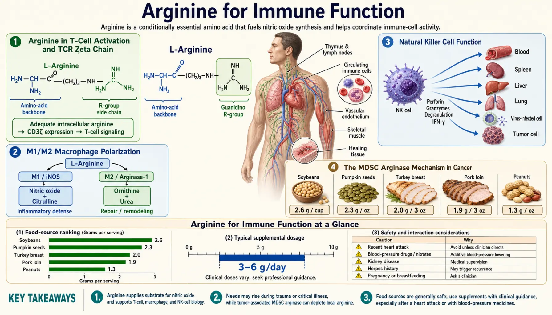
Arginine sits at a branch point in the immune system. It is the substrate of inducible nitric oxide synthase (iNOS, NOS2), which macrophages use to make antimicrobial nitric oxide, and the enzyme arginase competes with nitric oxide synthase for the same arginine (Bronte and Zanovello 2005). In cultured T cells, removing arginine lowered expression of the CD3-zeta chain, the principal signalling element of the T-cell receptor, and slowed proliferation (Rodriguez 2002). Myeloid-derived suppressor cells (MDSCs) that make arginase I deplete arginine and inhibit T-cell responses in tumour-bearing mice and in cancer patients (Rodríguez and Ochoa 2008), and plasma arginine is lower in patients with sepsis than in controls (Davis and Anstey 2011). In the clinic, a meta-analysis of immune-modulating nutrient mixtures found fewer infections after major open gastrointestinal surgery (Marimuthu 2012), while in severe sepsis a planned interim analysis recorded higher intensive-care mortality with an arginine-containing enteral formula than with parenteral nutrition (Bertolini 2003). This deep-dive walks through each of these mechanisms and the clinical evidence connected to them.
Table of Contents
- Arginine in T-Cell Activation and TCR Zeta Chain
- M1/M2 Macrophage Polarization
- Natural Killer Cell Function
- The MDSC Arginase Mechanism in Cancer
- Targeting MDSC Arginase Therapeutically
- Septic Patient Arginine Deficiency
- ICU Immunonutrition Trials
- Elective Surgery Immunonutrition
- HIV and Chronic Viral Infection
- Sickle Cell Disease and Vaso-Occlusive Crisis
- Asthma and the Th2/Arginase Axis
- Key Research Papers
- Connections
- Featured Videos
Arginine in T-Cell Activation and TCR Zeta Chain
One of the more counterintuitive findings in arginine immunology is that T-cell receptor signalling depends on the arginine supply around the cell. The finding concerns the T-cell receptor zeta chain (CD3-zeta, CD247), the principal signal-transduction element of the receptor (Rodriguez 2002).
The TCR is not a single molecule but a complex of multiple subunits: the antigen-recognizing alpha-beta or gamma-delta TCR heterodimer, the CD3 complex of gamma, delta, and epsilon chains, and the homodimeric CD3-zeta chain. Of these, CD3-zeta contains three immunoreceptor tyrosine-based activation motifs (ITAMs) that are the proximal docking sites for the kinase ZAP-70 upon TCR engagement. Loss of CD3-zeta therefore weakens the link between antigen recognition and the downstream signalling, IL-2 production and proliferation that follow it.
Work by Rodriguez, Ochoa and colleagues at Louisiana State University showed that Jurkat T cells (a human T-cell line) cultured in arginine-free medium rapidly lost CD3-zeta expression and proliferated less. Adding back arginine, but not other amino acids, completely reversed the change, and it was not caused by cell death. The fall in CD3-zeta protein was paralleled by a fall in CD3-zeta mRNA, and this was not due to slower transcription but to a significantly shorter CD3-zeta mRNA half-life — a post-transcriptional effect on mRNA stability (Rodriguez 2002).
The authors concluded that the arginine concentration in the microenvironment could be an important regulator of CD3-zeta and therefore of T-cell function (Rodriguez 2002). A 2007 review lists T-cell dependence on arginine for proliferation, zeta-chain and T-cell receptor expression and the development of memory, and notes that decreased proliferation and loss of the zeta chain are observed in cancer and after trauma (Popovic 2007). In human T cells, a 2016 study found that activation lowers intracellular arginine, and that raising arginine levels shifted activated T cells toward oxidative phosphorylation, favoured central memory-like cells with better survival and, in a mouse model, improved anti-tumour activity (Geiger 2016).
Low arginine in the local T-cell environment is therefore proposed as one cause of impaired T-cell function even where lymphocyte counts look normal. Arginase-expressing myeloid suppressor cells have been described in trauma, intra-abdominal sepsis, certain infections and, most prominently, cancer (Popovic 2007); the tumour setting is the most studied and is discussed below.
M1/M2 Macrophage Polarization
Macrophages are not a single cell type but a continuum of activation states. The two extreme phenotypes are:
- M1 (classically activated, pro-inflammatory) — induced by Th1 signals such as interferon-gamma and by microbial products such as LPS. High iNOS expression, high NO production, high pro-inflammatory cytokine output (IL-1, IL-6, IL-12, TNF), antimicrobial and anti-tumor effector function. Uses arginine via iNOS to produce NO.
- M2 (alternatively activated, pro-resolving) — induced by Th2 cytokines (IL-4, IL-13). High arginase expression, high ornithine/polyamine/proline production, tissue repair and angiogenesis functions, often immunosuppressive. Uses arginine via arginase to produce ornithine.
In myeloid cells, arginine is mainly metabolized either by iNOS or by arginase 1, enzymes stimulated by T helper 1 or T helper 2 cytokines respectively, so which enzyme is active reflects the type of inflammatory response (Popovic 2007). Both routes consume arginine but lead to different products: NO, which is cytotoxic to microbes, or ornithine, the precursor of proline (used for collagen) and of polyamines (used for cell division) (Morris 2009).
This split is the basis for arginine's different roles in different inflammatory states:
- In an acute infection with an M1-dominant macrophage response, arginine is the substrate for iNOS-driven antimicrobial NO production.
- In chronic Th2-driven inflammation such as allergic asthma, raised arginase is proposed to divert arginine away from NO synthesis and toward ornithine-driven tissue remodelling (Maarsingh 2009; Munder 2009); whether extra arginine helps or harms in that setting is not settled.
- In tumours with arginase-expressing MDSCs, local arginine depletion suppresses T cells (Rodríguez and Ochoa 2008), and reviews describe pharmacological interference with arginine metabolism, such as arginase inhibition, as the approach under study (Munder 2009).
Because arginase and nitric oxide synthase compete for the same substrate (Bronte and Zanovello 2005), reviews describe arginine's immune effects as context-dependent rather than a general immune “boost”: the result of more arginine depends on which immune cells are limited by arginine supply, and which enzymes are competing for it, in a given condition.
Natural Killer Cell Function
Natural killer (NK) cells are the principal innate-immune defense against virally infected cells and tumor cells. They detect "missing self" (cells that have downregulated MHC class I, often as a viral or tumor immune-evasion strategy) and kill targets via perforin-granzyme release and Fas-FasL interactions. NK cells are an important component of cancer immunosurveillance and contribute to viral immunity.
Human NK cells also depend on arginine, though not for every function. In a 2009 laboratory study, arginine depletion by granulocyte arginase severely reduced NK-cell proliferation and IL-12/IL-18-induced interferon-gamma secretion, while NK-cell viability, granule release and cytotoxicity did not depend on extracellular arginine. The suppression acted after transcription, since mRNA levels were unaffected, and human purulent exudate (pus) inhibited NK-cell functions ex vivo entirely through its released arginase (Oberlies 2009). In mouse tumour models, the arginase inhibitor CB-1158 increased the numbers of tumour-infiltrating NK cells and CD8+ T cells (Steggerda 2017).
Whether arginine supplementation changes NK-cell function, infection rates or tumour response in cancer patients, older adults or people with chronic viral infection is not established by the studies cited on this page.
The MDSC Arginase Mechanism in Cancer
Myeloid-derived suppressor cells (MDSCs) are a heterogeneous population of immature myeloid cells (granulocytic and monocytic subtypes) that accumulate in cancer patients and in tumors. Abnormal dendritic-cell differentiation and the accumulation of these immature myeloid suppressor cells have been described as one of the major mechanisms of tumour escape from the immune system (Mirza 2006).
Two arginine-related mechanisms by which MDSCs suppress anti-tumor immunity are described:
- Arginase 1 — increased metabolism of arginine by MDSCs producing arginase I inhibits T-lymphocyte responses in tumour-bearing mice and cancer patients (Rodríguez and Ochoa 2008). Human tumour samples of several types contained abundant arginase-1-expressing myeloid cells, and cancer patients' plasma had more arginase 1 and less arginine than that of healthy volunteers (Steggerda 2017). The depletion lowers CD3-zeta on T cells through the mechanism described above.
- Nitric oxide synthase — arginase and nitric oxide synthase, which compete for arginine, are both described as crucial components of the myeloid lymphocyte-suppression pathway, and the products of both enzymes moderate T-cell function (Bronte and Zanovello 2005).
Reviews describe the result as an impaired T-cell response in cancer patients, which can reduce the potential benefit of cancer vaccines and other forms of immunotherapy (Rodríguez and Ochoa 2008).
For immune checkpoint inhibitors (drugs such as pembrolizumab and nivolumab), the evidence is mainly from animals: in mouse models, the arginase inhibitor CB-1158 reduced tumour growth both alone and in combination with checkpoint blockade (Steggerda 2017). The sources cited here do not show that MDSC arginase explains resistance to checkpoint inhibitors in patients, and in the first human trial of the drug (below), arginase-1 expression in the tumour did not correlate with response (Naing 2024).
Targeting MDSC Arginase Therapeutically
The recognition of MDSC arginase as a mechanism of tumor immunosuppression has led to several approaches being studied:
- Arginase inhibitors — CB-1158 (numidargistat, INCB001158), an oral small-molecule arginase inhibitor, blocked myeloid-cell suppression of T-cell proliferation in vitro and reduced tumour growth in several mouse cancer models, alone and combined with checkpoint blockade, adoptive T-cell or NK-cell therapy, or gemcitabine (Steggerda 2017).
- Raising arginine — in a mouse model, T cells with raised arginine levels had greater anti-tumour activity (Geiger 2016). Trials of arginine supplementation for this purpose in cancer patients are not among the studies cited on this page.
- MDSC differentiation — in 18 patients with metastatic renal cell carcinoma, all-trans retinoic acid reduced immature myeloid suppressor cells, but only in patients who reached high plasma concentrations of the drug; it also improved dendritic-cell function and antigen-specific T-cell responses (Mirza 2006).
- iNOS inhibitors — aimed at the parallel NO-driven suppression. NO made by the endothelial enzyme (NOS3) also regulates blood vessels (Luiking 2012), so selectivity for the inducible enzyme matters.
In the first-in-human phase 1 study of INCB001158, 107 patients with advanced solid tumours received the drug alone and 153 received it with pembrolizumab. Plasma arginine rose dose-dependently, consistent with arginase inhibition. Grade 3 or higher treatment-emergent adverse events occurred in 45.8% of patients on the drug alone and 51.7% on the combination. The highest response rate, 19.2%, was in a head and neck squamous cell carcinoma cohort not previously treated with anti-PD-1/PD-L1 drugs. The authors concluded that response rates did not exceed background for the tumour types despite the drug's measurable pharmacodynamic activity, and that the role of arginine depletion in cancer is multifaceted (Naing 2024).
Septic Patient Arginine Deficiency
Sepsis is a syndrome of life-threatening organ dysfunction caused by dysregulated host response to infection. Surgery, trauma, burns and injury can induce an inflammatory response that becomes excessive, followed by an immunosuppressed state that increases susceptibility to infection (Calder 2007). Arginine depletion has been linked to the T-cell side of this dysfunction (Darcy 2014, Crit Care).
A 2011 systematic review and meta-analysis found plasma arginine lower in sepsis patients than in controls in all eight studies of predominantly medical sepsis. In the pooled analysis, mean plasma arginine was 33.9 µmol/L lower than in concurrent non-septic controls, a relative decrease of 41%. There were not enough good-quality studies to say whether the same holds for sepsis after trauma or surgery (Davis and Anstey 2011). Contributing mechanisms described in the research include:
- Raised arginase activity — plasma arginase activity was higher in 44 sepsis patients than in 25 controls and correlated with neutrophil count; patients with high neutrophil counts had higher arginase activity and lower plasma arginine (Darcy 2014, Clin Chem Lab Med).
- Reduced endogenous synthesis — de novo arginine production depends on citrulline, its immediate precursor, and reduced de novo production together with raised arginase activity has been reported in various conditions of acute and chronic stress (Luiking 2012).
- Increased consumption by activated immune cells — the same stress conditions are often characterized by increased NOS2 activity (Luiking 2012), which uses arginine to make NO.
- Reduced dietary intake — critically ill patients often have impaired nutritional intake, compounding the substrate deficit.
In septic shock, arginase-expressing neutrophils with suppressor function circulated in proportion to disease severity; outside the body they metabolised arginine and suppressed T-cell proliferation and CD3-zeta expression, and depleting or inhibiting them restored zeta-chain expression and T-cell function in vitro (Darcy 2014, Crit Care). Impaired availability of arginine as the substrate for NO synthesis has also been linked to sepsis severity (Darcy 2014, Clin Chem Lab Med).
Whether to add arginine in sepsis is therefore unresolved. A 2007 review notes that some studies of arginine-containing immunonutrient mixtures in critically ill patients showed increased mortality, and that one view attributes this to the high arginine content driving nitric oxide production (Calder 2007); the Bertolini trial is discussed below. Supplementing citrulline, the precursor the body converts to arginine, has been described as one way to raise arginine availability (Luiking 2012). In the IMMUNOCITRE trial of 120 mechanically ventilated intensive-care patients without sepsis or septic shock, enteral citrulline 5 g every 12 hours for 5 days raised plasma arginine on day 3 but did not change the day-7 SOFA organ-dysfunction score or the immune markers measured, compared with placebo (Tadié 2023).
ICU Immunonutrition Trials
The history of arginine-containing immunonutrition in the ICU shows how results from elective surgery may not carry over to critical illness. Enteral immunonutrient mixtures, usually including arginine, nucleotides and long-chain omega-3 fatty acids, have been used widely in surgical and critically ill patients; a 2007 review describes the evidence of efficacy as good in surgical patients, but whether the same mixtures are beneficial in critically ill patients as controversial (Calder 2007).
The best-known negative result is Bertolini et al. (2003), an unblinded randomised multicentre trial in 33 Italian intensive care units comparing enteral feeding with an immune-enhancing formula (containing extra arginine, omega-3 fatty acids, vitamin E, beta-carotene, zinc and selenium) against total parenteral nutrition. Of 237 patients recruited, 39 had severe sepsis or septic shock. A planned interim analysis of that subgroup — brought forward after a meta-analysis suggested excess mortality with enteral immunonutrition in severe sepsis — found intensive-care mortality of 44.4% with the enteral formula versus 14.3% with parenteral nutrition (p=0.039), and recruitment of patients with severe sepsis was stopped. The authors concluded that enteral immunonutrition, compared with parenteral nutrition, may be associated with excess mortality in patients with severe sepsis (Bertolini 2003).
The comparison in that trial was enteral feeding with a multi-nutrient formula against parenteral nutrition, not arginine against no arginine. One proposed explanation for the mortality signal is that extra arginine drives nitric oxide production in septic patients (Calder 2007). A 2005 commentary by Heyland and Dhaliwal, titled “Immunonutrition in the critically ill: from old approaches to new paradigms”, discusses the field's shift after these results (Heyland and Dhaliwal 2005).
The resulting picture in the reviews cited here is a split by setting: evidence of benefit in elective surgical patients (next section), and continuing controversy in critical illness, especially severe sepsis (Calder 2007). Decisions about nutrition in intensive care belong with the clinical team.
Elective Surgery Immunonutrition
The elective-surgery literature is more favourable. A 2012 meta-analysis of 26 randomised trials enrolling 2,496 patients undergoing major open gastrointestinal surgery compared immune-modulating nutrient combinations with a standard diet. It found strong evidence of fewer postoperative infectious complications (risk ratio 0.64, 95% CI 0.55–0.74) and a shorter hospital stay (mean difference −1.88 days); non-infectious complications were also lower but the quality of that evidence was low, and there was no statistically significant effect on mortality (Marimuthu 2012). A 2011 systematic review covers perioperative arginine-supplemented diets specifically (Drover 2011). The surgical evidence is discussed in more detail on the Wound Healing page.
Proposed immune mechanisms for the surgical findings include:
- Preservation of T-cell function — arginase 1 expression has been detected in mononuclear cells after trauma or surgery, and loss of the zeta chain is observed after trauma (Popovic 2007).
- Wound antimicrobial defense — via iNOS-driven NO production by wound-recruited macrophages.
- Gut barrier function — a proposed role for glutamine and arginine in enterocyte integrity and bacterial translocation; not tested separately in the trials above.
- Modulation of the inflammatory response — the formulas also contain long-chain omega-3 fatty acids, among the nutrients able to modulate inflammation (Calder 2007).
Because the formulas combine several nutrients, the trials do not show which component, if any single one, accounts for the effect.
HIV and Chronic Viral Infection
In HIV, arginase has been studied as a marker of immune suppression. Blood mononuclear cells from 23 HIV-positive patients with CD4 counts of 350 cells/µL or below expressed significantly more arginase than cells from 21 patients with higher counts, and arginase activity was associated with CD4 count and plasma viral load in patients not yet on antiretroviral therapy, but not in patients receiving therapy (Cloke 2010).
Antiretroviral therapy, which suppresses viral replication, is the established treatment for HIV; in the study above, the link between arginase and disease severity was not seen in treated patients (Cloke 2010). Clinical trials of arginine supplementation in HIV are not among the studies cited on this page.
Sickle Cell Disease and Vaso-Occlusive Crisis
Sickle cell disease (SCD) is an inherited hemoglobinopathy in which a single amino acid substitution in the beta-globin chain produces hemoglobin S, which polymerizes under low-oxygen conditions and deforms red blood cells into the characteristic sickle shape. The sickled cells are rigid, fragile, and prone to occluding small vessels, producing the painful vaso-occlusive crises that characterize the disease.
In a study of 228 adults with sickle cell disease, plasma arginase activity was raised, highest in those with secondary pulmonary hypertension, and correlated with the rate of intravascular hemolysis. A lower arginine-to-ornithine ratio was associated with more severe pulmonary hypertension and with mortality (risk ratio 2.5, 95% CI 1.2–5.2). The authors proposed that hemolysis lowers NO bioavailability by releasing erythrocyte arginase, which limits arginine, and hemoglobin, which scavenges NO (Morris 2005).
Clinical studies of arginine in SCD include:
- Vaso-occlusive pain in children — in a double-blind, placebo-controlled trial, 38 children hospitalized for 56 pain episodes received L-arginine 100 mg/kg three times daily or placebo for 5 days or until discharge. Total parenteral opioid use was 54% lower in the arginine arm (1.9 versus 4.1 mg/kg) and pain scores at discharge were lower; hospital length of stay did not differ significantly (4.1 versus 4.8 days). No drug-related adverse events were observed, and the authors called for a large multicentre trial to confirm the findings (Morris 2013).
- Pulmonary hypertension — in 10 patients with pulmonary hypertension, 5 days of oral arginine was followed by a 15.2% mean reduction in estimated pulmonary artery systolic pressure (63.9 to 54.2 mm Hg); the report describes a before-and-after measurement, and the authors called the result one that warrants further investigation (Morris 2003).
Asthma and the Th2/Arginase Axis
Asthma is a chronic Th2-driven inflammatory disease of the airways characterized by bronchospasm, airway hyperresponsiveness, mucus hypersecretion, and eosinophilic inflammation. The role of arginine in asthma is one of the more surprising chapters in arginine immunology because it does not fit the simple "more arginine is better for immune function" narrative.
In mouse myeloid cells, arginase is induced mainly by Th2 cytokines (Munder 2009). A 2009 review drawing on animal models and asthmatic patients proposes that increased airway arginase reduces the arginine available to the constitutive and inducible NO synthases, causing a deficiency of bronchodilating and anti-inflammatory NO and increased formation of peroxynitrite; through reduced NO and extra ornithine, it may also promote airway remodelling by cell proliferation and collagen deposition (Maarsingh 2009).
Findings on the two main approaches:
- Arginine supplementation — in a single-centre, double-blind crossover trial in 50 people with severe asthma, oral L-arginine did not significantly reduce moderate asthma exacerbations in the overall cohort; a higher arginine availability index was associated with fewer exacerbations (Liao 2020).
- Arginase inhibition — the 2009 review describes arginase inhibitors as having therapeutic potential in acute and chronic asthma (Maarsingh 2009).
The asthma findings illustrate the competition between arginase and nitric oxide synthase described above: adding substrate does not by itself determine which enzyme uses it.
Key Research Papers
- Rodríguez PC, Ochoa AC (2008). Arginine regulation by myeloid derived suppressor cells and tolerance in cancer: mechanisms and therapeutic perspectives. Immunol Rev 222:180-91. — PubMed PMID: 18364002
- Rodriguez PC, Zea AH, Culotta KS, Zabaleta J, Ochoa JB, Ochoa AC (2002). Regulation of T cell receptor CD3zeta chain expression by L-arginine. J Biol Chem 277(24):21123-9. — PubMed PMID: 11950832
- Bronte V, Zanovello P (2005). Regulation of immune responses by L-arginine metabolism. Nat Rev Immunol 5(8):641-54. — PubMed PMID: 16056256
- Geiger R, Rieckmann JC, Wolf T, Basso C, Feng Y, Fuhrer T et al. (2016). L-Arginine Modulates T Cell Metabolism and Enhances Survival and Anti-tumor Activity. Cell 167(3):829-842.e13. — PubMed PMID: 27745970
- Bertolini G, Iapichino G, Radrizzani D, Facchini R, Simini B, Bruzzone P et al. (2003). Early enteral immunonutrition in patients with severe sepsis: results of an interim analysis of a randomized multicentre clinical trial. Intensive Care Med 29(5):834-40. — PubMed PMID: 12684745
- Morris CR, Kuypers FA, Lavrisha L, Ansari M, Sweeters N, Stewart M et al. (2013). A randomized, placebo-controlled trial of arginine therapy for the treatment of children with sickle cell disease hospitalized with vaso-occlusive pain episodes. Haematologica 98(9):1375-82. — PubMed PMID: 23645695
- Morris SM Jr (2009). Recent advances in arginine metabolism: roles and regulation of the arginases. Br J Pharmacol 157(6):922-30. — PubMed PMID: 19508396
- Steggerda SM, Bennett MK, Chen J, Emberley E, Huang T, Janes JR et al. (2017). Inhibition of arginase by CB-1158 blocks myeloid cell-mediated immune suppression in the tumor microenvironment. J Immunother Cancer 5(1):101. — PubMed PMID: 29254508
- Maarsingh H, Zaagsma J, Meurs H (2009). Arginase: a key enzyme in the pathophysiology of allergic asthma opening novel therapeutic perspectives. Br J Pharmacol 158(3):652-64. — PubMed PMID: 19703164
- Calder PC (2007). Immunonutrition in surgical and critically ill patients. Br J Nutr 98 Suppl 1:S133-9. — PubMed PMID: 17922951
- Popovic PJ, Zeh HJ 3rd, Ochoa JB (2007). Arginine and immunity. J Nutr 137(6 Suppl 2):1681S-1686S. — PubMed PMID: 17513447
- Munder M (2009). Arginase: an emerging key player in the mammalian immune system. Br J Pharmacol 158(3):638-51. — PubMed PMID: 19764983
- Luiking YC, Ten Have GA, Wolfe RR, Deutz NE (2012). Arginine de novo and nitric oxide production in disease states. Am J Physiol Endocrinol Metab 303(10):E1177-89. — PubMed PMID: 23011059
- Heyland D, Dhaliwal R (2005). Immunonutrition in the critically ill: from old approaches to new paradigms. Intensive Care Med 31(4):501-3. — PubMed PMID: 15754194
- Oberlies J, Watzl C, Giese T, Luckner C, Kropf P, Müller I et al. (2009). Regulation of NK cell function by human granulocyte arginase. J Immunol 182(9):5259-67. — PubMed PMID: 19380772
- Mirza N, Fishman M, Fricke I, Dunn M, Neuger AM, Frost TJ et al. (2006). All-trans-retinoic acid improves differentiation of myeloid cells and immune response in cancer patients. Cancer Res 66(18):9299-307. — PubMed PMID: 16982775
- Naing A, Papadopoulos KP, Pishvaian MJ, Rahma O, Hanna GJ, Garralda E et al. (2024). First-in-human phase 1 study of the arginase inhibitor INCB001158 alone or combined with pembrolizumab in patients with advanced or metastatic solid tumours. BMJ Oncol 3(1):e000249. — PubMed PMID: 39886141
- Davis JS, Anstey NM (2011). Is plasma arginine concentration decreased in patients with sepsis? A systematic review and meta-analysis. Crit Care Med 39(2):380-5. — PubMed PMID: 21150584
- Darcy CJ, Minigo G, Piera KA, Davis JS, McNeil YR, Chen Y et al. (2014). Neutrophils with myeloid derived suppressor function deplete arginine and constrain T cell function in septic shock patients. Crit Care 18(4):R163. — PubMed PMID: 25084831
- Darcy CJ, Woodberry T, Davis JS, Piera KA, McNeil YR, Chen Y et al. (2014). Increased plasma arginase activity in human sepsis: association with increased circulating neutrophils. Clin Chem Lab Med 52(4):573-81. — PubMed PMID: 24166672
- Tadié JM, Locher C, Maamar A, Reignier J, Asfar P, Commereuc M et al. (2023). Enteral citrulline supplementation versus placebo on SOFA score on day 7 in mechanically ventilated critically ill patients: the IMMUNOCITRE randomized clinical trial. Crit Care 27(1):381. — PubMed PMID: 37784110
- Marimuthu K, Varadhan KK, Ljungqvist O, Lobo DN (2012). A meta-analysis of the effect of combinations of immune modulating nutrients on outcome in patients undergoing major open gastrointestinal surgery. Ann Surg 255(6):1060-8. — PubMed PMID: 22549749
- Drover JW, Dhaliwal R, Weitzel L, Wischmeyer PE, Ochoa JB, Heyland DK (2011). Perioperative use of arginine-supplemented diets: a systematic review of the evidence. J Am Coll Surg 212(3):385-99, 399.e1. — PubMed PMID: 21247782
- Cloke TE, Abebe T, Hailu A, Munder M, Taylor GP, Müller I et al. (2010). Antiretroviral therapy abrogates association between arginase activity and HIV disease severity. Trans R Soc Trop Med Hyg 104(11):746-8. — PubMed PMID: 20843532
- Morris CR, Kato GJ, Poljakovic M, Wang X, Blackwelder WC, Sachdev V et al. (2005). Dysregulated arginine metabolism, hemolysis-associated pulmonary hypertension, and mortality in sickle cell disease. JAMA 294(1):81-90. — PubMed PMID: 15998894
- Morris CR, Morris SM Jr, Hagar W, Van Warmerdam J, Claster S, Kepka-Lenhart D et al. (2003). Arginine therapy: a new treatment for pulmonary hypertension in sickle cell disease? Am J Respir Crit Care Med 168(1):63-9. — PubMed PMID: 12626350
- Liao SY, Showalter MR, Linderholm AL, Franzi L, Kivler C, Li Y et al. (2020). l-Arginine supplementation in severe asthma. JCI Insight 5(13):e137777. — PubMed PMID: 32497023