Vitamin K2 and Bone Health
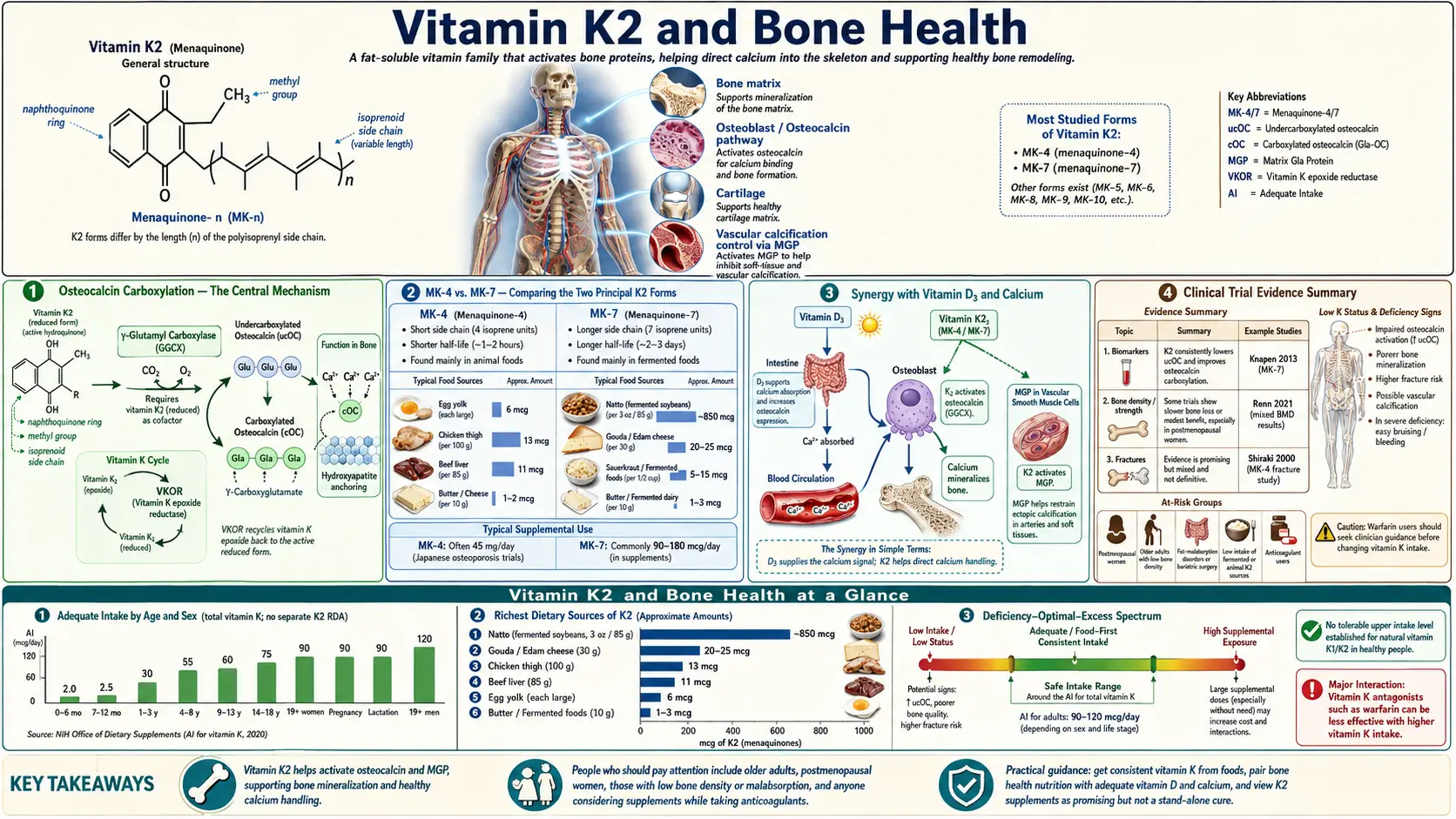
Vitamin K acts on bone mainly through osteocalcin, a protein made by osteoblasts whose three vitamin K-dependent Gla residues are critical for its structure, which appears to regulate the maturation of bone mineral (Gundberg 2012). The two natural forms behave differently in the body: in a comparison of supplements, vitamin K1 (phylloquinone) and the long-chain menaquinone MK-7, one of the vitamin K2 forms, were both absorbed well, but MK-7 had a very long half-life and accumulated in the blood during daily intake (Schurgers 2007). The clinical picture is mixed. In Japan, MK-4 (menatetrenone) at a pharmacological 45 mg a day is used to treat osteoporosis, with trial reports of fewer fractures, but several papers from that evidence base have since been retracted, among them the Yamaguchi Osteoporosis Prevention Study, and the largest Japanese trial, the OF study, found no significant reduction in new vertebral fractures overall (Inoue 2009). A 2019 systematic review found no evidence that vitamin K affects bone mineral density or vertebral fractures in postmenopausal or osteoporotic patients, and judged the evidence that it reduces clinical fractures insufficient (Mott 2019). In Western populations, nutritional-dose MK-7 produced one positive three-year bone-density result and two neutral results (one year in Norway, three years in Denmark), and high-dose K1 did not slow bone-density loss. This page sets out the mechanism, the Japanese and Western data, and the cautions side by side.
Table of Contents
- Osteocalcin Carboxylation — The Central Mechanism
- K2 vs. K1 for Bone Health
- MK-4 vs. MK-7 — Comparing the Two Principal K2 Forms
- Osteoporosis and Fracture Evidence
- Vitamin D3, Calcium and Vitamin K Together
- Japanese Natto Studies and Population Evidence
- Clinical Trial Evidence Summary
- Cautions & Contraindications
- Key Research Papers
- Connections
- Featured Videos
1. Osteocalcin Carboxylation — The Central Mechanism
Osteocalcin (also called bone Gla protein, BGP) is the principal vitamin K-dependent protein in bone and the protein through which most of vitamin K's proposed bone effects are explained.
- Osteocalcin Structure and Synthesis: Osteocalcin is a small protein made by osteoblasts (bone-building cells). It is deposited into bone or released into the circulation, where its level correlates with histological measures of bone formation (Gundberg 2012). In rat osteosarcoma cells, physiological levels of the active form of vitamin D (1,25-dihydroxyvitamin D3) raised osteocalcin production about 6-fold (Price & Baukol 1980), a biochemical link between the two vitamins. Osteocalcin contains three glutamic acid residues that require vitamin K-dependent gamma-carboxylation (Hauschka 1986).
- Gamma-Carboxylation of Osteocalcin: Using the reduced form of vitamin K, the enzyme gamma-glutamyl carboxylase converts glutamate (Glu) residues in vitamin K-dependent proteins to gamma-carboxyglutamate (Gla) residues (Cirilli 2022). In osteocalcin, the three Gla residues line up in a calcium-dependent helix that helps the protein adsorb to hydroxyapatite, the mineral of bone (Hauschka 1986).
- Calcium Binding to Hydroxyapatite: Carboxylated osteocalcin binds calcium and hydroxyapatite, and its structure appears to regulate the maturation of bone mineral (Gundberg 2012). How this works in living bone is not fully worked out: a 2012 review concluded that the specific function of osteocalcin in bone and glucose metabolism has yet to be elucidated (Gundberg 2012).
- Undercarboxylated Osteocalcin (ucOC): Osteocalcin that has not been fully carboxylated is called undercarboxylated osteocalcin (ucOC); in rats given the vitamin K antagonist warfarin, the non-carboxylated osteocalcin that was secreted could not bind hydroxyapatite (Price 1989). The percentage of circulating osteocalcin that is not carboxylated (percent ucOC) is used as a biomarker of vitamin K status; when ucOC is not corrected for total osteocalcin, it also reflects bone-formation activity independent of vitamin K (Gundberg 2012). In elderly French women, ucOC also tracked poor vitamin D status (Szulc 1993).
- ucOC/OC Ratio: No single biomarker is considered a gold standard for vitamin K status; the undercarboxylated fractions of three vitamin K-dependent proteins can be measured in blood, change with supplementation and are only modestly correlated with each other (Shea & Booth 2016). Vitamin K supplements lower ucOC dependably, including MK-7 at 100 micrograms a day or more (Inaba 2015; Emaus 2010). Observational studies using percent ucOC led to the conclusion that vitamin K insufficiency leads to age-related bone loss, but clinical trials do not provide overall support for the suggestion that vitamin K supplementation of the general population will reduce bone loss or fracture risk (Gundberg 2012).
2. K2 vs. K1 for Bone Health
Vitamin K1 and K2 share the same carboxylation chemistry but differ in where they go in the body and how long they stay there.
- Absorption of K1 and MK-7: Vitamin K is a cofactor in the production of blood coagulation factors in the liver and of osteocalcin in bone (Schurgers 2007). In a comparison in healthy volunteers, K1 and MK-7 were both absorbed well, with peak serum concentrations about 4 hours after intake (Schurgers 2007).
- Half-Life of MK-7: In the same comparison, MK-7 had a very long half-life, giving much more stable serum levels than K1 and 7- to 8-fold higher levels during prolonged intake, and it produced more complete carboxylation of osteocalcin (Schurgers 2007). After a single 420-microgram dose, MK-7 was still detectable in serum up to 48 hours later; the authors suggested that MK-7 may therefore be of particular importance for tissues outside the liver (Sato 2012).
- Direct Osteoblast Effects: In laboratory studies, vitamin K2 bound to and activated the steroid and xenobiotic receptor (SXR, called PXR in mice), a nuclear receptor, and increased osteoblast marker genes in osteosarcoma cells; it induced bone markers in bone cells from normal mice but not in cells from mice lacking PXR (Tabb 2003). These are cell and mouse findings, separate from osteocalcin carboxylation; whether they matter in people at the doses studied is not established.
- Osteoclast Inhibition: In cell studies, vitamin K2 stimulated osteoblast formation and suppressed osteoclast formation by down-regulating NF-κB activation, in a way that did not depend on gamma-carboxylation (Yamaguchi 2011). This dual action has been described in cultured cells, not demonstrated in human bone.
- Epidemiological Evidence: Within Japan, serum MK-7 in postmenopausal women was higher in Tokyo, where natto (rich in MK-7) is eaten frequently, than in Hiroshima, where it is seldom eaten, and hip-fracture incidence in women was inversely correlated with natto consumption across the country's prefectures (Kaneki 2001). This compares regions rather than individuals, so it cannot show that natto or K2 caused the difference (see section 6).
3. MK-4 vs. MK-7 — Comparing the Two Principal K2 Forms
MK-4 and MK-7 are the two most studied forms of vitamin K2, and they behave so differently that their doses cannot be compared directly.
- MK-4 Characteristics: MK-4 (menaquinone-4) is a short-chain menaquinone measured in US meat, eggs and dairy foods, none of which was a rich source of it (Elder 2006). Human tissues also make it from other forms of vitamin K; the enzyme responsible, UBIAD1, was identified in 2010 and is not blocked by warfarin (Nakagawa 2010). At nutritional doses MK-4 barely reaches the blood: a single 420-microgram dose was not detectable in serum at any time point, and 60 micrograms daily for 7 days did not raise serum MK-4 (Sato 2012). In Japan, MK-4 is used as an osteoporosis drug at 45 mg/day (Shiraki 2000); the Japanese OF study used 15-mg capsules (Inoue 2009).
- MK-7 Characteristics: MK-7 (menaquinone-7) is a long-chain menaquinone produced by fermentation or chemically (Cirilli 2022); natto, a Japanese fermented soybean food, contains a large amount of it (Kaneki 2001). Because of its long half-life, daily intake builds up serum levels (Schurgers 2007). In Japanese dose-finding trials, MK-7 increased the ratio of carboxylated to undercarboxylated osteocalcin dose-dependently, with significant effects at 100 and 200 micrograms a day; the authors suggested that 100 micrograms a day or more improves osteocalcin carboxylation (Inaba 2015).
- Osteocalcin Carboxylation Efficiency: Head-to-head trials of MK-7 and MK-4 at equal doses with osteocalcin as the outcome are scarce. In the bioavailability comparison, MK-7 at nutritional doses raised serum MK-7 in all subjects, while MK-4 at the same doses did not appear in serum (Sato 2012). At the pharmacological 45 mg/day dose, MK-4 lowered undercarboxylated osteocalcin compared with no treatment (Shiraki 2000).
- Dose Ranges Studied: The two forms are studied at very different doses: MK-7 in the microgram range (50 to 200 micrograms a day in the Japanese carboxylation dose-finding trial, 180 to 375 micrograms a day in the bone trials on this page), MK-4 in the milligram range (45 mg a day in the Japanese regimen).
- Complementary Roles: The receptor-signalling findings (SXR/PXR) come from cell and mouse studies, and the osteocalcin-carboxylation findings from human trials; none of the trials on this page tested MK-4 and MK-7 in combination.
4. Osteoporosis and Fracture Evidence
The clinical evidence on vitamin K, osteoporosis and fractures spans Japanese drug-dose trials, Western nutritional-dose trials and observational cohorts, and the findings do not all agree.
- Japanese MK-4 Trials: In an open-label randomized trial, Shiraki and colleagues (2000) assigned 241 patients with osteoporosis to 45 mg/day MK-4 or no treatment for two years: at 24 months lumbar bone mineral density had changed by −0.5% in the treated group and −3.3% in the control group, and clinical fractures were less frequent with treatment. Cockayne and colleagues (2006) pooled the 7 randomized trials that reported fracture data — all of them Japanese and all using menaquinone — and reported substantially lower odds of vertebral, hip and nonvertebral fractures. In 2018 the authors and the journal published a caution to readers: 3 of the included articles came from a single centre whose trial data have since been found to be problematic or retracted, and readers were asked to take into account only a sensitivity analysis that excluded them; the original pooled fracture figures therefore carry an expression of concern (notice PubMed PMID: 29582046). The same group's 2019 update (36 studies of oral vitamin K), written after the integrity of some of the earlier evidence had been questioned, concluded that for postmenopausal or osteoporotic patients there is no evidence that vitamin K affects bone mineral density or vertebral fractures, and that it may reduce clinical fractures but the evidence is insufficient to confirm this (Mott 2019). A 2020 correction removed the Yamaguchi Osteoporosis Prevention Study (YOPS; Ishida 2004), after concerns were raised about its integrity (Avenell 2020), and recalculated every estimate that had included it: the odds ratio for any clinical fracture became 0.74 (95% CI 0.56–0.97), the low-risk-of-bias estimate stayed at 0.76 (0.58–1.01), and vertebral fractures showed no difference (0.98, 0.85–1.14); the authors stated that the correction did not change their conclusions (Mott 2020 correction). The YOPS report itself was retracted in 2025 (notice PubMed PMID: 40222753). Several other vitamin K2 bone papers from one Japanese author group have also been retracted: a 2005 trial of menatetrenone with vitamin D2 for nonvertebral fractures in elderly women with Alzheimer's disease (Sato 2005, Bone, retracted 2018), a 2011 trial of menatetrenone with risedronate for hip-fracture prevention in elderly patients with Alzheimer disease, retracted “because of scientific misconduct” (Sato 2011, Kurume Medical Journal), and a 2004 review of vitamin K2 and osteoporosis, retracted in 2021 because its text, data and some figures came from sources that had been retracted or were under investigation for data fabrication and falsification, among other reasons (Iwamoto 2004). None of these retracted papers is relied on here. The largest menatetrenone trial, the multicentre Japanese OF study, randomized osteoporotic postmenopausal women aged 50 or older to calcium alone (2,193 women) or calcium plus menatetrenone (2,185 women): the rate of new vertebral fractures over 36 months, the primary endpoint, did not differ significantly, and the lower 48-month rate of new clinical fractures with menatetrenone was not significant; a lower risk of new vertebral fractures was seen in the subgroup with at least five vertebral fractures at baseline (Inoue 2009).
- MK-7 and Bone Loss: In a three-year randomized, placebo-controlled trial, 244 healthy postmenopausal women took 180 micrograms of MK-7 a day or placebo. MK-7 improved vitamin K status and significantly decreased the age-related decline in bone mineral content and density at the lumbar spine and femoral neck, but not at the total hip; femoral-neck bone-strength indices were favourably affected, and loss of vertebral height in the lower thoracic spine was smaller (Knapen 2013). A one-year trial of 360 micrograms of MK-7 a day (as natto capsules) in 334 early-menopausal Norwegian women lowered ucOC but found no difference in bone loss rates at the total hip or any other site (Emaus 2010). An earlier three-year trial by Knapen's group used the pharmacological MK-4 dose (45 mg/day) in 325 postmenopausal women: it did not change DXA bone density, but bone mineral content and femoral-neck width increased relative to placebo, and hip bone strength was maintained while it declined in the placebo group (Knapen 2007). In Denmark, 375 micrograms of MK-7 a day, added to calcium and vitamin D in 148 postmenopausal women with osteopenia, preserved trabecular bone microarchitecture at the tibia over 12 months compared with placebo, with no difference in bone density at any site (Rønn 2016); over 3 years, in 142 such women, bone density fell at all sites with no difference between the MK-7 and placebo groups, and bone turnover markers and microarchitecture changed similarly in both (Rønn 2021).
- Epidemiological Fracture Data: Two large US cohorts reported an inverse association between dietary vitamin K intake (mostly K1) and hip fracture. In the Nurses' Health Study (72,327 women aged 38–63, 10 years, 270 hip fractures), women in quintiles 2–5 of vitamin K intake had a lower age-adjusted relative risk of hip fracture (RR 0.70; 95% CI 0.53–0.93) than women in the lowest quintile (below 109 micrograms a day); risk did not decrease further between quintiles 2 and 5 (Feskanich 1999). In the Framingham Heart Study (335 men and 553 women, average age 75), people in the highest quartile of vitamin K intake (median 254 micrograms a day) had a lower fully adjusted relative risk of hip fracture (RR 0.35; 95% CI 0.13–0.94) than those in the lowest quartile (median 56 micrograms a day); vitamin K intake was not associated with bone mineral density (Booth 2000). These are observational associations and do not show that vitamin K caused the difference. In the ECKO trial, 440 postmenopausal women with osteopenia took 5 mg/day of vitamin K1 or placebo for 2 to 4 years: bone mineral density, the primary outcome, did not differ between the groups; fewer women in the vitamin K group had clinical fractures (9 versus 20), a secondary outcome the trial was not powered to examine (Cheung 2008).
- Bone Quality vs. Bone Density: In some MK-4 trials, effects on fractures or on bone-strength indices were reported without an increase in measured bone mineral density (Shiraki 2000; Knapen 2007). Knapen and colleagues concluded that MK-4 helped maintain femoral-neck bone strength by improving bone mineral content and femoral-neck width while having little effect on DXA bone density (Knapen 2007). In the Danish MK-7 trial, the microarchitecture difference seen at 12 months (Rønn 2016) was not present at 3 years (Rønn 2021).
- Combination with Standard Therapy: None of the trials described here tested vitamin K2 as a replacement for standard osteoporosis drugs such as bisphosphonates, denosumab or teriparatide. The trials enrolled postmenopausal women (healthy, osteopenic or osteoporotic) and Japanese osteoporosis patients, and the Japanese MK-4 regimen is itself a prescription drug.
5. Vitamin D3, Calcium and Vitamin K Together
Vitamin D, calcium and vitamin K act at different steps of the same mineralization pathway, which is why they are often studied together.
- Vitamin D3 Stimulates Osteocalcin Production: The active form of vitamin D3, 1,25-dihydroxyvitamin D3, stimulates the synthesis of both osteocalcin and matrix Gla protein in osteoblastic cells (Price 1989); in rat osteosarcoma cells, physiological levels raised osteocalcin production about 6-fold (Price & Baukol 1980).
- Vitamin K Activates Osteocalcin: Osteocalcin needs vitamin K-dependent gamma-carboxylation to bind hydroxyapatite: the non-carboxylated osteocalcin made by warfarin-treated rats could not bind it (Price 1989).
- Calcium Provides the Mineral Substrate: Osteocalcin is a calcium-binding protein of the bone matrix (Hauschka 1986). Several trials on this page gave calcium (with or without vitamin D) to both groups, so vitamin K was tested as an addition to it (Inoue 2009; Rønn 2021).
- The Complete Pathway: In the mechanistic model, (1) vitamin D3 stimulates osteocalcin production, (2) vitamin K activates osteocalcin through gamma-carboxylation, and (3) carboxylated osteocalcin binds hydroxyapatite in bone. This is a description of the biochemistry; the trials in sections 4 and 7 test whether adding vitamin K changes bone outcomes.
- The Vitamin D Without Vitamin K Hypothesis: A 2007 paper in Medical Hypotheses proposed that vitamin D toxicity works by inducing a vitamin K deficiency: vitamin D increases the production of proteins that need vitamin K-dependent carboxylation, and as that demand rises the vitamin K pool is depleted; the author cited animal studies in which soft-tissue calcification and other effects of excess vitamin D could be separated from high blood calcium, and noted that the idea still needed confirmation in animal experiments (Masterjohn 2007). Matrix Gla protein is one of the vitamin K-dependent proteins that inhibit vascular calcification, and warfarin leaves it uncarboxylated (Tesfamariam 2019). None of the trials on this page tested the hypothesis.
- Combined-Therapy Trial Data: In a two-year randomized trial in 172 Japanese postmenopausal women with osteopenia or osteoporosis, vertebral bone density rose 4.92% with combined vitamin K2 and D3 therapy, compared with 0.135% with K2 alone, and no adverse reactions were observed (Ushiroyama 2002). The ECKO participants were vitamin D replete at baseline (mean 25-hydroxyvitamin D 77 nmol/L), and high-dose K1 still did not change their bone density (Cheung 2008).
6. Japanese Natto Studies and Population Evidence
Much of the interest in vitamin K2 for bone grew out of population observations in Japan centred on natto.
- Natto and MK-7: Natto is a Japanese fermented soybean food that contains a large amount of MK-7. It is eaten frequently in eastern Japan (Tokyo) but seldom in western Japan (Hiroshima) (Kaneki 2001).
- Geographic Fracture Correlation: Kaneki and colleagues measured serum MK-7 of 5.26 ng/mL in postmenopausal women in Tokyo, 1.22 ng/mL in Hiroshima and 0.37 ng/mL in British women. Frequent natto eaters had significantly higher serum MK-7, and eating natto produced a marked, sustained rise. Across Japan's prefectures, hip-fracture incidence in women was inversely correlated with natto consumption. The authors suggested that higher MK-7 from natto may contribute to the relatively lower fracture risk of Japanese women (Kaneki 2001). A correlation between regions cannot separate natto from the other ways those regions differ.
- Serum ucOC and Fracture Risk: The Kaneki study measured serum MK-7, not ucOC. The link between ucOC and fracture comes from prospective studies in elderly French women: higher ucOC predicted hip fracture in institutionalized women (Szulc 1993), and in the EPIDOS cohort of independently living women over 75 it still predicted hip fracture after adjustment for femoral-neck bone density and gait speed (odds ratio 1.8; 95% CI 1.0–3.0) (Vergnaud 1997).
- International Comparisons: Japan and Western countries differ in many ways besides natto, so comparisons between whole populations cannot isolate the contribution of MK-7 from natto.
- Clinical Relevance: MK-7 supplements contain the same menaquinone found in natto. When it was tested directly, MK-7 given as natto capsules (360 micrograms a day) to Norwegian women for one year lowered ucOC but did not change bone loss rates (Emaus 2010), the three-year Dutch MK-7 trial found less bone loss at the spine and femoral neck (Knapen 2013), and a three-year Danish MK-7 trial found no effect on bone density (Rønn 2021).
7. Clinical Trial Evidence Summary
The clinical trial evidence for vitamin K in bone health is not consistent; it differs by vitamin K form, dose, population and outcome.
- MK-4 Trials: All 7 trials with fracture data in the 2006 meta-analysis were Japanese and used menaquinone (Cockayne 2006). That meta-analysis reported large fracture reductions, but a 2018 notice cautioned that it had included problematic trials, and the 2019 update found no difference in vertebral fractures and judged the evidence on clinical fractures insufficient (see section 4). The largest menatetrenone trial, the OF study, found no significant reduction in new vertebral fractures overall (Inoue 2009), and the Yamaguchi Osteoporosis Prevention Study and several papers from one Japanese author group have been retracted (see section 4). A 2013 review of menatetrenone by Iwamoto and Sato (Expert Opinion on Pharmacotherapy 14(4): 449–458) was retracted in 2019 and is not relied on here.
- MK-7 Trials: The three-year Knapen trial (180 micrograms a day) found less decline in bone mineral content and density at the lumbar spine and femoral neck, but not the total hip (Knapen 2013); the one-year Emaus trial (360 micrograms a day) found no effect on bone loss (Emaus 2010), and the three-year Danish trial (375 micrograms a day with calcium and vitamin D) found no effect on bone density, bone turnover markers or microarchitecture (Rønn 2021). Shorter trials found that MK-7 lowers ucOC and improves osteocalcin carboxylation (Inaba 2015). None of these MK-7 trials reported fewer fractures.
- K1 Evidence: The ECKO trial of vitamin K1 (5 mg/day) did not protect bone mineral density, its primary outcome; fewer women in the vitamin K group had clinical fractures (9 versus 20), a secondary outcome the authors said the study was not powered to examine (Cheung 2008).
- Other Meta-Analyses: A 2015 meta-analysis of 19 randomized trials (6,759 participants) of vitamin K2 in postmenopausal women found improved vertebral bone density in the subgroup with osteoporosis but no significant difference in the subgroup without it. Pooled across seven studies, fracture incidence did not differ significantly (RR 0.63, p = 0.08); after excluding the study causing heterogeneity, the difference became significant (RR 0.50). The vitamin K2 groups had a higher rate of adverse reactions that did not reach statistical significance (RR 1.22, p = 0.06), and the authors called for further high-quality, large randomized trials (Huang 2015). That analysis predates the 2018 caution and the 2020 correction described in section 4.
- Gaps in the Evidence: The 2019 review found too few trials to draw conclusions for patient groups other than postmenopausal or osteoporotic patients (Mott 2019), and the authors of the three-year MK-7 trial noted that whether their results apply to other populations, such as children and men, needs further investigation (Knapen 2013).
- Summary: The research supports vitamin K's role in osteocalcin carboxylation and shows that supplements lower ucOC. Fracture prevention by nutritional-dose K2 has not been demonstrated; the fracture data come mainly from pharmacological-dose MK-4 trials in Japan, part of which has since been retracted or flagged, and the largest of which found no overall reduction in new vertebral fractures (Inoue 2009). The trials enrolled mainly postmenopausal women.
Cautions & Contraindications
- Warfarin users: Warfarin is a vitamin K epoxide reductase inhibitor (Grysiewicz 2014), and acenocoumarol is another vitamin K antagonist anticoagulant; supplemental vitamin K2 can lower the INR of people taking these drugs (Theuwissen 2013). The authors of a K1 and MK-7 comparison noted that preparations supplying 50 micrograms a day or more of MK-7 may interfere with oral anticoagulant treatment in a clinically relevant way (Schurgers 2007). In 15 healthy volunteers stabilized on acenocoumarol, 45 micrograms of MK-7 a day lowered the mean INR by about 40%, and 10 and 20 micrograms a day were judged to cause a clinically relevant lowering of the INR in at least 40% and 60% of subjects; the authors concluded that MK-7 supplements need to be avoided in patients receiving vitamin K antagonist therapy (Theuwissen 2013). The direct oral anticoagulants work differently: dabigatran is a direct thrombin inhibitor, and rivaroxaban and apixaban are factor Xa inhibitors (Grysiewicz 2014). Decisions about anticoagulants and supplements belong with the prescribing clinician.
- Active bisphosphonate or denosumab therapy: No trial on this page tested vitamin K2 as a substitute for prescribed osteoporosis drugs, and the evidence does not establish K2 as a stand-alone fracture-prevention therapy for people with diagnosed osteoporosis or a prior fragility fracture; at most it has been studied alongside standard care.
- Vitamin D status: Vitamin D increases osteocalcin production, the substrate that vitamin K acts on. Low vitamin D status was associated with higher ucOC in elderly women (Szulc 1993). The trials on this page did not establish a vitamin D level below which K2 has no effect on bone.
- Cis-isomer MK-7: MK-7 exists as trans and cis isomers. In a laboratory study, trans MK-7 showed greater carboxylating activity, and cis MK-7 only slight activity; the authors noted that the impact of the isomers on humans is unknown (Cirilli 2022). In a crossover study in healthy adults, a synthetic MK-7 was bioequivalent to fermentation-derived MK-7 (Møller 2017).
- Soy allergy: Natto and natto-derived MK-7 come from fermented soybeans. The studies on this page did not report on soy-allergic people; MK-7 can also be made chemically (Cirilli 2022), and a synthetic MK-7 was bioequivalent to fermentation-derived MK-7 in the crossover study above (Møller 2017).
- Calcium supplementation context: The idea that calcium supplements taken without extra vitamin K2 favour arterial calcification rests on the mechanism described in section 5 (matrix Gla protein, a vitamin K-dependent inhibitor of vascular calcification; Tesfamariam 2019); none of the trials on this page tested it. Food sources of calcium are covered on the Calcium page.
- High-dose MK-4 (45 mg/day Japanese osteoporosis protocol): This is the drug dose used in the Japanese trials (Shiraki 2000), far above the microgram doses of MK-4 studied as nutrition (Sato 2012).
- Tolerability: In ECKO, there were no significant differences in adverse effects between 5 mg/day of vitamin K1 and placebo over 2 to 4 years (Cheung 2008); the 2015 meta-analysis found a higher adverse-reaction rate with K2 that did not reach statistical significance (RR 1.22, p = 0.06) (Huang 2015); and in the OF study, adverse events and adverse reactions were more frequent with calcium plus menatetrenone than with calcium alone (Inoue 2009). The interaction with vitamin K antagonist anticoagulants is described above.
- Pregnancy: The bone trials described on this page enrolled postmenopausal women and osteoporosis patients, so they give no information on K2 supplements in pregnancy or breastfeeding.
Key Research Papers
- Knapen MHJ, Drummen NE, Smit E, Vermeer C, Theuwissen E (2013). Three-year low-dose menaquinone-7 supplementation helps decrease bone loss in healthy postmenopausal women. Osteoporosis International 24(9): 2499–2507. — doi:10.1007/s00198-013-2325-6
- Shiraki M, Shiraki Y, Aoki C, Miura M (2000). Vitamin K2 (menatetrenone) effectively prevents fractures and sustains lumbar bone mineral density in osteoporosis. Journal of Bone and Mineral Research 15(3): 515–521. — doi:10.1359/jbmr.2000.15.3.515
- Knapen MHJ, Schurgers LJ, Vermeer C (2007). Vitamin K2 supplementation improves hip bone geometry and bone strength indices in postmenopausal women. Osteoporosis International 18(7): 963–972. — doi:10.1007/s00198-007-0337-9
- Cockayne S, Adamson J, Lanham-New S, Shearer MJ, Gilbody S, Torgerson DJ (2006). Vitamin K and the prevention of fractures: systematic review and meta-analysis of randomized controlled trials. Archives of Internal Medicine 166(12): 1256–1261. — doi:10.1001/archinte.166.12.1256 — Expression of concern (JAMA Intern Med 2018: PubMed PMID: 29582046)
- Torgerson DJ (2018). Caution to readers about systematic review on vitamin K and prevention of fractures that included problematic trials. JAMA Internal Medicine 178(6): 863–864. — PubMed PMID: 29582044
- Mott A, Bradley T, Wright K, Cockayne ES, Shearer MJ, Adamson J, Lanham-New SA, Torgerson DJ (2019). Effect of vitamin K on bone mineral density and fractures in adults: an updated systematic review and meta-analysis of randomised controlled trials. Osteoporosis International 30(8): 1543–1559. — PubMed PMID: 31076817
- Mott A, Bradley T, Wright K, Cockayne ES, Shearer MJ, Adamson J, Lanham-New SA, Torgerson DJ (2020). Correction to: Effect of vitamin K on bone mineral density and fractures in adults: an updated systematic review and meta-analysis of randomised controlled trials. Osteoporosis International 31(11): 2269–2270. — PubMed PMID: 32965511
- Kaneki M, Hodges SJ, Hosoi T, Fujiwara S, Lyons A, Crean SJ, Ishida N, Nakagawa M, Takechi M, Sano Y, Mizuno Y, Hoshino S, Miyao M, Inoue S, Horiki K, Shiraki M, Ouchi Y, Orimo H (2001). Japanese fermented soybean food as the major determinant of the large geographic difference in circulating levels of vitamin K2: possible implications for hip-fracture risk. Nutrition 17(4): 315–321. — doi:10.1016/s0899-9007(00)00554-2
- Feskanich D, Weber P, Willett WC, Rockett H, Booth SL, Colditz GA (1999). Vitamin K intake and hip fractures in women: a prospective study. American Journal of Clinical Nutrition 69(1): 74–79. — doi:10.1093/ajcn/69.1.74
- Cheung AM, Tile L, Lee Y, Tomlinson G, Hawker G, Scher J, Hu H, Vieth R, Thompson L, Jamal S, Josse R (2008). Vitamin K supplementation in postmenopausal women with osteopenia (ECKO trial): a randomized controlled trial. PLoS Medicine 5(10): e196. — doi:10.1371/journal.pmed.0050196
- Sato T, Schurgers LJ, Uenishi K (2012). Comparison of menaquinone-4 and menaquinone-7 bioavailability in healthy women. Nutrition Journal 11: 93. — doi:10.1186/1475-2891-11-93
- Inaba N, Sato T, Yamashita T (2015). Low-dose daily intake of vitamin K2 (menaquinone-7) improves osteocalcin gamma-carboxylation: a double-blind, randomized controlled trial. Journal of Nutritional Science and Vitaminology 61(6): 471–480. — doi:10.3177/jnsv.61.471
- Yamaguchi M, Weitzmann MN (2011). Vitamin K2 stimulates osteoblastogenesis and suppresses osteoclastogenesis by suppressing NF-κB activation. International Journal of Molecular Medicine 27(1): 3–14. — doi:10.3892/ijmm.2010.562
- Iwamoto J, Sato Y (2013). Menatetrenone for the treatment of osteoporosis. Expert Opinion on Pharmacotherapy 14(4): 449–458. — PubMed PMID: 23346882 — Retracted (retraction notice: PubMed PMID: 31437064)
- Booth SL, Tucker KL, Chen H, Hannan MT, Gagnon DR, Cupples LA, Wilson PWF, Ordovas J, Schaefer EJ, Dawson-Hughes B, Kiel DP (2000). Dietary vitamin K intakes are associated with hip fracture but not with bone mineral density in elderly men and women. American Journal of Clinical Nutrition 71(5): 1201–1208. — doi:10.1093/ajcn/71.5.1201
- Huang ZB, Wan SL, Lu YJ, Ning L, Liu C, Fan SW (2015). Does vitamin K2 play a role in the prevention and treatment of osteoporosis for postmenopausal women: a meta-analysis of randomized controlled trials. Osteoporosis International 26(3): 1175–1186. — doi:10.1007/s00198-014-2989-6
- Tabb MM, Sun A, Zhou C, Grun F, Errandi J, Romero K, Pham H, Inoue S, Mallick S, Lin M, Forman BM, Blumberg B (2003). Vitamin K2 regulation of bone homeostasis is mediated by the steroid and xenobiotic receptor SXR. Journal of Biological Chemistry 278(45): 43919–43927. — doi:10.1074/jbc.M303136200
- Gundberg CM, Lian JB, Booth SL (2012). Vitamin K-dependent carboxylation of osteocalcin: friend or foe? Advances in Nutrition 3(2): 149–157. — PubMed PMID: 22516722
- Shea MK, Booth SL (2016). Concepts and controversies in evaluating vitamin K status in population-based studies. Nutrients 8(1): 8. — PubMed PMID: 26729160
- Schurgers LJ, Teunissen KJ, Hamulyák K, Knapen MH, Vik H, Vermeer C (2007). Vitamin K-containing dietary supplements: comparison of synthetic vitamin K1 and natto-derived menaquinone-7. Blood 109(8): 3279–3283. — PubMed PMID: 17158229
- Emaus N, Gjesdal CG, Almås B, Christensen M, Grimsgaard AS, Berntsen GK, Salomonsen L, Fønnebø V (2010). Vitamin K2 supplementation does not influence bone loss in early menopausal women: a randomised double-blind placebo-controlled trial. Osteoporosis International 21(10): 1731–1740. — PubMed PMID: 19937427
- Nakagawa K, Hirota Y, Sawada N, Yuge N, Watanabe M, Uchino Y, Okuda N, Shimomura Y, Suhara Y, Okano T (2010). Identification of UBIAD1 as a novel human menaquinone-4 biosynthetic enzyme. Nature 468(7320): 117–121. — PubMed PMID: 20953171
- Ushiroyama T, Ikeda A, Ueki M (2002). Effect of continuous combined therapy with vitamin K2 and vitamin D3 on bone mineral density and coagulofibrinolysis function in postmenopausal women. Maturitas 41(3): 211–221. — PubMed PMID: 11886767
- Szulc P, Chapuy MC, Meunier PJ, Delmas PD (1993). Serum undercarboxylated osteocalcin is a marker of the risk of hip fracture in elderly women. Journal of Clinical Investigation 91(4): 1769–1774. — PubMed PMID: 8473517
- Vergnaud P, Garnero P, Meunier PJ, Bréart G, Kamihagi K, Delmas PD (1997). Undercarboxylated osteocalcin measured with a specific immunoassay predicts hip fracture in elderly women: the EPIDOS Study. Journal of Clinical Endocrinology and Metabolism 82(3): 719–724. — PubMed PMID: 9062471
- Theuwissen E, Teunissen KJ, Spronk HM, Hamulyák K, Ten Cate H, Shearer MJ, Vermeer C, Schurgers LJ (2013). Effect of low-dose supplements of menaquinone-7 (vitamin K2) on the stability of oral anticoagulant treatment: dose-response relationship in healthy volunteers. Journal of Thrombosis and Haemostasis 11(6): 1085–1092. — PubMed PMID: 23530987
- Cirilli I, Orlando P, Silvestri S, Marcheggiani F, Dludla PV, Kaesler N, Tiano L (2022). Carboxylative efficacy of trans and cis MK7 and comparison with other vitamin K isomers. BioFactors 48(5): 1129–1136. — PubMed PMID: 35583412
- Møller M, Gjelstad IMF, Baksaas I, Grande T, Aukrust IR, Drevon CA (2017). Bioavailability and chemical/functional aspects of synthetic MK-7 vs fermentation-derived MK-7 in randomised controlled trials. International Journal for Vitamin and Nutrition Research 87(5–6): 1–15. — PubMed PMID: 27374276
- Ishida Y, Kawai S (2004). Comparative efficacy of hormone replacement therapy, etidronate, calcitonin, alfacalcidol, and vitamin K in postmenopausal women with osteoporosis: the Yamaguchi Osteoporosis Prevention Study. American Journal of Medicine 117(8): 549–555. — PubMed PMID: 15465502 — Retracted (retraction notice: PubMed PMID: 40222753)
- Avenell A, Grey A, Gamble GD, Bolland MJ (2020). Concerns about the integrity of the Yamaguchi Osteoporosis Prevention Study (YOPS) report, Am J Med. 2004;117:549-555. American Journal of Medicine 133(6): e311–e314. — PubMed PMID: 32532372
- Sato Y, Kanoko T, Satoh K, Iwamoto J (2005). Menatetrenone and vitamin D2 with calcium supplements prevent nonvertebral fracture in elderly women with Alzheimer's disease. Bone 36(1): 61–68. — PubMed PMID: 15664003 — Retracted (retraction notice: PubMed PMID: 29150013)
- Sato Y, Honda Y, Umeno K, Hayashida N, Iwamoto J, Takeda T, Matsumoto H (2011). The prevention of hip fracture with menatetrenone and risedronate plus calcium supplementation in elderly patients with Alzheimer disease: a randomized controlled trial. Kurume Medical Journal 57(4): 117–124. — PubMed PMID: 21778673 — Retracted (retraction notice: PubMed PMID: 28408729)
- Iwamoto J, Takeda T, Sato Y (2004). Effects of vitamin K2 on osteoporosis. Current Pharmaceutical Design 10(21): 2557–2576. — PubMed PMID: 15320745 — Retracted (retraction notice: PubMed PMID: 34264184)
- Inoue T, Fujita T, Kishimoto H, Makino T, Nakamura T, Nakamura T, Sato T, Yamazaki K (2009). Randomized controlled study on the prevention of osteoporotic fractures (OF study): a phase IV clinical study of 15-mg menatetrenone capsules. Journal of Bone and Mineral Metabolism 27(1): 66–75. — PubMed PMID: 19082528
- Rønn SH, Harsløf T, Pedersen SB, Langdahl BL (2016). Vitamin K2 (menaquinone-7) prevents age-related deterioration of trabecular bone microarchitecture at the tibia in postmenopausal women. European Journal of Endocrinology 175(6): 541–549. — PubMed PMID: 27625301
- Rønn SH, Harsløf T, Oei L, Pedersen SB, Langdahl BL (2021). The effect of vitamin MK-7 on bone mineral density and microarchitecture in postmenopausal women with osteopenia, a 3-year randomized, placebo-controlled clinical trial. Osteoporosis International 32(1): 185–191. — PubMed PMID: 33030563
- Price PA, Baukol SA (1980). 1,25-Dihydroxyvitamin D3 increases synthesis of the vitamin K-dependent bone protein by osteosarcoma cells. Journal of Biological Chemistry 255(24): 11660–11663. — PubMed PMID: 6969260
- Hauschka PV (1986). Osteocalcin: the vitamin K-dependent Ca2+-binding protein of bone matrix. Haemostasis 16(3–4): 258–272. — PubMed PMID: 3530901
- Price PA (1989). Gla-containing proteins of bone. Connective Tissue Research 21(1–4): 51–57. — PubMed PMID: 2691199
- Masterjohn C (2007). Vitamin D toxicity redefined: vitamin K and the molecular mechanism. Medical Hypotheses 68(5): 1026–1034. — PubMed PMID: 17145139
- Tesfamariam B (2019). Involvement of vitamin K-dependent proteins in vascular calcification. Journal of Cardiovascular Pharmacology and Therapeutics 24(4): 323–333. — PubMed PMID: 30905168
- Elder SJ, Haytowitz DB, Howe J, Peterson JW, Booth SL (2006). Vitamin K contents of meat, dairy, and fast food in the U.S. diet. Journal of Agricultural and Food Chemistry 54(2): 463–467. — PubMed PMID: 16417305
- Grysiewicz R, Gorelick PB (2014). Incidence, mortality, and risk factors for oral anticoagulant-associated intracranial hemorrhage in patients with atrial fibrillation. Journal of Stroke and Cerebrovascular Diseases 23(10): 2479–2488. — PubMed PMID: 25440361
PubMed Topic Searches
- PubMed: Vitamin K2 and bone health
- PubMed: Osteocalcin carboxylation and Vitamin K
- PubMed: MK-7 and bone mineral density
- PubMed: MK-4 and osteoporosis
- PubMed: ucOC and fracture risk
- PubMed: Natto, K2, and fracture prevention
- PubMed: K2 + D3 + calcium for bone
- PubMed: Vitamin K in postmenopausal osteoporosis
- PubMed: Vitamin K2 and bone quality
- PubMed: Vitamin K and hip fracture risk
- PubMed: Knapen MK-7 bone trials
- PubMed: K2 effects on osteoblasts and osteoclasts
- PubMed: Menatetrenone 45 mg fracture (Japan)
- PubMed: SXR/PXR and Vitamin K2 in bone
Connections
- All Vitamins
- Vitamin K (Main Page)
- Vitamin K Benefits Hub
- K2 and Arterial Calcification
- MK-7 vs MK-4
- K2 and Insulin Sensitivity
- Vitamin K and Blood Clotting
- Vitamin D3
- Vitamin D3 and Bone Health
- Calcium
- Magnesium
- Osteoporosis
- Bisphosphonates
- Fermented Foods
- Collagen
- Natto
- Bacillus Subtilis
- Deficiency
- Toxicity
- History
- Sources