Vitamin D3 for Immune Function & Respiratory Infection
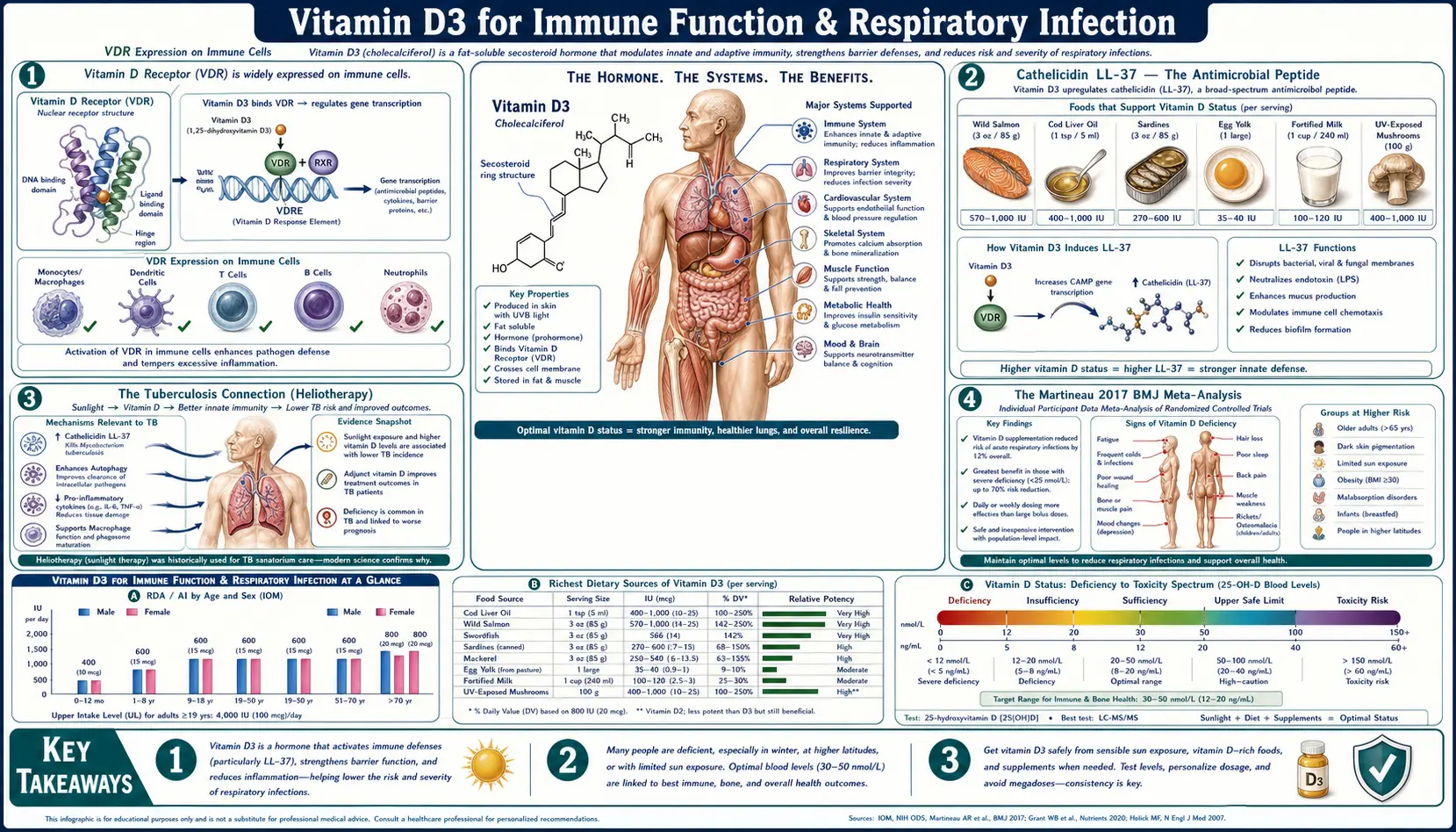
Vitamin D3 is studied for its role in immune defence against respiratory infection. In laboratory work, human macrophages activated through Toll-like receptors switched on the vitamin D receptor and the vitamin D-activating enzyme, produced the antimicrobial peptide cathelicidin (LL-37) and killed intracellular Mycobacterium tuberculosis. In trials, the 2017 Martineau individual-participant-data meta-analysis in BMJ (25 trials, 11,321 participants) found that vitamin D reduced the risk of acute respiratory infection overall (adjusted odds ratio 0.88), with the strongest protection among people on daily or weekly doses who started below 25 nmol/L. A 2021 update of 46 trials found a smaller reduction (odds ratio 0.92) and no significant effect in any subgroup defined by baseline vitamin D level. For COVID-19, a 76-patient pilot trial of calcifediol in Córdoba, Spain reported fewer intensive-care admissions, while a 240-patient double-blind trial of a single 200,000 IU dose and the 6,200-person CORONAVIT trial found no significant benefit.
Table of Contents
- VDR Expression on Immune Cells
- Cathelicidin LL-37 — The Antimicrobial Peptide
- The Tuberculosis Connection
- The Martineau 2017 and Jolliffe 2021 Meta-Analyses
- COVID-19 Trials: Calcifediol and Vitamin D3
- T-Cell Regulation
- Influenza, RSV, and Asthma
- 25(OH)D Levels: What Trials and Guidelines Report
- Bolus vs Daily Dosing in the Trials
- Doses Used in Trials and Guideline Statements
- Safety Findings
- Key Research Papers
- Connections
- Featured Videos
VDR Expression on Immune Cells
The vitamin D receptor (VDR) is not confined to the gut, kidney and bone. A 2011 review states that it is expressed on immune cells — B cells, T cells and antigen-presenting cells — and that these cells can all make the active vitamin D hormone themselves, so vitamin D can act in an autocrine way in a local immune setting. Specific findings:
- Macrophages and monocytes — when human macrophages were activated through Toll-like receptors, they up-regulated both the VDR and the vitamin D 1-hydroxylase gene, leading to cathelicidin production (Liu 2006)
- Antigen-presenting cells — a 2011 review covers vitamin D effects on antigen presentation as part of innate immunity (Hewison 2011)
- T cells — in human and mouse T cells, VDR expression was highest after about 72 hours of activation, and activated T cells are themselves potential sources of the active hormone 48–72 hours after activation (Cantorna 2015)
- B cells — listed among VDR-expressing immune cells, with B-lymphocyte function covered as part of adaptive immunity (Aranow 2011; Hewison 2011)
The activating enzyme is 1α-hydroxylase (CYP27B1). Hewison's 2011 review describes the local conversion of circulating 25-hydroxyvitamin D (25(OH)D) to active 1,25-dihydroxyvitamin D inside immune tissue as a mechanism for maintaining antibacterial activity in humans, through coordinated intracrine and paracrine actions of CYP27B1 and the VDR.
The same review frames the clinical question as a hypothesis: low vitamin D status “may lead to dysregulation of human immune responses” and may be an underlying cause of infectious and immune disorders. Whether immune outcomes need a higher blood level than bone outcomes has not been settled by trials: the 2021 meta-analysis found no significant effect in any subgroup defined by baseline 25(OH)D, and the 2024 Endocrine Society guideline found no clear evidence defining the optimal 25(OH)D level for disease prevention (see 25(OH)D Levels).
Cathelicidin LL-37 — The Antimicrobial Peptide
LL-37 is the only cathelicidin-derived antimicrobial peptide found in humans. A 2006 review describes it as a 37-residue, amphipathic, helical peptide with broad-spectrum antimicrobial activity, expressed in epithelial cells of the skin, gut, respiratory tract and testis and in white blood cells including monocytes, neutrophils, T cells, NK cells and B cells. Beyond direct antimicrobial action, the review lists roles in regulating the inflammatory response, attracting adaptive immune cells to wound or infection sites, binding and neutralizing bacterial lipopolysaccharide (endotoxin), and promoting wound closure (Dürr 2006). A 2012 review summarizes antibacterial, antifungal and antiviral functions, plus chemotactic and immune-modulating effects (Vandamme 2012).
The cathelicidin gene (CAMP) is a direct target of the vitamin D receptor. In a 2005 study, active vitamin D and three of its analogs induced CAMP expression in human myeloid, keratinocyte and colon cell lines and in normal human bone-marrow-derived macrophages, through a consensus vitamin D response element (VDRE) in the CAMP promoter that the VDR bound. The induction was not seen in mouse cells: the VDRE is conserved in primates but absent from the mouse, rat and dog genomes (Gombart 2005) — one reason mouse studies are a poor guide to this pathway.
Liu and colleagues (2006, Science) linked the pieces: Toll-like receptor activation of human macrophages up-regulated the VDR and the 1-hydroxylase, induced cathelicidin, and led to killing of intracellular M. tuberculosis. Sera from African-American individuals, who had low 25(OH)D, were inefficient at supporting cathelicidin messenger RNA induction. The authors suggested that differences in the ability of human populations to produce vitamin D may contribute to susceptibility to microbial infection. These are laboratory findings; they do not by themselves show that supplements change infection rates, which is what the trials below test.
The Tuberculosis Connection
Vitamin D was used to treat tuberculosis in the pre-antibiotic era, and its metabolites induce antimycobacterial immunity in the laboratory, as the authors of the first trial of adjunctive vitamin D on sputum culture conversion noted (Martineau 2011). The historical treatments included sunlight exposure (heliotherapy) and cod liver oil. Per-serving vitamin D amounts for cod liver oil from USDA FoodData Central are listed on the Vitamin D3 Sources page.
Modern trials give a mixed picture:
- Treatment, London (Martineau 2011, Lancet): 146 adults with smear-positive pulmonary tuberculosis were allocated to four doses of 2.5 mg vitamin D3 or placebo alongside standard treatment; 126 were in the primary analysis. Median time to sputum culture conversion was 36.0 days with vitamin D and 43.5 days with placebo, a difference that was not statistically significant (adjusted hazard ratio 1.39, 95% CI 0.90–2.16; p=0.14). Conversion was faster only in participants with the tt genotype of the TaqI vitamin D receptor polymorphism.
- Treatment, pooled (Jolliffe 2019, Eur Respir J): an individual-participant-data meta-analysis of eight trials (1,850 participants) found that vitamin D did not influence time to sputum culture conversion overall (adjusted HR 1.06, 95% CI 0.91–1.23). It did accelerate culture conversion in multidrug-resistant tuberculosis (adjusted HR 13.44, 95% CI 2.96–60.90) and accelerated sputum smear conversion overall (adjusted HR 1.15, 95% CI 1.01–1.31).
- Prevention, Mongolia (Ganmaa 2020, N Engl J Med): 8,851 schoolchildren, 95.6% with baseline 25(OH)D below 20 ng/mL, received 14,000 IU of vitamin D3 weekly or placebo for 3 years. Vitamin D did not lower the risk of tuberculosis infection (adjusted risk ratio 1.10, 95% CI 0.87–1.38), tuberculosis disease (0.87, 0.49–1.55) or hospitalization for acute respiratory infection (0.86, 0.52–1.40).
The Martineau 2017 and Jolliffe 2021 Meta-Analyses
Adrian Martineau and colleagues at Queen Mary University of London led an individual-participant-data (IPD) meta-analysis of double-blind, placebo-controlled trials in which acute respiratory infection was a prespecified outcome. It identified 25 eligible trials with 11,321 participants aged 0 to 95, and obtained raw data for 10,933 of them (96.6%). It was published in BMJ in 2017.
2017 findings
- Overall: vitamin D reduced the risk of acute respiratory tract infection (adjusted odds ratio 0.88, 95% CI 0.81–0.96), with significant heterogeneity between trials (P<0.001)
- Daily or weekly dosing without bolus doses: protective (adjusted OR 0.81, 95% CI 0.72–0.91)
- One or more bolus doses: no protection (adjusted OR 0.97, 95% CI 0.86–1.10; P for interaction 0.05)
- Baseline 25(OH)D below 25 nmol/L, on daily or weekly dosing: the strongest effect (adjusted OR 0.30, 95% CI 0.17–0.53)
- Baseline 25(OH)D of 25 nmol/L or above, on daily or weekly dosing: a smaller effect (adjusted OR 0.75, 95% CI 0.60–0.95; P for interaction 0.006)
- Safety: no effect on the proportion of participants with at least one serious adverse event (adjusted OR 0.98, 95% CI 0.80–1.20)
The authors assessed the body of evidence as high quality and concluded that people who were very vitamin D deficient and those not receiving bolus doses benefited most.
The 2021 update
Jolliffe and colleagues (Lancet Diabetes & Endocrinology, 2021) repeated the analysis with aggregate data from 46 trials (75,541 participants), with primary-outcome data for 48,488 people in 43 trials. One or more acute respiratory infections occurred in 61.3% of the vitamin D groups and 62.3% of the placebo groups (OR 0.92, 95% CI 0.86–0.99). Unlike the 2017 analysis, it found no significant effect in any subgroup defined by baseline 25(OH)D. Protection was seen with daily dosing (OR 0.78), with daily-dose equivalents of 400–1,000 IU (OR 0.70), with trials lasting 12 months or less (OR 0.82), and in participants aged 1 to 15 at enrolment (OR 0.71), although no significant interaction was found between vitamin D and dose, dose frequency, study duration or age. The authors described the overall risk reduction as small, found no difference in serious adverse events (OR 0.97), and stated that the relevance of the findings to COVID-19 was not known.
COVID-19 Trials: Calcifediol and Vitamin D3
The Córdoba pilot trial (Entrenas Castillo 2020)
This pilot trial, published in the Journal of Steroid Biochemistry and Molecular Biology, enrolled 76 consecutive patients hospitalized with COVID-19 pneumonia in Córdoba, Spain. All received the hospital's standard care at the time (hydroxychloroquine and azithromycin). Patients were allocated 2:1 by electronic randomization on the day of admission to oral calcifediol (25-hydroxyvitamin D3) or no calcifediol; the trial was described as open label. The regimen was 0.532 mg on day 1, then 0.266 mg on days 3 and 7, then weekly until discharge or ICU admission.
- ICU admission: 1 of 50 (2%) with calcifediol vs 13 of 26 (50%) without (p<0.001)
- Odds ratio for ICU admission adjusted for hypertension and type 2 diabetes: 0.03 (95% CI 0.003–0.25)
- Deaths: none in the calcifediol group; in the control group, 2 of the 13 patients admitted to the ICU died
The authors called it a pilot study and wrote that “larger trials with groups properly matched will be required to show a definitive answer.”
Barcelona cohort (Nogués 2021)
An observational cohort study, not a randomized trial, at Hospital del Mar in Barcelona (J Clin Endocrinol Metab) included 930 patients admitted to COVID-19 wards from March to May 2020; 92 were excluded for earlier calcifediol use, leaving 838. Of 447 given calcifediol at admission (532 μg on day 1 plus 266 μg on days 3, 7, 15 and 30), 20 (4.5%) needed the ICU, compared with 82 of 391 (21%) not treated (adjusted OR 0.13, 95% CI 0.07–0.23). Deaths were 4.7% vs 15.9% (adjusted OR 0.21, 95% CI 0.10–0.43); in a second analysis that moved 53 patients treated later in the ICU into the treated group, the mortality OR was 0.52 (95% CI 0.27–0.99). Observational designs cannot rule out differences between the groups other than the treatment.
Randomized trials that found no significant benefit
- São Paulo, Brazil (Murai 2021, JAMA): 240 moderately to severely ill hospitalized patients received a single oral dose of 200,000 IU vitamin D3 or placebo in a double-blind trial. Median hospital stay was 7.0 days in both groups (P=0.59). In-hospital mortality (7.6% vs 5.1%), ICU admission (16.0% vs 21.2%) and need for mechanical ventilation (7.6% vs 14.4%) did not differ significantly, although mean 25(OH)D rose to 44.4 ng/mL vs 19.8 ng/mL. The authors concluded that the findings do not support a high dose of vitamin D3 for treating moderate to severe COVID-19.
- United Kingdom (CORONAVIT, Jolliffe 2022, BMJ): 6,200 people aged 16 and over were randomized to an offer of a finger-prick 25(OH)D test, with 6 months of 800 IU/day or 3,200 IU/day for those below 75 nmol/L, or to no offer. Neither dose reduced acute respiratory infection of any cause (OR 1.26, 95% CI 0.96–1.66 and 1.09, 0.82–1.46) or swab-confirmed COVID-19 (OR 1.39, 0.98–1.97 and 1.13, 0.78–1.63).
The trials differ in the form given (calcifediol vs vitamin D3), timing, dose pattern and the patients studied, so they do not answer the same question. Calcifediol raises serum 25(OH)D rapidly, which is why the Córdoba investigators chose it, and in healthy people it is about 3.2 times more potent by weight than cholecalciferol (Nogués 2021).
T-Cell Regulation
Vitamin D acts on T cells directly and indirectly. A 2015 review with new primary data reports:
- In mice, vitamin D inhibited T-cell proliferation, inhibited interferon-γ (IFN-γ) and IL-17, and induced IL-4
- In both mouse and human T cells, active vitamin D inhibited IL-17 and IFN-γ and induced regulatory T cells and IL-4
- IL-4 was induced in mouse and human invariant natural killer T (iNKT) cells
- In mice, the effect of active vitamin D required NKT cells, IL-10, the IL-10 receptor and IL-4
The review's authors linked these effects mainly to immune-mediated diseases such as inflammatory bowel disease and multiple sclerosis, where reducing IL-17 and IFN-γ while inducing IL-4 and IL-10 would be beneficial (Cantorna 2015; see the Autoimmune page). For infection, a 2006 review proposed that active vitamin D acts as an immune modulator that prevents excessive expression of inflammatory cytokines while increasing the “oxidative burst” potential of macrophages (Cannell 2006). Whether this translates into fewer severe infections in people is the question the trials above address.
Influenza, RSV, and Asthma
The Urashima 2010 trial (American Journal of Clinical Nutrition) randomized schoolchildren to vitamin D3 1,200 IU/day or placebo from December 2008 through March 2009 in a double-blind design. Influenza A, diagnosed by antigen testing of a nasal swab, occurred in 18 of 167 children (10.8%) on vitamin D3 and 31 of 167 (18.6%) on placebo (relative risk 0.58, 95% CI 0.34–0.99; P=0.04). The reduction was more marked in children not taking other vitamin D supplements and in those who started nursery school after age 3. Among children with asthma, asthma attacks (a secondary outcome) occurred in 2 children on vitamin D3 and 12 on placebo.
The seasonality of influenza is the basis of a hypothesis rather than a trial result. In 1981 R. Edgar Hope-Simpson proposed that a “seasonal stimulus” linked to solar radiation explained the seasonality of epidemic influenza; a 2006 review argued that vitamin D, or lack of it, may be that stimulus, noting that vitamin D deficiency is common in winter (Cannell 2006).
For RSV (respiratory syncytial virus), a prospective birth cohort of 156 healthy newborns found that cord-blood 25(OH)D was lower in the 18 infants who later developed RSV lower respiratory tract infection (65 vs 84 nmol/L). Newborns below 50 nmol/L had a sixfold higher risk (95% CI 1.6–24.9) than those at 75 nmol/L or above. This was an observational association, not a test of supplements (Belderbos 2011).
For asthma, the 2016 Cochrane review (seven trials in children and two in adults in its primary analysis) found that vitamin D reduced the rate of exacerbations needing systemic corticosteroids (rate ratio 0.63, 95% CI 0.45–0.88; 3 studies, 680 participants). Its 2023 update (20 studies) did not confirm this: vitamin D or its hydroxylated metabolites did not reduce or increase the proportion of people with one or more such exacerbations (OR 1.04, 95% CI 0.81–1.34; 14 studies, 1,778 participants). The update noted that profound deficiency (below 25 nmol/L) was rare at baseline.
25(OH)D Levels: What Trials and Guidelines Report
No trial has established a separate “immune” target range for blood 25(OH)D. What the main sources state:
| Source | 25(OH)D level | What it states |
|---|---|---|
| Institute of Medicine, 2011 (Ross 2011) | At least 20 ng/mL (50 nmol/L) | Meets the needs of at least 97.5% of the population, based on bone health; higher values were not consistently associated with greater benefit, and some outcomes showed U-shaped associations with risk at both low and high levels |
| Martineau 2017 meta-analysis | Below 25 nmol/L at baseline | Strongest reduction in respiratory infection with daily or weekly dosing (adjusted OR 0.30) |
| Jolliffe 2021 meta-analysis | Four baseline bands, from below 25 to above 75 nmol/L | No significant effect in any baseline subgroup |
| Endocrine Society guideline, 2024 (Demay 2024) | No target set | No clear evidence defining the optimal 25(OH)D level for disease prevention; the panel suggests against routine 25(OH)D testing in the populations it considered |
| Vitamin D toxicity review (Marcinowska-Suchowierska 2018) | Above 150 ng/mL (375 nmol/L) | The hallmark of vitamin D toxicity from overdosing |
The 2024 Endocrine Society panel suggests empiric vitamin D (intake above the Dietary Reference Intakes, without testing) for children and adolescents aged 1 to 18, partly because of its potential to lower the risk of respiratory tract infections, as well as for adults over 75, during pregnancy, and in high-risk prediabetes. It suggests against empiric supplementation above the Dietary Reference Intakes for healthy adults younger than 75, and states that the optimal doses remain unclear.
Bolus vs Daily Dosing in the Trials
Several analyses have compared dosing patterns:
- Bergman 2013 (PLoS One, 11 placebo-controlled trials, 5,660 patients): vitamin D was protective overall (OR 0.64, 95% CI 0.49–0.84), and the effect was larger with once-daily dosing than with bolus doses (OR 0.51 vs 0.86, p=0.01). Heterogeneity was significant (I² 72%), and the authors found some evidence of publication bias and urged caution.
- Martineau 2017: protection with daily or weekly dosing (adjusted OR 0.81) but not with one or more bolus doses (adjusted OR 0.97; P for interaction 0.05).
- Jolliffe 2021: protection in trials of daily dosing (OR 0.78), but no significant interaction between vitamin D and dose frequency.
- Endocrine Society 2024: for non-pregnant people older than 50 for whom vitamin D is indicated, the panel suggests daily vitamin D rather than intermittent high doses.
A proposed explanation comes from a 2013 review by Hollis and Wagner: the parent compound, vitamin D itself, has a short circulating half-life and may act as a substrate in many tissues, so the authors argued that vitamin D should be available daily to keep blood levels stable, and that differences in dosing schedules could have profound effects on trial outcomes. This is a hypothesis drawn from laboratory data, trials and decades of 25(OH)D measurements; it has not been tested directly in a trial.
Doses Used in Trials and Guideline Statements
Doses in the trials on this page
- Urashima 2010 (influenza A, schoolchildren): vitamin D3 1,200 IU/day from December to March
- Jolliffe 2021 (pooled respiratory infection): protection associated with daily doses of 400–1,000 IU for up to 12 months
- CORONAVIT 2022 (UK adults): 800 or 3,200 IU/day for 6 months after a test below 75 nmol/L; no reduction in respiratory infection or COVID-19
- Ganmaa 2020 (Mongolian schoolchildren): 14,000 IU weekly for 3 years; no reduction in tuberculosis infection
- Martineau 2011 (tuberculosis treatment): 2.5 mg vitamin D3 at baseline and at 14, 28 and 42 days
- Entrenas Castillo 2020 (hospitalized COVID-19): calcifediol 0.532 mg on day 1, 0.266 mg on days 3 and 7, then weekly
- Murai 2021 (hospitalized COVID-19): a single 200,000 IU dose of vitamin D3; no reduction in hospital stay
Guideline statements
- Institute of Medicine (2011): Recommended Dietary Allowances of 600 IU/day for ages 1–70 and 800 IU/day from age 71, set from bone health under conditions of minimal sun exposure. The committee found the evidence for extraskeletal outcomes inconsistent and insufficient to set requirements (Ross 2011).
- Endocrine Society (2024): suggests empiric supplementation for the groups listed above, daily rather than intermittent high doses for people over 50, and no routine 25(OH)D testing; the optimal doses remain unclear (Demay 2024).
Other nutrients
The trials summarized here tested vitamin D or calcifediol against placebo or no treatment. None of their abstracts describes co-supplementation with magnesium, zinc, vitamin A, vitamin K2, selenium, vitamin C or quercetin, so they report no doses for those nutrients. A 2018 review states that all the enzymes that metabolize vitamin D seem to need magnesium as a cofactor in the liver and kidneys (Uwitonze 2018). Intake reference values for each of these nutrients are covered on their own pages (see Connections). Decisions about supplement doses belong with a clinician.
Safety Findings
- Serious adverse events in trials — neither respiratory-infection meta-analysis found a difference between vitamin D and placebo (adjusted OR 0.98 in 2017; OR 0.97 in 2021).
- Vitamin D toxicity — a 2018 clinical review lists confusion, apathy, recurrent vomiting, abdominal pain, polyuria, polydipsia and dehydration as the most often noted symptoms; its clinical expression is severe hypercalcemia, and 25(OH)D above 150 ng/mL (375 nmol/L) is the hallmark of toxicity from overdosing. The review also describes “hypersensitivity to vitamin D”: asymptomatic hypercalcemia found on routine testing in some people taking doses recommended for the general population (Marcinowska-Suchowierska 2018).
- Sarcoidosis and other granulomatous disorders — a 2000 clinical review reports high serum calcium in about 10% of patients with sarcoidosis, with high urinary calcium about three times as common, caused by unregulated calcitriol production by activated macrophages in granulomas. Tuberculosis, fungal granulomas, berylliosis and lymphomas are also associated with these calcium disorders, and undetected hypercalcemia can cause nephrocalcinosis, kidney stones and kidney failure (Sharma 2000).
- Drug interactions — a 2012 review states that several drugs interfere with vitamin D and bone metabolism, and that drugs which activate the pregnane X receptor can disrupt vitamin D metabolism and function (Gröber 2012).
- Magnesium — the vitamin D-metabolizing enzymes appear to require magnesium as a cofactor (Uwitonze 2018).
- Form in acute illness — calcifediol raises serum 25(OH)D rapidly (Entrenas Castillo 2020); after a single 200,000 IU dose of vitamin D3, mean 25(OH)D was 44.4 ng/mL vs 19.8 ng/mL with placebo in hospitalized COVID-19 patients, without a significant change in their outcomes (Murai 2021).
Key Research Papers
- Martineau AR, Jolliffe DA, Hooper RL, et al. (2017). Vitamin D supplementation to prevent acute respiratory tract infections: systematic review and meta-analysis of individual participant data. BMJ 356:i6583. — PubMed PMID: 28202713
- Entrenas Castillo M, Entrenas Costa LM, Vaquero Barrios JM, et al. (2020). Effect of calcifediol treatment and best available therapy versus best available therapy on intensive care unit admission and mortality among patients hospitalized for COVID-19: a pilot randomized clinical study. J Steroid Biochem Mol Biol 203:105751. — PubMed PMID: 32871238
- Liu PT, Stenger S, Li H, et al. (2006). Toll-like receptor triggering of a vitamin D-mediated human antimicrobial response. Science 311:1770-1773. (The seminal cathelicidin/macrophage paper) — PubMed PMID: 16497887
- Urashima M, Segawa T, Okazaki M, et al. (2010). Randomized trial of vitamin D supplementation to prevent seasonal influenza A in schoolchildren. Am J Clin Nutr 91:1255-1260. — PubMed PMID: 20219962
- Hewison M (2011). Vitamin D and innate and adaptive immunity. Vitamins & Hormones 86:23-62. — PubMed PMID: 21419266
- Cantorna MT, Snyder L, Lin YD, Yang L (2015). Vitamin D and 1,25(OH)2D regulation of T cells. Nutrients 7:3011-3021. — PubMed PMID: 25912039
- Aranow C (2011). Vitamin D and the immune system. J Investig Med 59:881-886. — PubMed PMID: 21527855
- Nogues X, Ovejero D, Pineda-Moncusi M, et al. (2021). Calcifediol treatment and COVID-19-related outcomes. J Clin Endocrinol Metab 106:e4017-e4027. (Observational cohort study.) — PubMed PMID: 34097036
- Bergman P, Lindh AU, Björkhem-Bergman L, Lindh JD (2013). Vitamin D and respiratory tract infections: a systematic review and meta-analysis of randomized controlled trials. PLoS One 8:e65835. — PubMed PMID: 23840373
- Wei R, Christakos S (2015). Mechanisms underlying the regulation of innate and adaptive immunity by vitamin D. Nutrients 7:8251-8260. — PubMed PMID: 26404359
- Martineau AR, Cates CJ, Urashima M, et al. (2016). Vitamin D for the management of asthma. Cochrane Database Syst Rev 9:CD011511. — PubMed PMID: 27595415 — superseded by the 2023 update (PMID 36744416, below)
- Cannell JJ, Vieth R, Umhau JC, et al. (2006). Epidemic influenza and vitamin D. Epidemiol Infect 134:1129-1140. (Review of the Hope-Simpson seasonal-stimulus hypothesis.) — PubMed PMID: 16959053
- Jolliffe DA, Camargo CA Jr, Sluyter JD, et al. (2021). Vitamin D supplementation to prevent acute respiratory infections: a systematic review and meta-analysis of aggregate data from randomised controlled trials. Lancet Diabetes Endocrinol 9:276-292. — PubMed PMID: 33798465
- Murai IH, Fernandes AL, Sales LP, et al. (2021). Effect of a single high dose of vitamin D3 on hospital length of stay in patients with moderate to severe COVID-19: a randomized clinical trial. JAMA 325:1053-1060. — PubMed PMID: 33595634
- Jolliffe DA, Holt H, Greenig M, et al. (2022). Effect of a test-and-treat approach to vitamin D supplementation on risk of all cause acute respiratory tract infection and covid-19: phase 3 randomised controlled trial (CORONAVIT). BMJ 378:e071230. — PubMed PMID: 36215226
- Ganmaa D, Uyanga B, Zhou X, et al. (2020). Vitamin D supplements for prevention of tuberculosis infection and disease. N Engl J Med 383:359-368. — PubMed PMID: 32706534
- Martineau AR, Timms PM, Bothamley GH, et al. (2011). High-dose vitamin D3 during intensive-phase antimicrobial treatment of pulmonary tuberculosis: a double-blind randomised controlled trial. Lancet 377:242-250. — PubMed PMID: 21215445
- Jolliffe DA, Ganmaa D, Wejse C, et al. (2019). Adjunctive vitamin D in tuberculosis treatment: meta-analysis of individual participant data. Eur Respir J 53:1802003. — PubMed PMID: 30728208
- Gombart AF, Borregaard N, Koeffler HP (2005). Human cathelicidin antimicrobial peptide (CAMP) gene is a direct target of the vitamin D receptor and is strongly up-regulated in myeloid cells by 1,25-dihydroxyvitamin D3. FASEB J 19:1067-1077. — PubMed PMID: 15985530
- Dürr UH, Sudheendra US, Ramamoorthy A (2006). LL-37, the only human member of the cathelicidin family of antimicrobial peptides. Biochim Biophys Acta 1758:1408-1425. — PubMed PMID: 16716248
- Vandamme D, Landuyt B, Luyten W, Schoofs L (2012). A comprehensive summary of LL-37, the factotum human cathelicidin peptide. Cell Immunol 280:22-35. — PubMed PMID: 23246832
- Williamson A, Martineau AR, Sheikh A, et al. (2023). Vitamin D for the management of asthma. Cochrane Database Syst Rev 2:CD011511. — PubMed PMID: 36744416
- Belderbos ME, Houben ML, Wilbrink B, et al. (2011). Cord blood vitamin D deficiency is associated with respiratory syncytial virus bronchiolitis. Pediatrics 127:e1513-e1520. — PubMed PMID: 21555499
- Ross AC, Manson JE, Abrams SA, et al. (2011). The 2011 report on dietary reference intakes for calcium and vitamin D from the Institute of Medicine: what clinicians need to know. J Clin Endocrinol Metab 96:53-58. — PubMed PMID: 21118827
- Demay MB, Pittas AG, Bikle DD, et al. (2024). Vitamin D for the prevention of disease: an Endocrine Society clinical practice guideline. J Clin Endocrinol Metab 109:1907-1947. — PubMed PMID: 38828931
- Hollis BW, Wagner CL (2013). Clinical review: The role of the parent compound vitamin D with respect to metabolism and function: why clinical dose intervals can affect clinical outcomes. J Clin Endocrinol Metab 98:4619-4628. — PubMed PMID: 24106283
- Marcinowska-Suchowierska E, Kupisz-Urbańska M, Łukaszkiewicz J, et al. (2018). Vitamin D toxicity — a clinical perspective. Front Endocrinol (Lausanne) 9:550. — PubMed PMID: 30294301
- Sharma OP (2000). Hypercalcemia in granulomatous disorders: a clinical review. Curr Opin Pulm Med 6:442-447. — PubMed PMID: 10958237
- Gröber U, Kisters K (2012). Influence of drugs on vitamin D and calcium metabolism. Dermatoendocrinol 4:158-166. — PubMed PMID: 22928072
- Uwitonze AM, Razzaque MS (2018). Role of magnesium in vitamin D activation and function. J Am Osteopath Assoc 118:181-189. — PubMed PMID: 29480918