⚠ Evidence warning: Clinical trials or other evidence do not support the claims that tauroursodeoxycholic acid (TUDCA) restores bile flow in a sluggish gallbladder, that bitter herbs raise cholecystokinin and gallbladder contraction, that beet kvass and betaine help, and that optimized bile flow benefits metabolic syndrome, diabetes and obesity. This page keeps it as a report of what people, websites or other media say.
Liver Cleansing — Bile Flow and TUDCA
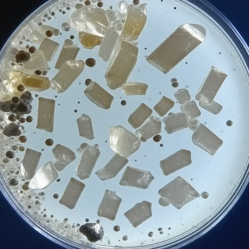
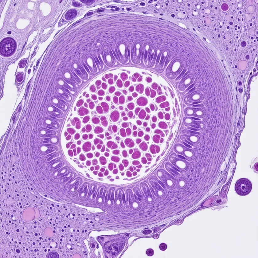
Bile flow is the forgotten third phase of detoxification. After hepatocytes have completed Phase I oxidation and Phase II conjugation, the water-soluble conjugates must physically exit the cell — transported into bile canaliculi by the multidrug resistance-associated protein 2 (MRP2) and breast cancer resistance protein (BCRP) pumps, mixed with primary and secondary bile acids, concentrated in the gallbladder, and dumped into the duodenum on meal stimulation. When bile flow stalls (cholestasis, sluggish gallbladder, gallstones, opioid-induced biliary sphincter spasm, post-cholecystectomy state), Phase II conjugates accumulate in hepatocytes, back up into systemic circulation, get deconjugated by intestinal beta-glucuronidase, and recirculate via enterohepatic circulation. Tauroursodeoxycholic acid (TUDCA) is the most clinically useful intervention for restoring bile flow — a hydrophilic bile acid that displaces toxic hydrophobic bile acids, reduces endoplasmic reticulum (ER) stress, and is approved for cholestatic conditions in Europe.
Table of Contents
- Why Bile Flow Is the Phase III Detoxification Bottleneck
- The Bile Acid Pool: Primary, Secondary, Hydrophobic vs Hydrophilic
- Enterohepatic Circulation and Beta-Glucuronidase Recirculation
- TUDCA Mechanism: Membrane Stabilization and ER Chaperoning
- UDCA vs TUDCA: Choice, Dosing, Cost
- Sluggish Gallbladder and Functional Cholestasis
- After Gallbladder Removal: The Constant-Trickle Problem
- FXR and TGR5 Signaling: Bile Acids as Hormones
- Cautions and Drug Interactions
- Key Research Papers
- Connections
- Featured Videos
Why Bile Flow Is the Phase III Detoxification Bottleneck
Detoxification education historically focused on Phase I (cytochrome P450 oxidation) and Phase II (glucuronidation, sulfation, glutathione conjugation), as if excretion were automatic. It is not. The hepatocyte must actively transport the Phase II conjugate across its canalicular membrane into the bile canaliculus — an energy-requiring step mediated by adenosine triphosphate-binding cassette transporters (primarily MRP2/ABCC2 for glucuronide and sulfate conjugates, BCRP/ABCG2 for various drug metabolites, BSEP/ABCB11 for bile salts themselves, and MDR1/P-glycoprotein for many xenobiotic conjugates).
If these canalicular transporters are inhibited (many drugs do this — cyclosporine, troglitazone, bosentan, estradiol-17beta-glucuronide itself), or if downstream bile flow is mechanically obstructed (gallstone, ampullary stricture, pancreatic head tumor), or if the gallbladder is sluggish (functional biliary disorder, post-bariatric surgery, opioid-induced sphincter of Oddi spasm), the Phase II conjugates back up into the hepatocyte. When intracellular concentration rises high enough, the conjugates leak back into sinusoidal blood and reach the systemic circulation. This is one mechanism of conjugated hyperbilirubinemia in cholestasis and a mechanism by which patients with sluggish bile flow can experience apparent "detox reactions" disproportionate to their actual chemical exposure.
Practitioners who use this framing say that optimizing bile flow is not just about digesting fats: it is about completing the third phase of detoxification.
The Bile Acid Pool: Primary, Secondary, Hydrophobic vs Hydrophilic
The human bile acid pool is a mixture of related amphipathic molecules synthesized from cholesterol in hepatocytes. Understanding the pool composition matters because individual bile acids vary in hydrophobicity, detergent strength, and cytotoxicity to the hepatocyte that produces them.
The two primary bile acids synthesized in the liver are cholic acid (CA, trihydroxy, less hydrophobic) and chenodeoxycholic acid (CDCA, dihydroxy, more hydrophobic). Both are conjugated with glycine or taurine before secretion into bile, increasing their solubility and reducing passive reabsorption in the small bowel. Once in the small intestine, gut bacteria (primarily species of Bacteroides, Clostridium, and Eubacterium) deconjugate the bile salts via bile salt hydrolase and 7-alpha-dehydroxylase, converting CA to deoxycholic acid (DCA) and CDCA to lithocholic acid (LCA). DCA and LCA are the secondary bile acids; LCA in particular is highly hydrophobic and cytotoxic.
The bile acid pool in healthy individuals contains roughly 30-40% cholic, 30-40% chenodeoxycholic, 20-25% deoxycholic, and a small percentage of lithocholic and ursodeoxycholic acids. In cholestatic disease, the pool shifts toward more hydrophobic species, which themselves further damage the hepatocyte canalicular membrane and worsen cholestasis — a positive feedback loop.
Ursodeoxycholic acid (UDCA) is the most hydrophilic of the naturally occurring bile acids, present at only about 1-3% of the normal human pool. It does not damage the hepatocyte membrane. When taken orally at therapeutic doses (13-15 mg/kg/day), UDCA expands to roughly 40-60% of the pool, displacing the toxic hydrophobic bile acids. This is the reasoning behind its use in primary biliary cholangitis and its trials in primary sclerosing cholangitis. In primary biliary cholangitis (then called primary biliary cirrhosis), a two-year double-blind trial of 146 patients at 13-15 mg/kg/day found treatment failure in 6 on ursodeoxycholic acid and 13 on placebo. In primary sclerosing cholangitis, the authors of a long-term double-blind trial of 150 patients at a higher dose (28-30 mg/kg/day) described earlier controlled trials as inconclusive. Their own trial was stopped after 6 years for futility: liver blood tests improved, yet 39% on ursodeoxycholic acid versus 26% on placebo reached a clinical endpoint (cirrhosis, varices, bile duct cancer, transplant or death; adjusted risk 2.3 times greater), and serious adverse events were 63% versus 37%. It did not improve survival. Tauroursodeoxycholic acid (TUDCA) is the taurine-conjugated form of UDCA — better water solubility, more efficient enterohepatic recirculation, and direct chemical chaperone effects on the endoplasmic reticulum.
Enterohepatic Circulation and Beta-Glucuronidase Recirculation
The body recycles its bile acid pool with extraordinary efficiency. Approximately 95% of the bile acids dumped into the duodenum after a meal are actively reabsorbed in the terminal ileum (via ASBT, the apical sodium-dependent bile acid transporter), returned to the liver via the portal vein, and re-secreted into bile. The total bile acid pool is approximately 3-5 grams, but it cycles 6-10 times per day, delivering roughly 20-30 grams of bile acids to the gut daily. Only 5% (about 0.5 g) is lost in feces and replaced by de novo hepatic synthesis from cholesterol.
The same enterohepatic loop is exploited by Phase II conjugates of xenobiotics and steroid hormones — they are secreted in bile, reach the gut, get partially deconjugated by intestinal beta-glucuronidase (an enzyme produced by gut bacteria, particularly elevated in dysbiosis), and the deconjugated parent compound is reabsorbed. Estradiol is the textbook example: estradiol-17beta-glucuronide is secreted in bile, deconjugated by gut beta-glucuronidase, and reabsorbed as free estradiol, contributing to estrogen recirculation. This is one mechanism behind the proposed estrogen-detoxification role of cruciferous vegetables and calcium-D-glucarate, which inhibit beta-glucuronidase. See our Cruciferous Vegetables page for the dietary side of this.
The clinical implication: a sluggish gut with constipation and dysbiosis amplifies enterohepatic recirculation of conjugated toxins. Daily bowel movements and a healthy gut microbiome are integral to liver detoxification, not separate concerns. Hydration, soluble fiber, and magnesium for regularity are part of a real liver cleanse.
TUDCA Mechanism: Membrane Stabilization and ER Chaperoning
TUDCA works through several overlapping mechanisms that together restore hepatocyte health in cholestatic and ER-stressed states:
- Membrane stabilization — TUDCA partitions into the hepatocyte canalicular membrane and physically displaces the more toxic hydrophobic bile acids. This reduces membrane permeabilization, prevents mitochondrial swelling, and lowers cytochrome c release.
- Chemical chaperone for the endoplasmic reticulum — TUDCA reduces ER stress and the unfolded protein response (UPR). Misfolded proteins accumulating in the ER trigger PERK, IRE1, and ATF6 stress pathways that culminate in CHOP-mediated apoptosis. TUDCA stabilizes nascent proteins during folding, reducing CHOP induction and protecting against ER-stress-driven apoptosis.
- Anti-apoptotic via mitochondrial pathway — TUDCA prevents Bax translocation to mitochondria, reducing cytochrome c release and downstream caspase-9 activation.
- Bile acid receptor signaling (FXR, TGR5) — TUDCA modulates FXR (farnesoid X receptor) signaling to suppress CYP7A1 (the rate-limiting bile acid synthesis enzyme), reducing toxic bile acid load.
Two of the cited studies behind these mechanisms were not done in patients. The protection against harsher bile salts was shown in rat liver cells and human red blood cells in the laboratory, at millimolar concentrations (Heuman 1991). The restored blood sugar came from obese diabetic mice (Ozcan 2006).
These mechanisms make TUDCA useful well beyond cholestasis — emerging evidence supports its use in retinal protection (retinitis pigmentosa, age-related macular degeneration, AMD), neurodegeneration (Huntington's, amyotrophic lateral sclerosis, ALS), and pancreatic beta-cell protection in type 1 diabetes. The pharmacology underlying all of these applications is the same: TUDCA reduces ER stress and apoptosis in vulnerable cell populations.
For amyotrophic lateral sclerosis, TUDCA (also called taurursodiol) combined with sodium phenylbutyrate was approved in the United States in 2022 on the results of a phase 2 trial, then voluntarily withdrawn from the market in 2024 after the phase 3 PHOENIX trial showed no change in the total score of the standard function scale at 48 weeks. That result is for the combination, not TUDCA alone.
UDCA vs TUDCA: Choice, Dosing, Cost
UDCA (ursodeoxycholic acid, also called ursodiol) is U.S. Food and Drug Administration-approved for primary biliary cholangitis and gallstone dissolution. Standard dose for primary biliary cholangitis is 13-15 mg/kg/day (typically 250-500 mg twice daily). It is prescription-only in the United States. Cost is moderate; generics are widely available. Side effects are mild — transient diarrhea in some patients.
TUDCA (tauroursodeoxycholic acid) is the taurine-conjugated form. Available over-the-counter as a dietary supplement in the U.S. (typically 250 mg or 500 mg capsules). The functional medicine community uses it at 250-1000 mg/day for sluggish bile flow, fatty liver, and "estrogen detox" support, divided into 1-3 doses with meals. TUDCA is generally well tolerated; some patients report transient loose stools when starting.
Choice between them depends on indication and access. For a formal diagnosis of primary biliary cholangitis, UDCA at standard prescription dose is the approved treatment. For primary sclerosing cholangitis, the high-dose trial described above did not improve survival, and its authors called earlier controlled trials inconclusive. For functional/lifestyle support of bile flow in patients without a primary cholestatic disease, TUDCA at 250-500 mg twice daily is the typical regimen used in integrative practice.
A direct comparison exists: in a crossover study of 23 people with primary biliary cirrhosis, 500 mg a day of each for 6 months with a 3-month break between, liver enzymes improved on both with no significant difference between them, and both were well tolerated. The authors called the result short-term.
Integrative practitioners describe taking TUDCA with the largest meal of the day (when endogenous bile is being released anyway) and cycling with breaks (4 weeks on, 1 week off is a common pattern). They describe reassessing every 3 months, with the stated goal of restoring bile flow rather than chronic dependence. By their account, patients with documented sluggish flow may continue indefinitely, and many taper after 6-12 months as digestion and stool quality normalize.
Sluggish Gallbladder and Functional Cholestasis
Many patients with right upper quadrant discomfort, post-prandial bloating, fatty food intolerance, and intermittent nausea undergo workup for gallstones, find no stones on ultrasound, and are told nothing is wrong. The underlying problem in many of these patients is functional biliary disorder — sluggish gallbladder emptying (low ejection fraction on a hepatobiliary iminodiacetic acid scan with cholecystokinin, or HIDA-CCK scan, typically defined as <35-40%) without obstructing stones. The clinical syndrome was previously called "biliary dyskinesia" and the standard surgical answer was cholecystectomy, often with limited symptom resolution.
The functional-medicine approach to sluggish bile flow combines several interventions:
- TUDCA 500 mg twice daily with meals — restores hydrophilic bile acid pool
- Bitter herbs before meals — dandelion root, gentian, artichoke leaf, milk thistle (see our Milk Thistle page) all stimulate vagal afferents and cholecystokinin release, triggering gallbladder contraction
- Beet kvass, fresh beet juice, or supplemental betaine — traditional remedies with some mechanistic plausibility
- Adequate dietary fat at each meal — counterintuitive to many people. Very low-fat diets reduce gallbladder ejection signal (CCK release) and predispose to gallstone formation and bile stasis. Adequate dietary fat (avocado, olive oil, fatty fish, eggs) maintains regular gallbladder contraction.
- The sphincter of Oddi — opioid use, including codeine and tramadol, causes sphincter of Oddi spasm and worsens biliary flow. Practitioners replace these drugs where clinically feasible.
- Magnesium — smooth muscle relaxant; oral or transdermal forms can reduce biliary smooth muscle spasm
After Gallbladder Removal: The Constant-Trickle Problem
Approximately 750,000 cholecystectomies are performed annually in the U.S. After removal of the gallbladder, bile no longer concentrates and stores between meals — it trickles continuously from the liver into the duodenum. This produces three downstream problems:
- Reduced fat digestion at meals — without the bolus of concentrated bile delivered at meal time, fat emulsification is incomplete. Steatorrhea, bloating, and fat-soluble vitamin deficiency are common.
- Bile acid diarrhea — the constant trickle of bile acids overwhelms ileal reabsorption capacity in a subset of patients, leading to colonic bile acid exposure and secretory diarrhea. This is treated with bile acid sequestrants (cholestyramine, colestipol, colesevelam).
- Increased colorectal cancer risk — some epidemiologic data suggest a modest increase in proximal colon cancer risk in cholecystectomy patients, attributed to chronic colonic exposure to secondary bile acids (DCA, LCA).
Practitioners describe post-cholecystectomy support that includes ox bile or bile salt supplementation (125-500 mg with each fatty meal), TUDCA to support remaining hepatic biliary function, pancreatic enzymes if steatorrhea is significant, and a moderate-fat-distributed-across-the-day eating pattern rather than large fatty meals.
FXR and TGR5 Signaling: Bile Acids as Hormones
The paradigm shift in bile acid biology over the past two decades is the recognition that bile acids are not just detergents — they are endogenous ligands for nuclear and membrane receptors that regulate glucose metabolism, lipid metabolism, energy expenditure, and inflammation throughout the body.
FXR (farnesoid X receptor, NR1H4) is a nuclear receptor expressed in liver, intestine, kidney, and adrenal gland. Bile acids bind FXR with the rank order CDCA > DCA > LCA > CA. FXR activation suppresses hepatic bile acid synthesis (via SHP-FGF19-FGFR4 feedback to CYP7A1), reduces hepatic gluconeogenesis, lowers triglycerides, and reduces inflammation. Obeticholic acid, a semi-synthetic FXR agonist, was approved by the U.S. Food and Drug Administration for primary biliary cholangitis. Its primary biliary cholangitis authorizations in the European Union and the United States have since been withdrawn, and it was voluntarily withdrawn from the U.S. market. A 2026 review lists itching (pruritus), adverse lipoprotein changes and hepatic safety concerns, and says the COBALT confirmatory trial failed to verify benefit in its randomized analysis. In the earlier 12-month POISE trial (217 patients), the liver blood-test target was reached by 46-47% on the drug against 10% on placebo, with itching in 56-68% against 38% and serious adverse events in 11-16% against 4%. TUDCA modulates FXR, contributing to its bile-acid-pool-regulating effect.
TGR5 (GPBAR1) is a membrane G-protein-coupled receptor with widespread expression. TGR5 activation by bile acids in brown adipose tissue and skeletal muscle increases energy expenditure via deiodinase 2 (D2) activation of thyroid hormone, and in intestinal L cells stimulates glucagon-like peptide-1 (GLP-1) release — the same incretin pathway targeted by semaglutide. This is part of why bariatric surgery (which alters bile acid signaling by rerouting the duodenum) produces metabolic effects beyond what mechanical food restriction would predict.
The functional-medicine practical implication: bile flow is not just about Phase III detoxification. It is also a signal in glucose and energy metabolism. Patients with metabolic syndrome, type 2 diabetes, and obesity benefit from optimized bile flow as part of their broader metabolic care.
One small human trial tested the insulin side of this idea: 20 obese adults were randomized to 1,750 mg of TUDCA a day (above the 250-1000 mg/day practitioners describe) or placebo for 4 weeks. Liver and muscle insulin sensitivity rose about 30% on TUDCA and did not change on placebo, while markers of ER stress in muscle and fat did not change on either. The authors said more studies are needed to find the target cells and mechanisms.
Cautions and Drug Interactions
- Mechanical biliary obstruction is a contraindication — if a gallstone or stricture is obstructing the common bile duct, increasing bile flow upstream cannot help and can worsen pain or trigger cholangitis. Right upper quadrant pain with elevated alkaline phosphatase, gamma-glutamyl transferase (GGT), and bilirubin is a pattern clinicians image (ultrasound, magnetic resonance cholangiopancreatography) before TUDCA or bile-flow stimulants are started.
- Acute cholecystitis is a contraindication — acute gallbladder inflammation is a surgical emergency, not a TUDCA problem.
- Diarrhea is the most common side effect — practitioners describe starting at the low end of dosing (250 mg/day) and increasing gradually. Persistent diarrhea suggests bile acid malabsorption rather than tolerance issue.
- Pregnancy — UDCA is sometimes used (off-label) for intrahepatic cholestasis of pregnancy and has been considered safe in this setting. In the PITCHES trial (605 women, ursodeoxycholic acid versus placebo), perinatal death, preterm delivery or neonatal unit admission occurred in 23% versus 27% of infants (adjusted risk ratio 0.85, 95% confidence interval 0.62-1.15); no serious adverse events were regarded as related to treatment. The authors concluded that it does not reduce adverse perinatal outcomes and that its routine use for this condition should be reconsidered. TUDCA over-the-counter use in pregnancy lacks adequate safety data.
- Bile acid sequestrants reduce TUDCA absorption — cholestyramine and colesevelam will bind TUDCA in the gut. Practitioners describe separating the doses by 4 hours.
- Aluminum-containing antacids reduce absorption — similarly bind bile acids. Practitioners describe separating these doses too.
Key Research Papers
- Beuers U (2006). Drug insight: mechanisms and sites of action of ursodeoxycholic acid in cholestasis. Nat Clin Pract Gastroenterol Hepatol. — PubMed PMID: 16741551
- Heuman DM, Pandak WM, Hylemon PB, Vlahcevic ZR (1991). Conjugates of ursodeoxycholate protect against cytotoxicity of more hydrophobic bile salts: in vitro studies in rat hepatocytes and human erythrocytes. Hepatology. 14(5):920-6. — PubMed PMID: 1937396
- Ozcan U et al. (2006). Chemical chaperones reduce ER stress and restore glucose homeostasis in a mouse model of type 2 diabetes. Science. — PubMed PMID: 16931765
- Vang S et al. (2014). The unexpected uses of urso- and tauroursodeoxycholic acid in the treatment of non-liver diseases. Glob Adv Health Med. — PubMed PMID: 24891994
- Lindor KD et al. (2009). High-dose ursodeoxycholic acid for the treatment of primary sclerosing cholangitis. Hepatology. 50(3):808-14. — PubMed PMID: 19585548
- Poupon RE et al. (1991). A multicenter, controlled trial of ursodiol for the treatment of primary biliary cirrhosis. NEJM. — PubMed PMID: 1674105
- Hofmann AF, Hagey LR (2008). Bile acids: chemistry, pathochemistry, biology, pathobiology, and therapeutics. Cell Mol Life Sci. — PubMed PMID: 18488143
- Watanabe M et al. (2006). Bile acids induce energy expenditure by promoting intracellular thyroid hormone activation. Nature. (TGR5 mechanism) — PubMed PMID: 16400329
- Makishima M et al. (1999). Identification of a nuclear receptor for bile acids. Science. (FXR discovery) — PubMed PMID: 10334992
- Nevens F et al. (2016). A placebo-controlled trial of obeticholic acid in primary biliary cholangitis. NEJM. (POISE trial) — PubMed PMID: 27532829
- Boyer JL (2013). Bile formation and secretion. Compr Physiol. — PubMed PMID: 23897680
- Larghi A et al. (1997). Ursodeoxycholic and tauro-ursodeoxycholic acids for the treatment of primary biliary cirrhosis: a pilot crossover study. Aliment Pharmacol Ther. 11(2):409-14. — PubMed PMID: 9146783
- Kars M et al. (2010). Tauroursodeoxycholic Acid may improve liver and muscle but not adipose tissue insulin sensitivity in obese men and women. Diabetes. 59(8):1899-905. — PubMed PMID: 20522594
- Chappell LC et al. (2019). Ursodeoxycholic acid versus placebo in women with intrahepatic cholestasis of pregnancy (PITCHES): a randomised controlled trial. Lancet. 394(10201):849-860. — PubMed PMID: 31378395
- Ketabforoush A et al. (2024). Sodium Phenylbutyrate and Tauroursodeoxycholic Acid: A Story of Hope Turned to Disappointment in Amyotrophic Lateral Sclerosis Treatment. Clin Drug Investig. 44(7):495-512. — PubMed PMID: 38909349
- Calcagno N et al. (2025). Real-World Clinical Experience With Sodium Phenylbutyrate and Taurursodiol at a Single Amyotrophic Lateral Sclerosis Center in the United States. Eur J Neurol. 32(9):e70360. — PubMed PMID: 40965373
- Fiorucci S (2026). Obeticholic acid: discovery, clinical translation, and regulatory withdrawal. Eur Rev Med Pharmacol Sci. 30(10):375-395. — PubMed PMID: 42961163
- Dichter J (2026). Bile Acid Metabolism as a Unifying Readout for Diet, Microbiome Function, and Gastrointestinal Disease. FASEB J. 40(17):e72273. — PubMed PMID: 42687847