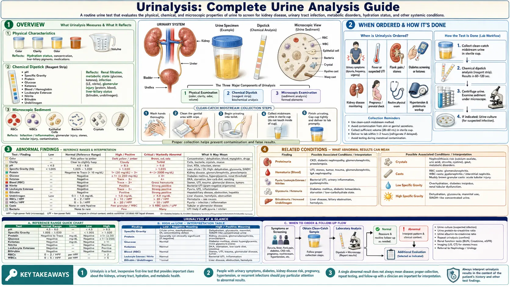
Urinalysis: Complete Urine Analysis Guide
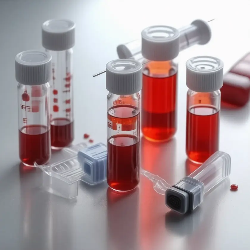

A urinalysis (UA) is the physical, chemical, and microscopic examination of urine (MedlinePlus). By looking at how the urine appears, what a chemical dipstick detects in it, and what is seen under the microscope, clinicians gather information about the kidneys, the urinary tract, the liver, and blood-sugar control. A StatPearls chapter describes urinalysis as the first laboratory test developed in the history of medicine, in use in some form for several thousand years (Queremel Milani 2023).
Table of Contents
- Overview
- When Ordered
- Components and Reference Ranges
- Dipstick Accuracy for Urinary Tract Infection
- Vitamin C and Other Interferences
- Bacteria Without Symptoms: What the Guidelines Say
- Interpretation Patterns
- Microscopic Findings: Casts and Crystals
- Related Conditions
- Key Research Papers
- Connections
- Featured Videos
Overview
Urine is produced by the kidneys as they filter blood, removing waste products, excess electrolytes, and water. A standard urinalysis has three parts (MedlinePlus): a visual inspection (color and clarity), a dipstick chemical analysis (a strip with chemical pads that change color, commonly reading pH, specific gravity, protein, glucose, blood, leukocyte esterase, nitrites, bilirubin, ketones, and urobilinogen), and a microscopic examination (cells, casts, crystals, mucus, bacteria, and other organisms).
MedlinePlus notes that normal urine varies in color from almost colorless to dark yellow, that some foods such as beets and blackberries can turn it red, and that several medicines change urine color without any disease being present.
About the ranges on this page. Unless another source is named, each range below is the example normal value given in the MedlinePlus Medical Encyclopedia (U.S. National Library of Medicine). MedlinePlus notes that normal value ranges may vary slightly among laboratories and that some labs use different measurements or test different samples; a result is read against the range printed by the laboratory that ran it. A published review of urinalysis (Simerville 2005) gives slightly different ranges for some values, and where they differ both are shown.
Collection. The usual routine sample is a midstream “clean catch” specimen, collected after cleaning the genital area so that skin germs do not get into the sample (MedlinePlus); the Simerville review describes a midstream clean-catch technique as usually adequate in men and women. For albuminuria testing, the KDIGO 2024 kidney guideline states that a first void in the morning midstream sample is preferred in adults and children. The Simerville review notes that urine is refrigerated if it cannot be examined promptly, because delays of more than two hours between collection and examination often cause unreliable results.
When Ordered
MedlinePlus lists these reasons for a urinalysis, and the specific-test pages add others:
- Routine medical exam — to screen for early signs of disease
- Suspected urinary tract infection (UTI) — MedlinePlus lists frequent urges to urinate, pain or burning when urinating, dark, cloudy, or reddish urine, bad-smelling urine, and, in older people, weakness, fatigue, or confusion
- Kidney disease — to detect it or to monitor people being treated for it
- Diabetes — signs of diabetes or monitoring of treatment; MedlinePlus describes yearly kidney testing with a urine albumin-to-creatinine ratio as important for people with diabetes
- Blood in the urine — visible or found on a previous test
- Liver disease or hemolytic anemia — through the bilirubin and urobilinogen pads
- Pregnancy — MedlinePlus lists preeclampsia among causes of protein in the urine; screening for bacteria in the urine in pregnancy is done with a urine culture rather than a dipstick (USPSTF 2019)
Components and Reference Ranges
In this section: pH · Specific Gravity · Protein and Albumin · Glucose · Blood (Hematuria) · White Blood Cells (Leukocyte Esterase) · Nitrites · Bilirubin · Ketones · Urobilinogen
pH
Urine pH reflects the kidney’s role in acid-base balance: the kidneys excrete acid or retain bicarbonate as needed. The Simerville review states that urinary pH can range from 4.5 to 8 but is normally slightly acidic (5.5 to 6.5), and that protein and acidic fruits such as cranberries can make urine more acidic while diets high in citrate can make it alkaline.
Urine pH (MedlinePlus example range)
Clinical notes: MedlinePlus lists renal tubular acidosis (kidneys that do not properly remove acids from the blood), kidney failure, gastric suction, urinary tract infection, and vomiting among causes of a high urine pH, and diarrhea, metabolic acidosis such as diabetic ketoacidosis, and starvation among causes of a low one. According to the Simerville review, alkaline urine in a person with a UTI suggests a urea-splitting organism, which may be associated with magnesium-ammonium phosphate (struvite) crystals and staghorn stones, while uric acid stones are associated with acidic urine.
Specific Gravity
Specific gravity compares the density of urine with that of water. It reflects urine concentration and the kidney’s concentrating ability, and so the body’s water balance. MedlinePlus notes that the dipstick gives only a rough result and that urine osmolality is a more specific and precise measure of urine concentration.
Specific Gravity (dimensionless ratio; MedlinePlus example range)
Clinical notes: The Simerville review gives a normal range of 1.003 to 1.030 and describes a value below 1.010 as indicating relative hydration and a value above 1.020 as indicating relative dehydration. In intrinsic kidney insufficiency, it states, specific gravity becomes fixed at 1.010, the specific gravity of the glomerular filtrate (isosthenuria). MedlinePlus lists dehydration, glucose in the urine, heart failure, high blood sodium, SIADH, adrenal insufficiency, renal artery stenosis, and shock among causes of increased specific gravity, and diabetes insipidus, drinking too much fluid, kidney failure, low blood sodium, damage to kidney tubule cells, and severe kidney infection among causes of decreased specific gravity.
Protein and Albumin
Healthy kidney filters hold back large protein molecules, so very little protein normally reaches the urine; protein that leaks through over time can be an early sign of kidney damage (MedlinePlus). The Simerville review notes that the reagent on most dipsticks is sensitive to albumin but may not detect low concentrations of gamma-globulins and Bence Jones proteins, and that trace results appear at protein concentrations of 5 to 10 mg per dL.
Urine Protein (random sample; MedlinePlus example range)
MedlinePlus gives 30 to 150 mg per 24 hours as the example normal value for a 24-hour collection. It lists heart failure, kidney damage, diabetic kidney disease, dehydration, multiple myeloma, preeclampsia or eclampsia, and urinary tract problems such as infection or a bladder tumor among causes of higher amounts.
Albuminuria categories (KDIGO 2024). The KDIGO 2024 chronic kidney disease guideline classifies albuminuria by the urine albumin-to-creatinine ratio (ACR), the same albuminuria category it uses with eGFR to classify chronic kidney disease (see the Comprehensive Metabolic Panel page):
- A1 — ACR below 30 mg/g (below 3 mg/mmol): Normal to mildly increased
- A2 — ACR 30 to 300 mg/g (3 to 30 mg/mmol): Moderately increased (relative to the young adult level; the range traditionally called “microalbuminuria”)
- A3 — ACR above 300 mg/g (above 30 mg/mmol): Severely increased
KDIGO lists the urine ACR first among initial tests for albuminuria, ahead of reagent-strip testing, and states that a positive reagent-strip result is confirmed by a quantitative laboratory measurement expressed as a ratio to urine creatinine, and that an ACR of 30 mg/g or more on a random sample is confirmed with a subsequent first-morning sample. Among factors that falsely raise urine albumin or protein, its guideline lists hematuria, menstruation, exercise, and symptomatic urinary infection.
Clinical notes: The Simerville review lists alkaline or concentrated urine and phenazopyridine among causes of false-positive dipstick protein, and describes orthostatic (postural) proteinuria as a benign condition that can result from prolonged standing, confirmed by a negative urinalysis after eight hours of lying down. In nephrotic syndrome, a 2016 review gives the proteinuria threshold as more than 3 to 3.5 g of protein in a 24-hour collection, or a spot protein-to-creatinine ratio above 3 to 3.5 mg/mg, together with edema and low serum albumin (Kodner 2016).
Glucose
Glucose filtered by the kidneys is normally reabsorbed by the renal tubules, so it is usually not detectable in urine (MedlinePlus). According to the Simerville review, glucose appears in the urine when the filtered load exceeds the ability of the tubules to reabsorb it, at a blood glucose of about 180 to 200 mg per dL.
Urine Glucose (qualitative)
Clinical notes: MedlinePlus lists diabetes, pregnancy (up to half of women have glucose in their urine at some time during pregnancy), gestational diabetes, and renal glycosuria (a rare condition in which glucose spills into the urine even when blood glucose is normal) among causes. It notes that a urine glucose test cannot diagnose diabetes on its own, that blood glucose tests have largely replaced it for that purpose, and that a normal urine glucose does not exclude diabetes. One class of diabetes medicines, the SGLT2 inhibitors, lowers blood sugar by causing the kidneys to get rid of more glucose in the urine, so a person taking one may test positive for urine glucose (MedlinePlus drug information, empagliflozin).
Blood (Hematuria)
The dipstick blood pad reacts to heme, so it is positive with intact red blood cells and also with free hemoglobin or myoglobin; the Simerville review lists hemoglobinuria, myoglobinuria, menstrual blood, dehydration, and exercise among causes of a positive dipstick, and microscopy is what counts the red cells themselves.
Urine Red Blood Cells (RBC/HPF on microscopy)
Two definitions that overlap. MedlinePlus gives 4 red blood cells per high power field (RBC/HPF) or fewer as a normal result. The 2020 American Urological Association / Society of Urodynamics, Female Pelvic Medicine & Urogenital Reconstruction (AUA/SUFU) guideline defines microhematuria as 3 or more red blood cells per high power field on microscopic examination of a single specimen (Barocas 2020); the older Simerville review gives the generally accepted definition as 3 or more RBC/HPF in two of three samples. A dipstick result alone does not meet the guideline definition, which rests on microscopy.
Evaluation. The AUA/SUFU guideline classifies people with microhematuria as low, intermediate, or high risk for a genitourinary cancer, using age, sex, smoking and other urothelial cancer risk factors, the degree and persistence of the microhematuria, and any prior visible blood, and it recommends evaluation with cystoscopy and imaging of the upper urinary tract according to that risk, with shared decision-making. Where a gynecologic or non-cancer urinary cause is found, the guideline states that urinalysis is repeated after that cause resolves (Barocas 2020). For visible (gross) hematuria, the Simerville review states that up to 20 percent of patients have a urinary tract cancer.
Clinical notes: MedlinePlus notes that many causes of blood in the urine are not serious, including certain medicines, intense exercise, sexual activity, and menstruation, and lists urinary tract infection, kidney or bladder stones, kidney injury, kidney inflammation, an enlarged prostate, polycystic kidney disease, blood disorders, and cancers of the bladder, kidney, or prostate among others. The Simerville review notes that dysmorphic red cells, misshapen from passing through an abnormal glomerulus, suggest glomerular disease.
White Blood Cells (Leukocyte Esterase)
The leukocyte esterase pad detects an enzyme released by white blood cells, so a positive result points to white cells in the urine (pyuria). MedlinePlus describes a negative result as normal and a positive one as a possible urinary tract infection.
White Blood Cells on Microscopy (WBC/HPF; Simerville review)
Clinical notes: MedlinePlus lists Trichomonas infection and vaginal secretions as causes of a positive test without a UTI, and a high level of protein or of vitamin C as factors that can hide a positive result when a UTI is present; the Simerville review adds contamination as a cause of false positives, and elevated specific gravity, glucose, ketones, protein, some oxidizing drugs, and vitamin C as causes of false negatives. White cells without a bacterial infection on culture (“sterile pyuria”) have many causes; the Simerville review lists balanitis, urethritis, tuberculosis, bladder tumors, viral infections, kidney stones, foreign bodies, exercise, glomerulonephritis, and corticosteroid or cyclophosphamide use. White blood cell casts on microscopy are covered below.
Nitrites
Many bacteria that cause urinary tract infections convert nitrates, normally present in urine, into nitrites (MedlinePlus). A positive nitrite test therefore suggests bacteria in the urine, but MedlinePlus notes that a negative result does not exclude infection because some types of bacteria do not make this conversion.
Urine Nitrites (qualitative)
Clinical notes: Gram-negative enteric bacteria, the most common uropathogens and the group that includes Escherichia coli, produce urinary nitrite, while Enterococcus does not; in a study of 7,599 young children evaluated for UTI, every enterococcal infection had a negative nitrite result, yet 95.6 percent of the nitrite-negative infections were still caused by gram-negative organisms (Chaudhari 2020). Time matters as well: in a primary-care study of 1,070 women with symptoms of a lower UTI, accuracy depended strongly on how long urine had been in the bladder, the optimal bladder incubation time was 4 hours or more, and the nitrite test’s sensitivity at that time was 66 percent (Ferry 2015). The Simerville review also lists elevated specific gravity, elevated urobilinogen, nitrate reductase-negative bacteria, a urine pH below 6.0, and vitamin C as causes of false-negative nitrite results, and contamination, exposure of the dipstick to air, and phenazopyridine as causes of false positives. Dipstick accuracy for UTI is covered in its own section below.
Bilirubin
Conjugated (direct) bilirubin is water-soluble and can pass into urine; unconjugated bilirubin, carried on albumin in the blood, does not. MedlinePlus states that bilirubin is not normally found in urine, and the Simerville review that urine normally contains no detectable bilirubin.
Urine Bilirubin (qualitative)
Clinical notes: MedlinePlus lists biliary tract disease including gallstones, cirrhosis, hepatitis and other liver disease, and tumors of the liver or gallbladder among causes of bilirubin in urine. Because the excess bilirubin in hemolysis is unconjugated, hemolytic jaundice does not by itself produce bilirubin in the urine. MedlinePlus notes that bilirubin breaks down in light, and the Simerville review lists phenazopyridine as a cause of a false-positive result.
Ketones
Ketones (acetoacetate, beta-hydroxybutyrate, and acetone) build up when the body breaks down fats and fatty acids for fuel, most often when it does not get enough sugar or carbohydrates (MedlinePlus). The urine dipstick uses a nitroprusside reaction, which measures acetoacetic acid but not beta-hydroxybutyrate, the main ketoacid produced in diabetic ketoacidosis (Umpierrez 2024).
Urine Ketones (qualitative)
Clinical notes: MedlinePlus describes diabetic ketoacidosis (DKA) as a life-threatening problem for people with diabetes and lists fasting or starvation, a high-protein or low-carbohydrate (ketogenic) diet, prolonged vomiting, acute or severe illness, high fever, hyperthyroidism, and SGLT2-inhibitor medicines among other causes of ketones in urine. The 2024 ADA/EASD consensus report on hyperglycemic crises states that urine ketone testing can underestimate severity early in DKA and overestimate it later, and recommends direct measurement of blood beta-hydroxybutyrate for diagnosis and monitoring; it also reports that SGLT2 inhibitors account for most recent cases of euglycemic DKA, with glucose below 200 mg/dL (Umpierrez 2024). The Simerville review lists delay in examining the urine as a cause of a false-negative ketone result.
Urobilinogen
Urobilinogen is made from bilirubin by bacteria in the intestine. Some leaves the body in stool, and some is absorbed into the blood and returns to the liver to be recycled into bile, so normal urine contains a small amount (MedlinePlus).
Urine Urobilinogen (qualitative)
Clinical notes: MedlinePlus lists hepatitis, cirrhosis, liver damage from medicines, and hemolytic anemia among causes of higher urobilinogen, and a blockage of the bile ducts, a blockage of blood flow through the liver, or other liver problems among causes of little or none. It notes that the test alone cannot diagnose a specific condition.
Dipstick Accuracy for Urinary Tract Infection
The two dipstick pads used for infection, leukocyte esterase and nitrite, behave very differently. In the ranges given by the Simerville review (2005), leukocyte esterase had a sensitivity of 72 to 97 percent and a specificity of 41 to 86 percent for urinary tract infection, while nitrite had a sensitivity of 19 to 48 percent and a specificity of 92 to 100 percent. An abnormal result on either pad had a sensitivity of 46 to 100 percent and a specificity of 42 to 98 percent.
Figure: the spread of published sensitivity and specificity values for each dipstick pad, from the Simerville 2005 review; the note on negative results is from the Devillé 2004 meta-analysis, and the bladder-time figure from Ferry 2015.
A meta-analysis of 70 publications from 1990 to 1999 (Devillé 2004) concluded that the urine dipstick alone seems useful in all populations to exclude infection when both nitrite and leukocyte esterase are negative. The combined sensitivity of the two tests ranged from 68 to 88 percent across patient groups, positive results had to be confirmed, and the authors judged the usefulness of the dipstick alone to rule an infection in to be doubtful, even when the pre-test probability was high. Nitrite accuracy was high in pregnant women and elderly people, and leukocyte esterase sensitivity was highest in studies in family medicine (86 percent).
The two factors that make a negative nitrite less reassuring are covered in the Nitrites section above: Enterococcus does not produce nitrite (Chaudhari 2020), and urine that has spent less than about 4 hours in the bladder gives less time for the conversion (Ferry 2015). MedlinePlus notes that home UTI test strips check for nitrites and white blood cells and do not always give accurate results.
Vitamin C and Other Interferences
Vitamin C (ascorbic acid) in the urine is a well-documented cause of falsely negative dipstick results. In a study of 4,379 routine urinalysis specimens tested with strips sensitive to ascorbic acid, 22.8 percent contained vitamin C, and specimens with vitamin C had a high rate of false-negative results for blood (hemoglobin); the highest false-negative rates were in samples with fewer than 50 red cells per high power field. When volunteers took vitamin C at 100, 250, 500, or 1,000 mg, or drank vitamin C-containing fruit juice, even the lowest doses produced enough urinary vitamin C to cause false-negative blood and glucose results; the authors called for dipstick-only screening protocols to include a check for urinary vitamin C or to use a strip not subject to vitamin C interference (Brigden 1992).
The Simerville review lists vitamin C as a cause of false-negative results for blood, glucose, leukocyte esterase, nitrite, and protein. Other interferences named in the sections above include phenazopyridine (false-positive protein, nitrite, and bilirubin), concentrated urine (false-positive protein; false-negative blood, leukocyte esterase, and nitrite), alkaline urine (false-positive protein), and delay before testing (false-negative ketones; unreliable results after more than two hours).
Bacteria Without Symptoms: What the Guidelines Say
Bacteria are often found in the urine of people who have no urinary symptoms. The Infectious Diseases Society of America (IDSA) defines asymptomatic bacteriuria as 105 colony-forming units (CFU) per mL or more in a voided urine specimen without signs or symptoms attributable to a urinary tract infection, and its 2019 guideline describes antibiotic treatment of asymptomatic bacteriuria as an important contributor to inappropriate antibiotic use, which promotes antimicrobial resistance (Nicolle 2019).
The 2019 IDSA guideline:
- recommends screening for and treating asymptomatic bacteriuria in pregnant women, and in people who will undergo endoscopic urologic procedures associated with mucosal trauma (screening and treatment before the procedure);
- recommends against screening for or treating it in healthy premenopausal non-pregnant women and healthy postmenopausal women, functionally impaired older adults living in the community, older residents of long-term care facilities, people with diabetes, kidney transplant recipients more than 1 month after surgery, people having elective non-urologic surgery, people with short-term or long-term indwelling urinary catheters, people with spinal cord injury, and infants and children (Nicolle 2019).
The U.S. Preventive Services Task Force reached a similar conclusion in 2019: it recommends screening pregnant persons for asymptomatic bacteriuria using a urine culture (a B recommendation) and recommends against screening non-pregnant adults (a D recommendation). It estimated asymptomatic bacteriuria to be present in about 1 to 6 percent of premenopausal women and 2 to 10 percent of pregnant women, and found the harms of treatment, including antibiotic side effects and changes in the microbiome, to be at least small (USPSTF 2019).
Interpretation Patterns
Urinalysis findings are read together, as patterns, rather than one pad at a time. Commonly described patterns include:
- Urinary tract infection: positive leukocyte esterase and nitrite, with white cells and bacteria on microscopy; positive dipstick results are confirmed by urine culture (Devillé 2004).
- Nephrotic syndrome: heavy proteinuria, with fatty casts reflecting lipids in the urine (MedlinePlus); the syndrome itself is defined by heavy proteinuria, edema, and low serum albumin (Kodner 2016).
- Glomerular bleeding (nephritic pattern): blood and protein in the urine, with dysmorphic red cells or red blood cell casts on microscopy pointing to the glomerulus (Simerville 2005).
- Diabetic kidney disease: increased urine albumin, measured as the albumin-to-creatinine ratio and graded A1 to A3 by KDIGO; glucose may also be present when blood sugar is high.
- Liver disease or blocked bile flow: bilirubin in the urine; urobilinogen tends to be higher in liver-cell disease and low or absent when bile flow into the intestine is blocked (MedlinePlus).
- Diabetic ketoacidosis: glucose and ketones in the urine with a low urine pH (MedlinePlus); the dipstick does not measure beta-hydroxybutyrate, the main ketoacid in DKA (Umpierrez 2024).
- Dehydration: high specific gravity and dark, concentrated urine; dehydration is also among MedlinePlus causes of protein in the urine.
- Kidney stones: blood in the urine, with crystals on microscopy (calcium oxalate, calcium phosphate, uric acid, or struvite) (MedlinePlus).
Microscopic Findings: Casts and Crystals
Casts are cylinders of protein, sometimes with cells trapped inside, that form in the kidney tubules and wash out in the urine, so they show that a finding comes from the kidney itself. A 2019 review describes urine sediment examination as a test that can alert clinicians to kidney disease and often identify which part of the kidney is injured (Cavanaugh 2019). MedlinePlus and the Simerville review describe:
- Hyaline casts: the most common type, made of the protein uromodulin (also called Tamm-Horsfall protein). A few are normal; they are often the result of dehydration or strenuous exercise and also occur with chronic kidney failure (MedlinePlus).
- Granular casts: a sign of many types of kidney disease (MedlinePlus); the Simerville review associates them with advanced kidney disease.
- Red blood cell casts: mean there is microscopic bleeding from within the kidney and are seen in many kidney diseases (MedlinePlus); the Simerville review lists glomerulonephritis as their associated condition.
- White blood cell casts: common with acute kidney infection (pyelonephritis) and interstitial nephritis (MedlinePlus); the Simerville review also lists glomerulonephritis.
- Waxy casts: found in advanced kidney disease and long-term kidney failure (MedlinePlus).
- Renal tubular epithelial cell casts: reflect damage to kidney tubule cells, as in renal tubular necrosis, viral disease such as cytomegalovirus nephritis, and kidney transplant rejection (MedlinePlus); the Simerville review adds interstitial nephritis, eclampsia, and heavy metal ingestion.
- Fatty casts: seen with lipids in the urine, most often as a complication of nephrotic syndrome (MedlinePlus).
- Crystals: the Simerville review describes calcium oxalate crystals as square “envelope” shapes, uric acid crystals as yellow to orange-brown diamonds or barrels, triple phosphate (struvite) crystals as “coffin lids”, and cystine crystals as hexagons. MedlinePlus notes that calcium oxalate and calcium phosphate crystals are the most common cause of kidney stones, that repeated urinary tract infections may produce large amounts of struvite crystals, and that crystals in urine do not always mean a medical condition, since some medicines, diet, and low fluid intake can produce them.
Related Conditions
Urinalysis findings take part in the diagnosis and monitoring of many conditions:
- Urinary tract infections — cystitis, pyelonephritis, urethritis
- Diabetes mellitus — glucose, ketones (DKA), and increased urine albumin (diabetic kidney disease)
- Chronic kidney disease (CKD) — albuminuria, persistent hematuria, and urine sediment abnormalities are among KDIGO’s markers of kidney damage
- Glomerulonephritis — IgA nephropathy, lupus nephritis, post-streptococcal glomerulonephritis
- Nephrotic syndrome — membranous nephropathy and focal segmental glomerulosclerosis are the most common types of primary nephrotic syndrome in adults (Kodner 2016)
- Kidney stones (nephrolithiasis) — blood in the urine, crystals
- Liver disease — hepatitis, cirrhosis, blocked bile ducts
- Bladder and kidney cancer — visible or microscopic blood in the urine (Barocas 2020)
- Preeclampsia — protein in the urine during pregnancy (MedlinePlus)
- Interstitial nephritis — white blood cells and white blood cell casts
Key Research Papers
- Devillé WL, Yzermans JC, van Duijn NP, Bezemer PD, van der Windt DA, Bouter LM (2004). The urine dipstick test useful to rule out infections. A meta-analysis of the accuracy. BMC Urology 4:4 — PubMed PMID: 15175113
- Simerville JA, Maxted WC, Pahira JJ (2005). Urinalysis: a comprehensive review. American Family Physician 71(6):1153-1162 — PubMed PMID: 15791892
- Nicolle LE, Gupta K, Bradley SF, et al. (2019). Clinical Practice Guideline for the Management of Asymptomatic Bacteriuria: 2019 Update by the Infectious Diseases Society of America. Clinical Infectious Diseases 68(10):e83-e110 — PubMed PMID: 30895288
- US Preventive Services Task Force; Owens DK, Davidson KW, Krist AH, et al. (2019). Screening for Asymptomatic Bacteriuria in Adults: US Preventive Services Task Force Recommendation Statement. JAMA 322(12):1188-1194 — PubMed PMID: 31550038
- Barocas DA, Boorjian SA, Alvarez RD, et al. (2020). Microhematuria: AUA/SUFU Guideline. Journal of Urology 204(4):778-786 — PubMed PMID: 32698717
- Kidney Disease: Improving Global Outcomes (KDIGO) CKD Work Group (2024). KDIGO 2024 Clinical Practice Guideline for the Evaluation and Management of Chronic Kidney Disease. Kidney International 105(4S):S117-S314 — PubMed PMID: 38490803
- Brigden ML, Edgell D, McPherson M, Leadbeater A, Hoag G (1992). High incidence of significant urinary ascorbic acid concentrations in a west coast population—implications for routine urinalysis. Clinical Chemistry 38(3):426-431 — PubMed PMID: 1547565
- Ferry SA, E Holm S, Ferry BM, Monsen TJ (2015). High Diagnostic Accuracy of Nitrite Test Paired with Urine Sediment can Reduce Unnecessary Antibiotic Therapy. Open Microbiology Journal 9:150-159 — PubMed PMID: 26668667
- Chaudhari PP, Monuteaux MC, Bachur RG (2020). Should the Absence of Urinary Nitrite Influence Empiric Antibiotics for Urinary Tract Infection in Young Children? Pediatric Emergency Care 36(10):481-485 — PubMed PMID: 29135902
- Umpierrez GE, Davis GM, ElSayed NA, et al. (2024). Hyperglycemic Crises in Adults With Diabetes: A Consensus Report. Diabetes Care 47(8):1257-1275 — PubMed PMID: 39052901
- Cavanaugh C, Perazella MA (2019). Urine Sediment Examination in the Diagnosis and Management of Kidney Disease: Core Curriculum 2019. American Journal of Kidney Diseases 73(2):258-272 — PubMed PMID: 30249419
- Kodner C (2016). Diagnosis and Management of Nephrotic Syndrome in Adults. American Family Physician 93(6):479-485 — PubMed PMID: 26977832
- Queremel Milani DA, Jialal I (2023). Urinalysis. In: StatPearls [Internet]. Treasure Island (FL): StatPearls Publishing; NCBI Bookshelf NBK557685 — PubMed PMID: 32491617
PubMed Topic Searches
- Urinalysis clinical interpretation — PubMed literature search
- Microalbuminuria kidney disease — PubMed literature search
- Urinary tract infection diagnosis — PubMed literature search
- Hematuria evaluation — PubMed literature search
- Proteinuria chronic kidney disease — PubMed literature search
- Urine specific gravity hydration — PubMed literature search
- Ketonuria diabetic ketoacidosis — PubMed literature search
- Urinary casts interpretation — PubMed literature search
- Urinalysis dipstick accuracy — PubMed literature search
- Glucosuria diabetes — PubMed literature search
External Authoritative Resources
- MedlinePlus Medical Encyclopedia — Urinalysis
- MedlinePlus Medical Encyclopedia — Urine pH test
- MedlinePlus Medical Encyclopedia — Urine specific gravity test
- MedlinePlus Medical Encyclopedia — Urine protein dipstick test
- MedlinePlus Medical Encyclopedia — Glucose urine test
- MedlinePlus Medical Encyclopedia — RBC urine test
- MedlinePlus Medical Encyclopedia — Leukocyte esterase urine test
- MedlinePlus Medical Encyclopedia — Ketones urine test
- MedlinePlus Medical Encyclopedia — Bilirubin, urine
- MedlinePlus Medical Encyclopedia — Urinary casts
- MedlinePlus — Nitrites in Urine (Medical Test)
- MedlinePlus — Blood in Urine (Medical Test)
- MedlinePlus — Protein in Urine (Medical Test)
- MedlinePlus — Glucose in Urine (Medical Test)
- MedlinePlus — Urobilinogen in Urine (Medical Test)
- MedlinePlus — Bilirubin in Urine (Medical Test)
- MedlinePlus — Ketones in Urine (Medical Test)
- MedlinePlus — Crystals in Urine (Medical Test)
- MedlinePlus — Microalbumin Creatinine Ratio (Medical Test)
- MedlinePlus Drug Information — Empagliflozin
- MedlinePlus — Laboratory Tests Reference
- MedlinePlus — Medical Tests (U.S. National Library of Medicine)
- MedlinePlus — Medical Tests (U.S. National Library of Medicine)
Connections
- All Lab Tests
- Comprehensive Metabolic Panel
- Kidney Function Tests
- Creatinine
- Uric Acid
- Kidney Disease
- Glomerulonephritis
- Nephrotic Syndrome
- Liver Disease
- Cirrhosis
- Urinary Tract Infection
- Kidney Stones
- Bladder Cancer
- Diabetes
- Diabetes Insipidus
- Hypertension
- Preeclampsia
- Jaundice
- Hepatitis
- Complete Blood Count
Featured Videos