aPTT Test: Activated Partial Thromboplastin Time
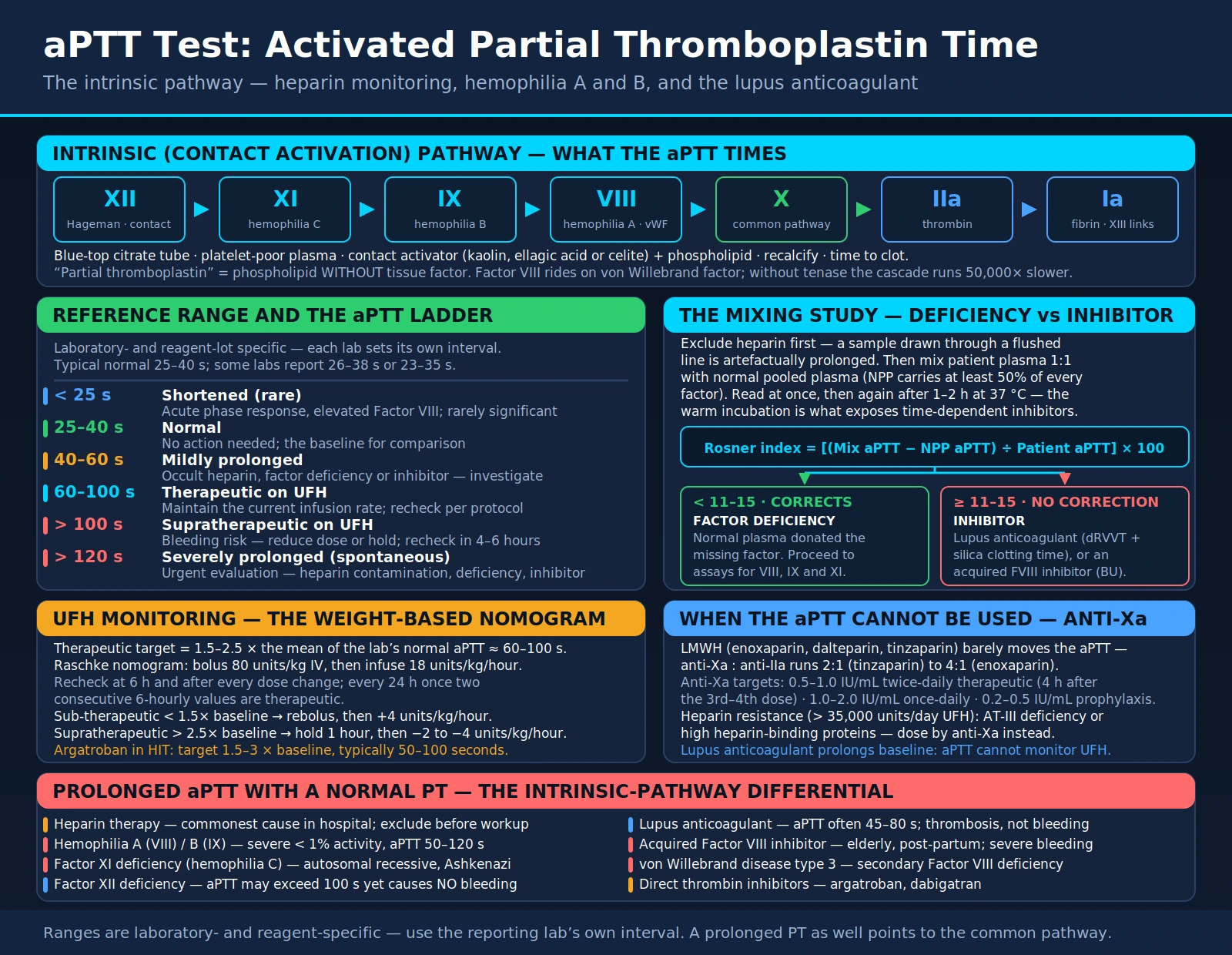
The activated partial thromboplastin time (aPTT) measures how long plasma takes to clot through the intrinsic (contact) and common pathways of coagulation. The StatPearls chapter on the test describes it as checking the function of every clotting factor except Factor VII and Factor XIII. It is used to monitor unfractionated heparin (UFH), as a screening test for factor deficiencies such as hemophilia A and B, and in the work-up for lupus anticoagulant. The same prolonged aPTT can come from very different causes: a missing factor, an antibody against a factor, a drug, or lupus anticoagulant, which prolongs the test in the tube yet is linked to clotting rather than bleeding in the body.
Table of Contents
- Overview — Intrinsic Coagulation Pathway
- How the aPTT Is Measured
- Normal Range and Therapeutic Targets
- Heparin Monitoring with aPTT
- Unfractionated vs. Low-Molecular-Weight Heparin
- Prolonged aPTT — Differential Diagnosis
- The Mixing Study — Factor Deficiency vs. Inhibitor
- Hemophilia A and B
- Lupus Anticoagulant and Antiphospholipid Syndrome
- Key Research Papers
- Connections
- Featured Videos
Overview — Intrinsic Coagulation Pathway
The intrinsic (contact activation) pathway begins when Factor XII is activated on a negatively charged surface; in the test tube, glass or an added activator supplies that surface (Winter 2017). Activation then runs from Factor XII to Factor XI to Factor IX, which, with Factor VIII as its cofactor, activates Factor X. Activated Factor X, together with Factor Va (the prothrombinase complex), converts prothrombin to thrombin, and thrombin cleaves fibrinogen to fibrin. Factor XIII then cross-links the fibrin into a stable clot; the aPTT does not measure Factor XIII.
Factor VIII circulates bound to von Willebrand factor (VWF), which acts as its carrier and maintains its level in the blood (StatPearls: Von Willebrand Disease). Activated Factor IX forms the tenase complex with activated Factor VIII, calcium and a phospholipid surface, and this complex greatly speeds the activation of Factor X. Losing either Factor VIII or Factor IX weakens thrombin generation and clot formation, which is why a deficiency of either one causes hemophilia (StatPearls: Hemophilia A).
Deficiency of Factor VIII causes hemophilia A; deficiency of Factor IX causes hemophilia B (Christmas disease). Both genes lie on the X chromosome and both conditions are inherited in an X-linked recessive pattern, so they mainly affect males. Female carriers have variable factor levels: the StatPearls chapter on hemophilia B notes that carriers with Factor IX activity of at least 50% of normal generally have no symptoms, while carriers with lower levels can have clinically significant bleeding. Both hemophilias prolong the aPTT and leave the prothrombin time (PT) normal, because the PT starts with tissue factor and Factor VII and does not depend on Factors VIII or IX.
The two pathways meet at Factor X, the start of the so-called common pathway of Factors X, V, II (prothrombin) and fibrinogen. A marked deficiency of a common-pathway factor can therefore prolong both the PT and the aPTT, a pattern that helps narrow the list of possible causes.
How the aPTT Is Measured
Blood is drawn into a light-blue-top tube containing sodium citrate, which binds calcium and stops clotting in the tube. The ratio of blood to citrate matters. An under-filled tube leaves extra citrate for the amount of plasma, and a very high hematocrit does the same because the sample holds less plasma. In a study of 28 patients with hematocrit values of 55% to 72%, PT and aPTT results in tubes with unadjusted citrate differed significantly from adjusted tubes and rose exponentially with the hematocrit; the authors concluded that citrate has to be adjusted in these patients for accurate results (Marlar 2006).
In the laboratory, platelet-poor plasma is separated by centrifugation and then incubated with a contact activator — kaolin (a silicate clay), ellagic acid, silica or celite (diatomaceous earth) — which provides the negatively charged surface that activates Factor XII. Phospholipid (a stand-in for platelet membranes) is added to support the tenase and prothrombinase complexes. This is the "partial thromboplastin" in the test name: phospholipid without tissue factor. The PT reagent, by contrast, contains tissue factor as well as phospholipid and starts the extrinsic pathway.
After incubation, the plasma mixture is recalcified with calcium chloride. A photometric detector or mechanical wire detects clot formation; the elapsed time from calcium addition to clot formation is reported as the aPTT in seconds. The "activated" part of the name refers to the added contact activator, which standardizes Factor XII activation and makes results reproducible within a laboratory.
The aPTT is much more sensitive to heparin than the PT. Heparin acts through antithrombin, which then inactivates thrombin and Factor Xa — both enzymes the aPTT depends on. The PT is relatively insensitive to heparin at usual therapeutic levels.
Preanalytical variables that affect the result include under-filled tubes; the temperature, timing and conditions of transport and storage; centrifugation that leaves platelets in the plasma (Adcock Funk 2012); hemolysis, icterus and lipemia (Lippi 2013); and a very high hematocrit (Marlar 2006).
Normal Range and Therapeutic Targets
MedlinePlus gives a general aPTT range of 25 to 35 seconds and notes that normal value ranges vary among laboratories. Each laboratory sets its own reference interval, which depends on its reagent and instrument, so a result is read against the range reported by the laboratory that ran it.
There is no fixed therapeutic aPTT in seconds for unfractionated heparin. The 2012 American College of Chest Physicians (ACCP/CHEST) review explains that a therapeutic range of 1.5 to 2.5 times control gained wide acceptance after a retrospective study in the 1970s, and that its clinical relevance is uncertain because randomized trials have not confirmed it. The review also notes that an aPTT ratio of 1.5 to 2.5 corresponded to a heparin level of 0.2 to 0.4 units/mL by protamine titration and 0.3 to 0.7 units/mL by an anti-Xa assay, and that aPTT reagents and instruments differ in their response to heparin (Garcia 2012). Hospitals therefore set their own therapeutic aPTT range for the reagent they use.
| Situation | What the sources describe |
|---|---|
| Within the laboratory's reference range | MedlinePlus example: 25 to 35 seconds; ranges vary among laboratories |
| Above the reference range, not on an anticoagulant | MedlinePlus lists bleeding disorders, disseminated intravascular coagulation, liver disease, malabsorption, low vitamin K and lupus anticoagulant (the last associated with clotting rather than bleeding); see the differential diagnosis below |
| On unfractionated heparin | Read against the hospital's own therapeutic range for its reagent (Garcia 2012); some hospitals measure anti-Xa activity instead |
| On argatroban | The US prescribing information sets the target at 1.5 to 3 times the initial baseline value, not to exceed 100 seconds |
| On low-molecular-weight heparin or emicizumab | The aPTT is not used to monitor either drug (enoxaparin and emicizumab prescribing information) |
In hemophilia, factor activity is measured directly rather than using the aPTT as a treatment target. The argatroban prescribing information gives its aPTT target for heparin-induced thrombocytopenia (HIT) as 1.5 to 3 times the initial baseline value, not to exceed 100 seconds. For bivalirudin, some protocols use the aPTT, while the activated clotting time (ACT) is used during procedures.
Heparin Monitoring with aPTT
Unfractionated heparin (UFH) is a mixture of polysaccharide chains ranging in molecular weight from 3,000 to 30,000 daltons, with a mean of about 15,000 (Garcia 2012). Chains that carry a specific pentasaccharide sequence bind antithrombin and convert it from a slow to a rapid inhibitor; the heparin–antithrombin complex inactivates thrombin (Factor IIa) and Factor Xa, along with other clotting enzymes.
Because heparin binds plasma proteins, endothelial cells and macrophages to differing degrees, its anticoagulant effect varies from patient to patient and cannot be predicted from the dose alone. Laboratory monitoring has therefore been standard practice, and the aPTT has been the usual monitoring test for decades (Garcia 2012).
Weight-based heparin dosing comes from a 1993 randomized trial of 115 patients (Raschke 1993). The weight-based arm started with an 80 units/kg bolus and an infusion of 18 units/kg per hour; the comparison ("standard care") arm started with a 5,000-unit bolus and 1,000 units per hour. The aPTT was measured every 6 hours and doses were adjusted by each nomogram, with a therapeutic range of 1.5 to 2.3 times control. Within 24 hours, 60 of 62 patients (97%) on the weight-based nomogram exceeded the therapeutic threshold, compared with 37 of 48 (77%) on standard care, and recurrent thromboembolism was more frequent with standard care (relative risk 5.0).
aPTT or Anti-Xa for Heparin Monitoring
The anti-Xa assay measures heparin's inhibition of Factor Xa rather than a clotting time. Patients who need unusually high doses of heparin to reach a therapeutic aPTT are described as heparin resistant. In a randomized trial of patients with acute venous thromboembolism who needed 35,000 units or more of heparin a day, monitoring by anti-Xa level (target 0.35 to 0.67 units/mL) or by aPTT (target 60 to 85 seconds) gave similar results: recurrent venous thromboembolism occurred in 3 of 65 patients (4.6%) in the anti-Xa group and 4 of 66 (6.1%) in the aPTT group, and the aPTT group received significantly more heparin (Levine 1994). The 2012 ACCP/CHEST review states that, given these results, it is reasonable to adjust heparin doses by anti-Xa level in such patients, and adds that anti-Xa assays, like aPTT assays, vary in their response to heparin (Garcia 2012). The American Society of Hematology (ASH) 2018 guideline on anticoagulation management addresses anti-Xa monitoring for low-molecular-weight heparin (below) (Witt 2018). A 2017 laboratory review describes the anti-Xa assay as now the preferred measure of the effect of unfractionated heparin (Winter 2017), and some hospitals use it in place of the aPTT.
Heparin-induced thrombocytopenia (HIT) is an adverse drug reaction in which antibodies against complexes of platelet factor 4 and heparin activate platelets; patients have a falling platelet count and a markedly increased risk of thrombosis (Cuker 2018). The ASH 2018 HIT guideline makes conditional recommendations on the choice among non-heparin anticoagulants — argatroban, bivalirudin, danaparoid, fondaparinux and direct oral anticoagulants — for treating acute HIT. The aPTT is used to monitor argatroban.
Unfractionated vs. Low-Molecular-Weight Heparin
Low-molecular-weight heparins (LMWHs), including enoxaparin, dalteparin and tinzaparin, are made by breaking UFH into shorter chains, with a mean molecular weight of 4,000 to 5,000 (Garcia 2012). Most of these chains still bind antithrombin but are too short to bridge antithrombin to thrombin, so LMWHs act more against Factor Xa than against thrombin, with anti-Xa to anti-IIa ratios between 2:1 and 4:1.
The bioavailability of LMWHs after subcutaneous injection is about 90%, whereas low and moderate subcutaneous doses of UFH have reduced plasma recovery (Garcia 2012). LMWH doses are based on body weight; for acute deep vein thrombosis the enoxaparin prescribing information lists 1 mg/kg every 12 hours, or 1.5 mg/kg once a day for inpatient treatment.
The enoxaparin prescribing information states that the PT and aPTT are not adequate for monitoring the anticoagulant effect of enoxaparin.
Anti-Xa measurement for LMWH comes up in a few settings, and the sources differ:
- Renal impairment: the enoxaparin prescribing information says anti-Xa levels may be used to monitor patients with renal impairment. For creatinine clearance below 30 mL/min, the ASH 2018 guideline suggests against using anti-Xa monitoring to guide LMWH dose adjustment (a conditional recommendation), pointing instead to labeled renal dose adjustment or a different anticoagulant (Witt 2018).
- Obesity: ASH 2018 suggests against anti-Xa monitoring to guide LMWH dosing and bases treatment doses on actual body weight (Witt 2018).
- Pregnancy: the ASH 2018 pregnancy guideline makes a conditional recommendation against routine anti-Xa monitoring to guide LMWH dosing for treatment of venous thromboembolism (Bates 2018).
- Abnormal coagulation or bleeding during therapy: the enoxaparin prescribing information says anti-Xa levels may be used.
Where anti-Xa is measured for twice-daily enoxaparin in venous thromboembolism, the ACCP/CHEST review gives a conservative peak target, measured 4 hours after a dose, of 0.6 to 1.0 units/mL, and states that the target for once-daily enoxaparin is likely to be above 1.0 units/mL. Fondaparinux, a synthetic pentasaccharide, inhibits Factor Xa through antithrombin and does not increase the rate of thrombin inhibition; routine monitoring is not recommended in that review, and a fondaparinux-calibrated anti-Xa assay is used when a measurement is needed (Garcia 2012).
Prolonged aPTT — Differential Diagnosis
A prolonged aPTT is a result above the laboratory's reference range. The clinical setting — anticoagulant use, a personal or family bleeding history, or a clotting event — and the PT result together narrow the possibilities considerably.
Isolated Prolonged aPTT (PT Normal)
This pattern points to the intrinsic pathway (Factors XII, XI, IX, VIII) or to a substance that interferes with it:
- Heparin: unfractionated heparin prolongs the aPTT, and MedlinePlus lists heparin among the causes of a long result; a sample drawn through a heparin-flushed line can be contaminated with it
- Hemophilia A (Factor VIII deficiency): X-linked; bleeding severity follows the residual factor level
- Hemophilia B (Factor IX deficiency, Christmas disease): clinically similar to hemophilia A; distinguished by factor assay
- Factor XI deficiency (sometimes called hemophilia C): an autosomal recessive, injury-related bleeding tendency that is common in Jewish people, particularly of Ashkenazi origin (Seligsohn 2009)
- Factor XII deficiency: prolongs the aPTT but does not cause bleeding. A review of surgical procedures in people with severe (homozygous) Factor XII deficiency, including major operations such as coronary bypass and gastrectomy, found neither bleeding nor thrombotic complications (Girolami 2004)
- Lupus anticoagulant: an antibody that interferes with phospholipid-dependent clotting tests in the tube; MedlinePlus notes that it is associated with a clotting problem rather than bleeding
- Von Willebrand disease (VWD): VWD lowers Factor VIII only when VWF is missing or cannot carry it, so the aPTT is often normal — in one clinic series all four women diagnosed with VWD had a normal aPTT (Hassan 2012). It is prolonged when Factor VIII falls far enough, as in type 3 VWD, where VWF is virtually absent, and in type 2N, where VWF cannot bind Factor VIII and the disorder is clinically similar to mild hemophilia (Mazurier 2001)
- Direct oral anticoagulants: dabigatran (a direct thrombin inhibitor) and the anti-Xa drugs rivaroxaban and apixaban have variable effects on clotting tests. For dabigatran, the aPTT and PT are less sensitive than drug-specific tests and may be normal at trough levels; for rivaroxaban and apixaban, the aPTT lacks the sensitivity and linearity to measure drug levels (Cuker 2014). Drug-specific anti-Xa or anti-IIa assays are the preferred methods when a quantitative result is useful (Douxfils 2018)
- Argatroban and bivalirudin: intravenous direct thrombin inhibitors that prolong the aPTT; argatroban is monitored with it
- Acquired Factor VIII inhibitor (acquired hemophilia A): autoantibodies against Factor VIII in men and women with no previous history of bleeding, typically presenting with an isolated prolonged aPTT; about 10% of patients do not present with bleeding (Tiede 2020). It tends to occur in elderly people with other illnesses, and bleeding is often spontaneous and severe (Kruse-Jarres 2017)
Both PT and aPTT Prolonged
This pattern suggests a common-pathway defect, a deficiency of several factors, or a broad anticoagulant effect:
- Severe liver disease or hepatic failure: the liver synthesizes Factors I, II, V, VII, IX, X and XI; severe dysfunction impairs synthesis of both vitamin K-dependent and other factors (MedlinePlus lists liver disease among causes of a long PTT)
- Disseminated intravascular coagulation (DIC): consumption of clotting factors lowers several at once; low platelets and a raised D-dimer accompany the coagulopathy
- Warfarin or vitamin K deficiency: lower Factors II, VII, IX and X and mainly prolong the PT; MedlinePlus also lists low vitamin K and malabsorption among causes of a long PTT
- Massive transfusion or dilutional coagulopathy: large-volume replacement without enough clotting-factor replacement
- Common pathway factor deficiency: rare isolated deficiency of Factor X, V, II or fibrinogen (afibrinogenemia/hypofibrinogenemia)
- Amyloidosis: acquired Factor X deficiency is described in systemic amyloidosis (Furie 1981)
The Mixing Study — Factor Deficiency vs. Inhibitor
When a prolonged aPTT is unexpected, heparin contamination — for example from a heparin flush in an indwelling catheter — is one of the first explanations laboratories look for, because a specimen drawn through a heparin-containing line can give an entirely artifactual result; a fresh sample from a peripheral vein settles the question. Once heparin is excluded, the usual next step is a mixing study.
A mixing test combines the patient's plasma with normal plasma and repeats the clotting test on the mixture (Kershaw 2013):
- If the result normalizes, the patient's plasma is likely deficient in one or more clotting factors that the normal plasma has supplied, and specific factor assays follow.
- If the result stays prolonged, an inhibitor or other interference is likely — for example lupus anticoagulant, a specific factor inhibitor, or a high dose of a heparin-like anticoagulant.
Typical steps:
- Patient plasma is mixed with normal pooled plasma (NPP) in a 1:1 ratio.
- The aPTT is measured on the mix straight away (immediate mix).
- The mix is incubated at 37°C for 1–2 hours and measured again (incubated mix).
- Both results are compared with the aPTT of NPP alone and of the patient's plasma alone.
Interpretation criteria have differed between laboratories; Kershaw and Orellana describe standardized procedures and validated cutoffs as the most favorable approach (Kershaw 2013), and the International Council for Standardization in Haematology (ICSH) published recommendations for performing and interpreting aPTT and PT mixing tests in 2024 (Adcock 2024).
The incubation step matters for time-dependent inhibitors — particularly the acquired Factor VIII inhibitor, an IgG antibody that acts slowly at 37°C, so the immediate mix may look corrected while the incubated mix fails to correct. Lupus anticoagulant, by contrast, typically fails to correct on both the immediate and the incubated mix.
One way to express the result is the Rosner index, also called the index of circulating anticoagulant (ICA), proposed by Rosner and colleagues in 1987 (Rosner 1987):
Rosner index = [(Mix aPTT − NPP aPTT) ÷ Patient aPTT] × 100
- Below the laboratory's cutoff: correction, pointing to a factor deficiency; individual factor assays (VIII, IX, XI) follow
- At or above the cutoff: non-correction, pointing to an inhibitor; inhibitor identification follows
- Cutoffs are specific to the reagent; one 2019 study used cutoffs of 10.4 to 13.6 depending on the reagent, and found the index more specific than a mixing-test-specific cutoff (Kumano 2019)
Two major inhibitor categories:
- Lupus anticoagulant (LA): a phospholipid-dependent antibody that is associated with thrombosis rather than bleeding. The 2020 International Society on Thrombosis and Haemostasis (ISTH) guidance keeps a three-step procedure — screening, mixing and confirmation, in which excess phospholipid shortens the clotting time — with two test systems, the dilute Russell's viper venom time (dRVVT) and an aPTT, with silica the preferred activator (Devreese 2020)
- Acquired Factor VIII inhibitor: an autoantibody against Factor VIII in a person with no previous bleeding history; Factor VIII activity is low, and the inhibitor is measured with the Nijmegen-modified Bethesda assay in Bethesda units. The 2020 international recommendations list bypassing agents (recombinant activated Factor VII or activated prothrombin complex concentrate) or recombinant porcine Factor VIII for bleeding, and immunosuppressive therapy to eradicate the antibody (Tiede 2020)
Hemophilia A and B
Hemophilia A (Factor VIII deficiency) and hemophilia B (Factor IX deficiency) are inherited bleeding disorders caused by variants in genes on the X chromosome (Factor VIII gene: Xq28; Factor IX gene: Xq27.1), with X-linked recessive inheritance. A 2019 meta-analysis of national registries estimated prevalence at birth at 24.6 per 100,000 males for hemophilia A and 5.0 per 100,000 males for hemophilia B (9.5 and 1.5 per 100,000 for the severe forms), and found that people with hemophilia in high-income countries still have a life-expectancy disadvantage (Iorio 2019). Some cases arise from new (de novo) mutations with no family history (StatPearls: Hemophilia B).
Severity Classification
The ISTH definitions classify severity by residual clotting factor activity (White 2001), and bleeding severity follows that level:
| Severity | Factor Activity | Clinical Manifestations |
|---|---|---|
| Severe | < 1% (< 0.01 IU/mL) | Spontaneous joint bleeding (hemarthrosis), deep muscle hematomas and life-threatening bleeding; in hemophilia B, spontaneous bleeding may begin in the newborn period or early infancy |
| Moderate | 1–5% (0.01–0.05 IU/mL) | Excessive bleeding mainly after trauma, dental procedures or surgery, with occasional spontaneous bleeding |
| Mild | 5–40% (0.05–0.40 IU/mL) | Bleeding mainly after trauma, dental procedures or surgery; may go unrecognized until later childhood or adulthood |
Recurrent joint bleeding can lead to chronic hemophilic arthropathy and long-term disability (StatPearls: Hemophilia A).
Laboratory Pattern
In both hemophilia A and B:
- aPTT: prolonged; how much depends on the factor level and on the reagent's sensitivity to that factor (Kershaw 2013)
- PT/INR: normal (extrinsic pathway unaffected)
- Platelet count: normal
- Thrombin time: normal
- Fibrinogen: normal
- Factor VIII level: reduced in hemophilia A; normal in hemophilia B
- Factor IX level: reduced in hemophilia B; normal in hemophilia A
- Mixing study: corrects (factor deficiency pattern), unless an inhibitor has developed
Treatment Advances
Treatment of hemophilia has changed considerably:
- Factor concentrates: plasma-derived or recombinant Factor VIII or IX, given to treat bleeding or as regular prophylaxis; their use is covered in the World Federation of Hemophilia (WFH) guidelines, 3rd edition (Srivastava 2020)
- Emicizumab: a bispecific monoclonal antibody that bridges activated Factor IX and Factor X to restore the function of missing activated Factor VIII; given by subcutaneous injection every week, every 2 weeks or every 4 weeks; labeled for prophylaxis in hemophilia A with or without Factor VIII inhibitors. Its prescribing information states that it affects the aPTT and all aPTT-based assays, which are not used to monitor it
- Gene therapy: the US Food and Drug Administration's list of approved cellular and gene therapy products includes valoctocogene roxaparvovec for hemophilia A, and etranacogene dezaparvovec and fidanacogene elaparvovec for hemophilia B
Some people with hemophilia develop alloantibodies (inhibitors) against infused factor concentrate. In a cohort of 574 previously untreated children with severe hemophilia A, inhibitors developed in 177 (cumulative incidence 32.4%), and 116 developed a high-titer inhibitor, defined as a peak titer of at least 5 Bethesda units per mL (cumulative incidence 22.4%) (Gouw 2013). Inhibitors are identified and quantified with the Bethesda assay. Immune tolerance induction (prolonged regular factor infusions intended to eradicate the inhibitor) and bypassing agents for acute bleeds are among the approaches the WFH guidelines cover (Srivastava 2020).
Lupus Anticoagulant and Antiphospholipid Syndrome
Lupus anticoagulant (LA) is one of the most counter-intuitive results in coagulation testing: it prolongs clotting tests in the tube, yet in the body it is a risk factor for arterial and venous thrombosis and pregnancy loss. The StatPearls chapter calls the name a misnomer, since LA is neither found only in lupus nor mainly associated with bleeding. The term was first used for plasma samples from people with systemic lupus erythematosus (SLE) that failed to clot in the expected time, and LA can be transient in the setting of certain medications or infections, so it is also found in people without symptoms (StatPearls: Biochemistry, Lupus Anticoagulant).
The lupus anticoagulant is a member of the broader antiphospholipid antibody (aPL) family, which also includes:
- Anticardiolipin antibodies (aCL): IgG or IgM antibodies against the phospholipid cardiolipin, measured by ELISA
- Anti-β2-glycoprotein I (anti-β2GPI) antibodies: IgG or IgM, measured by ELISA; β2GPI is a phospholipid-binding protein that is the target of many aPL
The in vitro mechanism of LA prolongation: LA antibodies bind to phospholipid-binding proteins (primarily β2GPI) sitting on phospholipid surfaces. This blocks the phospholipid platform required for the tenase and prothrombinase complexes in coagulation assays, slowing the cascade and prolonging the aPTT and other phospholipid-dependent tests. Adding excess phospholipid overcomes the antibody and shortens the clotting time, which is the basis of the confirmatory step in LA testing.
Proposed mechanisms of thrombosis in the body include activation of endothelial cells and platelets and interference with the protein C anticoagulant pathway. The result is venous and arterial thrombosis; StatPearls notes that the most common sites are the veins of the legs and the arteries of the brain, though thrombosis can occur in any organ (StatPearls: Antiphospholipid Syndrome).
Diagnosis of Antiphospholipid Syndrome
The revised Sapporo/Sydney criteria (Miyakis et al., 2006) require at least one clinical criterion AND at least one laboratory criterion confirmed on two occasions ≥ 12 weeks apart:
- Clinical criteria: (1) vascular thrombosis (any vessel, any organ, confirmed by imaging or histopathology); (2) obstetric morbidity (≥3 unexplained consecutive miscarriages before week 10; ≥1 unexplained fetal death at ≥10 weeks; ≥1 premature birth ≤34 weeks due to severe pre-eclampsia, eclampsia, or placental insufficiency)
- Laboratory criteria: LA detected by phospholipid-dependent coagulation tests; or medium-to-high titer anticardiolipin antibodies; or anti-β2GPI antibodies of IgG or IgM isotype
The 2023 ACR/EULAR classification criteria, built for research studies, use an entry criterion of at least one positive aPL test within 3 years of a qualifying clinical event, followed by weighted clinical and laboratory domains; a patient with at least 3 points from each is classified as having APS. In the validation cohort, the new criteria had a specificity of 99% versus 86% for the 2006 criteria, and a sensitivity of 84% versus 99% (Barbhaiya 2023).
aPTT in APS Diagnosis and Monitoring
The aPTT does not always detect LA. In a 1987 study of 66 patients with SLE, 49% had LA activity, and the aPTT gave false-negative results in 73% and 52% of cases with two different reagents (Rosner 1987). For this reason the ISTH guidance uses two test systems — the dRVVT and an LA-sensitive aPTT — each with screening, mixing and confirmation steps (Devreese 2020). When LA already prolongs a person's baseline aPTT, the aPTT no longer reflects heparin effect alone, which complicates monitoring of unfractionated heparin. ISTH guidance also covers LA testing in people already taking anticoagulants, including anti-Xa measurement for heparins and specific assays for direct oral anticoagulants (Devreese 2020; Tripodi 2020).
Treatment
The 2019 EULAR recommendations for adults with APS (Tektonidou 2019) include:
- First unprovoked venous thrombosis: long-term treatment with a vitamin K antagonist such as warfarin, target INR 2–3
- First arterial thrombosis: a vitamin K antagonist with INR 2–3 or INR 3–4, weighing the individual's bleeding and thrombosis risk
- Triple aPL positivity (LA, anticardiolipin and anti-β2GPI): the recommendations state that rivaroxaban should not be used. In the randomized TRAPS trial of 120 triple-positive patients, stopped early, events occurred in 11 of 59 (19%) on rivaroxaban and 2 of 61 (3%) on warfarin, including 7 thromboembolic events (12%) with rivaroxaban and none with warfarin (Pengo 2018)
- Recurrent thrombosis despite adequate treatment: adding low-dose aspirin, raising the INR target to 3–4 or switching to LMWH may be considered
- Prior obstetric APS: low-dose aspirin plus prophylactic-dose heparin during pregnancy; with recurrent pregnancy complications, a therapeutic heparin dose, hydroxychloroquine or low-dose prednisolone in the first trimester may be considered
Key Research Papers
- Raschke RA, Reilly BM, Guidry JR, Fontana JR, Srinivas S (1993). The weight-based heparin dosing nomogram compared with a "standard care" nomogram. A randomized controlled trial. Ann Intern Med. 119(9):874–881. — PubMed PMID: 8214998
- Garcia DA, Baglin TP, Weitz JI, Samama MM; American College of Chest Physicians (2012). Parenteral anticoagulants: antithrombotic therapy and prevention of thrombosis, 9th ed. Chest. 141(2 Suppl):e24S–e43S. — PubMed PMID: 22315264
- Miyakis S, Lockshin MD, Atsumi T, et al. (2006). International consensus statement on an update of the classification criteria for definite antiphospholipid syndrome (APS). J Thromb Haemost. 4(2):295–306. — PubMed PMID: 16420554
- Kershaw G, Orellana D (2013). Mixing tests: diagnostic aides in the investigation of prolonged prothrombin times and activated partial thromboplastin times. Semin Thromb Hemost. 39(3):283–290. — PubMed PMID: 23457048
- Lusher JM (2000). Inhibitor antibodies to factor VIII and factor IX: management. Semin Thromb Hemost. 26(2):179–188. — PubMed PMID: 10919411
- Ginsberg JS (1996). Management of venous thromboembolism. N Engl J Med. 335(24):1816–1828. — PubMed PMID: 8943165
- Favaloro EJ (2014). Diagnosing von Willebrand disease: a short history of laboratory milestones and innovations, plus current status, challenges, and solutions. Semin Thromb Hemost. 40(5):551–570. — PubMed PMID: 24978322
- Moake JL (2002). Thrombotic microangiopathies. N Engl J Med. 347(8):589–600. — PubMed PMID: 12192020
- Levine MN, Hirsh J, Gent M, et al. (1994). A randomized trial comparing activated thromboplastin time with heparin assay in patients with acute venous thromboembolism requiring large daily doses of heparin. Arch Intern Med. 154(1):49–56. — PubMed PMID: 8267489
- Witt DM, Nieuwlaat R, Clark NP, et al. (2018). American Society of Hematology 2018 guidelines for management of venous thromboembolism: optimal management of anticoagulation therapy. Blood Adv. 2(22):3257–3291. — PubMed PMID: 30482765
- Bates SM, Rajasekhar A, Middeldorp S, et al. (2018). American Society of Hematology 2018 guidelines for management of venous thromboembolism: venous thromboembolism in the context of pregnancy. Blood Adv. 2(22):3317–3359. — PubMed PMID: 30482767
- Cuker A, Arepally GM, Chong BH, et al. (2018). American Society of Hematology 2018 guidelines for management of venous thromboembolism: heparin-induced thrombocytopenia. Blood Adv. 2(22):3360–3392. — PubMed PMID: 30482768
- Winter WE, Flax SD, Harris NS (2017). Coagulation testing in the core laboratory. Lab Med. 48(4):295–313. — PubMed PMID: 29126301
- Cuker A, Siegal DM, Crowther MA, Garcia DA (2014). Laboratory measurement of the anticoagulant activity of the non-vitamin K oral anticoagulants. J Am Coll Cardiol. 64(11):1128–1139. — PubMed PMID: 25212648
- Douxfils J, Ageno W, Samama CM, et al. (2018). Laboratory testing in patients treated with direct oral anticoagulants: a practical guide for clinicians. J Thromb Haemost. 16(2):209–219. — PubMed PMID: 29193737
- Marlar RA, Potts RM, Marlar AA (2006). Effect on routine and special coagulation testing values of citrate anticoagulant adjustment in patients with high hematocrit values. Am J Clin Pathol. 126(3):400–405. — PubMed PMID: 16880137
- Adcock Funk DM, Lippi G, Favaloro EJ (2012). Quality standards for sample processing, transportation, and storage in hemostasis testing. Semin Thromb Hemost. 38(6):576–585. — PubMed PMID: 22706973
- Lippi G, Plebani M, Favaloro EJ (2013). Interference in coagulation testing: focus on spurious hemolysis, icterus, and lipemia. Semin Thromb Hemost. 39(3):258–266. — PubMed PMID: 23229354
- Adcock DM, Moore GW, Kershaw GW, Montalvao SAL, Gosselin RC (2024). International Council for Standardization in Haematology (ICSH) recommendations for the performance and interpretation of activated partial thromboplastin time and prothrombin time mixing tests. Int J Lab Hematol. 46(5):777–788. — PubMed PMID: 39009028
- Rosner E, Pauzner R, Lusky A, Modan M, Many A (1987). Detection and quantitative evaluation of lupus circulating anticoagulant activity. Thromb Haemost. 57(2):144–147. — PubMed PMID: 3110995
- Kumano O, Moore GW (2019). Ruling out lupus anticoagulants with mixing test-specific cutoff assessment and the index of circulating anticoagulant. Res Pract Thromb Haemost. 3(4):695–703. — PubMed PMID: 31624789
- Devreese KMJ, de Groot PG, de Laat B, et al. (2020). Guidance from the Scientific and Standardization Committee for lupus anticoagulant/antiphospholipid antibodies of the International Society on Thrombosis and Haemostasis: update of the guidelines for lupus anticoagulant detection and interpretation. J Thromb Haemost. 18(11):2828–2839. — PubMed PMID: 33462974
- Tripodi A, Cohen H, Devreese KMJ (2020). Lupus anticoagulant detection in anticoagulated patients. Guidance from the Scientific and Standardization Committee for lupus anticoagulant/antiphospholipid antibodies of the International Society on Thrombosis and Haemostasis. J Thromb Haemost. 18(7):1569–1575. — PubMed PMID: 32619349
- Girolami A, Ruzzon E, Lombardi AM, Cabrio L, Randi ML (2004). Thrombosis-free surgical procedures in severe (homozygote) factor XII deficiency: report of four additional cases and literature review. Clin Appl Thromb Hemost. 10(4):351–355. — PubMed PMID: 15497021
- Seligsohn U (2009). Factor XI deficiency in humans. J Thromb Haemost. 7 Suppl 1:84–87. — PubMed PMID: 19630775
- Hassan R, Yusof WA, Hussain NH, Abdullah WZ (2012). Single-center experience of von Willebrand disease (vWD) among patients with menorrhagia: a diagnosis which could be missed. Indian J Hematol Blood Transfus. 28(3):157–161. — PubMed PMID: 23997452
- Mazurier C, Goudemand J, Hilbert L, et al. (2001). Type 2N von Willebrand disease: clinical manifestations, pathophysiology, laboratory diagnosis and molecular biology. Best Pract Res Clin Haematol. 14(2):337–347. — PubMed PMID: 11686103
- Tiede A, Collins P, Knoebl P, et al. (2020). International recommendations on the diagnosis and treatment of acquired hemophilia A. Haematologica. 105(7):1791–1801. — PubMed PMID: 32381574
- Kruse-Jarres R, Kempton CL, Baudo F, et al. (2017). Acquired hemophilia A: updated review of evidence and treatment guidance. Am J Hematol. 92(7):695–705. — PubMed PMID: 28470674
- Furie B, Voo L, McAdam KP, Furie BC (1981). Mechanism of factor X deficiency in systemic amyloidosis. N Engl J Med. 304(14):827–830. — PubMed PMID: 7207512
- Iorio A, Stonebraker JS, Chambost H, et al. (2019). Establishing the prevalence and prevalence at birth of hemophilia in males: a meta-analytic approach using national registries. Ann Intern Med. 171(8):540–546. — PubMed PMID: 31499529
- White GC 2nd, Rosendaal F, Aledort LM, et al. (2001). Definitions in hemophilia. Recommendation of the scientific subcommittee on factor VIII and factor IX of the scientific and standardization committee of the International Society on Thrombosis and Haemostasis. Thromb Haemost. 85(3):560. — PubMed PMID: 11307831
- Srivastava A, Santagostino E, Dougall A, et al. (2020). WFH Guidelines for the Management of Hemophilia, 3rd edition. Haemophilia. 26 Suppl 6:1–158. — PubMed PMID: 32744769
- Gouw SC, van der Bom JG, Ljung R, et al. (2013). Factor VIII products and inhibitor development in severe hemophilia A. N Engl J Med. 368(3):231–239. — PubMed PMID: 23323899
- Barbhaiya M, Zuily S, Naden R, et al. (2023). 2023 ACR/EULAR antiphospholipid syndrome classification criteria. Ann Rheum Dis. 82(10):1258–1270. — PubMed PMID: 37640450
- Tektonidou MG, Andreoli L, Limper M, et al. (2019). EULAR recommendations for the management of antiphospholipid syndrome in adults. Ann Rheum Dis. 78(10):1296–1304. — PubMed PMID: 31092409
- Pengo V, Denas G, Zoppellaro G, et al. (2018). Rivaroxaban vs warfarin in high-risk patients with antiphospholipid syndrome. Blood. 132(13):1365–1371. — PubMed PMID: 30002145
PubMed Topic Searches
- APTT and heparin monitoring — PubMed
- Lupus anticoagulant and antiphospholipid syndrome — PubMed
- Hemophilia and Factor VIII inhibitor treatment — PubMed
- Mixing study for coagulation inhibitor vs. factor deficiency — PubMed
External Authoritative Resources
- MedlinePlus Medical Encyclopedia — Partial thromboplastin time (PTT)
- StatPearls / NCBI Bookshelf — Partial Thromboplastin Time
- StatPearls / NCBI Bookshelf — Interpretation of Blood Clotting Studies and Values (PT, PTT, aPTT, INR, Anti-Factor Xa, D-Dimer)
- StatPearls / NCBI Bookshelf — Von Willebrand Disease
- StatPearls / NCBI Bookshelf — Hemophilia A
- StatPearls / NCBI Bookshelf — Hemophilia B
- StatPearls / NCBI Bookshelf — Acquired Hemophilia
- StatPearls / NCBI Bookshelf — Biochemistry, Lupus Anticoagulant
- StatPearls / NCBI Bookshelf — Antiphospholipid Syndrome
- StatPearls / NCBI Bookshelf — Heparin
- StatPearls / NCBI Bookshelf — Low-Molecular-Weight Heparin (LMWH)
- StatPearls / NCBI Bookshelf — Heparin-Induced Thrombocytopenia
- DailyMed — argatroban injection prescribing information
- DailyMed — enoxaparin sodium injection prescribing information
- DailyMed — emicizumab-kxwh prescribing information
- US FDA — Approved Cellular and Gene Therapy Products