PT/INR Test: Prothrombin Time and International Normalized Ratio
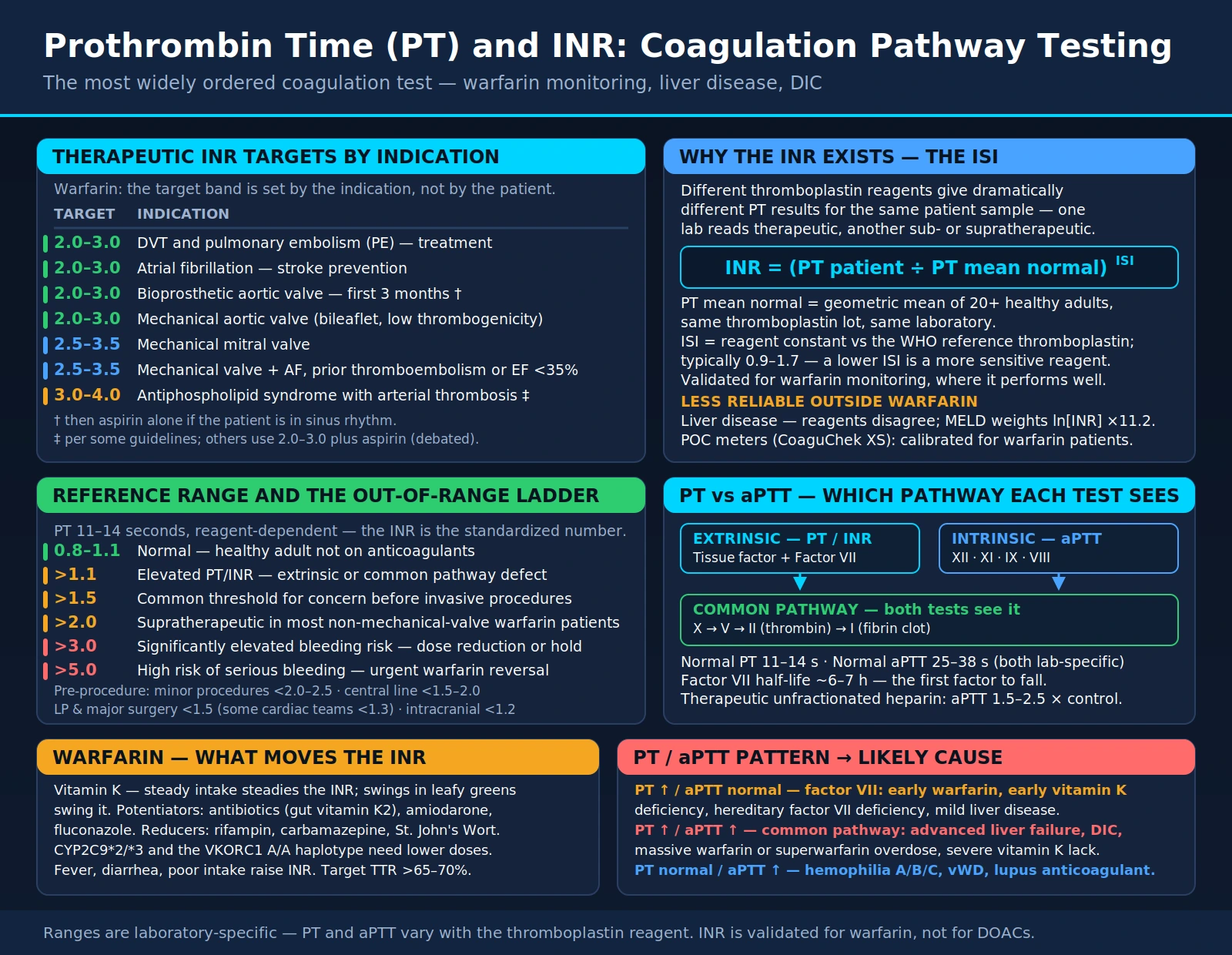
The prothrombin time (PT) measures how long plasma takes to clot through the extrinsic (tissue factor) and common pathways of coagulation. The international normalized ratio (INR) is a calculation from the PT that corrects for differences between laboratory reagents, so that results for people taking warfarin can be compared from one laboratory to another (Ageno 2012). The PT and INR are used to monitor warfarin and other vitamin K antagonists, as part of the assessment of liver synthetic function, and in the work-up of bleeding and clotting problems. MedlinePlus lists bleeding disorders, disseminated intravascular coagulation, liver disease and low vitamin K among the causes of a high INR in people who are not taking a blood thinner.
Table of Contents
- Overview — Extrinsic Coagulation Pathway
- How PT and INR Are Measured
- Normal and Therapeutic Ranges
- Pre-Procedure INR Thresholds
- Warfarin Therapy and INR Monitoring
- PT as a Liver Function Test
- Vitamin K and PT/INR
- Factor V Leiden and Coagulation Genetics
- Point-of-Care INR Testing
- Causes of Prolonged PT/INR
- aPTT: The Intrinsic Pathway Partner Test
- Urgent Reversal Strategies
- Key Research Papers
- Connections
- Featured Videos
Overview — Extrinsic Coagulation Pathway
The extrinsic pathway starts when tissue factor (Factor III), which sits on cells outside the bloodstream, is exposed to blood after a vessel is injured. Tissue factor binds Factor VII, and the tissue factor–Factor VIIa complex activates Factor X and Factor IX. Activated Factor X, together with its cofactor Factor Va on a phospholipid surface (the prothrombinase complex), converts prothrombin (Factor II) to thrombin far faster than Factor Xa can alone. Thrombin cleaves fibrinogen to fibrin, and Factor XIII, also activated by thrombin, cross-links the fibrin into a stable clot. The PT does not measure Factor XIII.
Factors II, VII, IX and X need vitamin K for a final chemical step, the gamma-carboxylation of glutamate residues, without which they cannot bind calcium and phospholipid membranes and do not work as clotting factors. Warfarin and other vitamin K antagonists act at this step (Ageno 2012). Most clotting factors are made in the liver, so the PT is sensitive both to vitamin K status and to liver disease.
Factors Measured by the PT
Despite its name, the prothrombin time does not measure prothrombin alone. It reflects the combined activity of Factor VII and the common-pathway factors X, V, II and fibrinogen (Factor I). A marked deficiency anywhere along that chain prolongs the result:
- Factor VII: vitamin K-dependent and the only factor unique to the extrinsic pathway. Its half-life is about 6 hours, the shortest of the vitamin K-dependent factors (Ageno 2012), which is why the PT is the first test to change when warfarin is started or vitamin K runs short.
- Factor X: vitamin K-dependent; the point where the extrinsic and intrinsic pathways meet. As Factor Xa it converts prothrombin to thrombin.
- Factor V: a cofactor that is not vitamin K-dependent. Because warfarin and vitamin K deficiency do not lower it, while liver failure does, a Factor V level helps separate the two. The Factor V Leiden variant is a clotting tendency, not a bleeding disorder, and does not prolong the PT.
- Factor II (prothrombin): vitamin K-dependent, with a long half-life of about 60 to 72 hours (Ageno 2012). It is converted to thrombin, which cleaves fibrinogen, activates platelets and feeds back to amplify the cascade.
- Factor I (fibrinogen): the protein thrombin converts to fibrin. A marked fall in fibrinogen can prolong the PT on its own; consumption (as in disseminated intravascular coagulation) and failing liver synthesis are two common causes.
Factor VII is the only factor the PT measures that the activated partial thromboplastin time (aPTT) does not, so a deficiency or depletion of Factor VII alone prolongs the PT while the aPTT stays normal. When the shared common-pathway factors (X, V, II or fibrinogen) fall far enough, as in severe liver failure or disseminated intravascular coagulation (DIC), both tests are prolonged.
This extrinsic–intrinsic–common cascade is a model for reading laboratory tests and simplifies what happens in the body. The cell-based model of coagulation describes clotting as starting on tissue factor-bearing cells, being amplified by platelets and propagating on the platelet surface. The cascade model remains the usual framework for interpreting PT and aPTT results.
How PT and INR Are Measured
The prothrombin time was described by the American physician Armand Quick in 1935 (Quick 1935): citrated plasma is recalcified in the presence of a tissue extract and the time to clot formation is measured. The reagents and the clot-detection instruments have changed since then; the principle has not.
Blood is drawn into a light-blue-top tube containing sodium citrate, which binds calcium and stops clotting in the tube. The ratio of blood to citrate matters. An under-filled tube leaves extra citrate for the amount of plasma, and a very high hematocrit does the same because the sample holds less plasma. In a study of 28 patients with hematocrit values of 55% to 72%, PT and aPTT results in tubes with unadjusted citrate differed significantly from adjusted tubes and rose exponentially with the hematocrit; the authors concluded that citrate has to be adjusted in these patients for accurate results (Marlar 2006).
In the laboratory, platelet-poor plasma is separated by centrifugation and warmed to 37°C. A thromboplastin reagent — tissue factor (recombinant or from tissue extracts) with phospholipid — and calcium are added, and an optical or mechanical detector records the time to clot formation. That time, in seconds, is the PT.
Thromboplastin reagents differ in how strongly they respond to the clotting-factor changes caused by vitamin K antagonists, so the same patient sample can give different PT results in different laboratories. To standardize the test, the World Health Organization adopted a scheme in 1977 for calibrating thromboplastins against an International Reference Preparation, and in 1982 a revised calibration model (Kirkwood 1983). That calibration gives each reagent an International Sensitivity Index (ISI), which is used in the INR formula (Ageno 2012):
INR = (Patient PT ÷ Mean Normal PT)ISI
The mean normal PT is established by each laboratory from healthy people tested with the same reagent and instrument. A reagent with an ISI of 1.0 gives an INR equal to the plain PT ratio; the 2012 American College of Chest Physicians (ACCP/CHEST) review notes that highly sensitive thromboplastins with an ISI of about 1.0 are now available (Ageno 2012).
The INR was calibrated with plasma from people taking vitamin K antagonists, and its standardization rests on that use. Liver disease lowers clotting factors in a different pattern, and in a study of 57 patients with cirrhosis tested with 7 thromboplastins, mean INR values differed significantly between reagents (Tripodi 2007). A systematic review found insufficient evidence that a prolonged PT or raised INR predicts bleeding during invasive procedures (Segal 2005). The INR is a clotting-time measurement; its link to bleeding is best established in people taking warfarin.
Point-of-care analyzers measure the INR in a drop of fingerstick blood with a dried thromboplastin reagent and a device-specific calibration. The ACCP/CHEST review describes greater differences from laboratory plasma-based methods as the INR rises above the therapeutic range, and cases of incorrect ISI calibration of point-of-care instruments (Ageno 2012). See Point-of-Care INR Testing below.
Normal and Therapeutic Ranges
MedlinePlus gives these general values for people who are not taking a blood thinner, and notes that normal ranges vary among laboratories:
- PT: 11 to 13.5 seconds; each laboratory reports its own reference range for its reagent and instrument
- INR: 0.8 to 1.1
For people taking warfarin, MedlinePlus states that the INR is most often kept from 2.0 to 3.0. The 2012 ACCP/CHEST guidelines make targeting an INR of 2.0 to 3.0 for patients on vitamin K antagonists one of their two strong recommendations (Holbrook 2012), and set higher targets for some mechanical heart valves:
- Venous thromboembolism (DVT or PE) and atrial fibrillation: INR 2.0 to 3.0 (Holbrook 2012)
- Mechanical aortic valve: target 2.5, range 2.0 to 3.0 (Whitlock 2012)
- Mechanical mitral valve, or mechanical valves in both positions: target 3.0, range 2.5 to 3.5 (Whitlock 2012)
- Bioprosthetic valve, first 3 months: for a bioprosthetic mitral valve, a vitamin K antagonist with a target of 2.5 (range 2.0 to 3.0); for a bioprosthetic aortic valve, the 2012 guideline suggests aspirin rather than a vitamin K antagonist (Whitlock 2012)
- Antiphospholipid syndrome with a first arterial thrombosis: the 2019 EULAR recommendations list a vitamin K antagonist with an INR of 2 to 3 or 3 to 4, weighing the individual's bleeding and thrombosis risk (Tektonidou 2019)
The 2020 American College of Cardiology/American Heart Association (ACC/AHA) valve guideline also addresses anticoagulation for prosthetic valves (Otto 2020); the full text was not available for this review, so its targets are not restated here.
How guidelines handle an INR above the target range (2012 ACCP/CHEST, Holbrook 2012):
- A single INR up to 0.5 above or below the range in a previously stable patient: the guideline suggests continuing the current dose and retesting within 1 to 2 weeks
- INR between 4.5 and 10 with no bleeding: the guideline suggests against routine use of vitamin K
- INR above 10 with no bleeding: the guideline suggests oral vitamin K
- Major bleeding on a vitamin K antagonist: the guideline suggests rapid reversal with 4-factor prothrombin complex concentrate rather than plasma, plus vitamin K 5 to 10 mg by slow intravenous injection (see Urgent Reversal Strategies)
The 2020 American College of Cardiology (ACC) Expert Consensus Decision Pathway covers the management of bleeding in people taking oral anticoagulants, including warfarin and the direct oral anticoagulants (Tomaselli 2020). Decisions about anticoagulant doses and reversal belong with the treating clinician.
The INR level is tied to bleeding risk. In a 1994 case-control study of 121 people taking warfarin who were hospitalized with intracranial hemorrhage, each 0.5 increase in the prothrombin time ratio (the study used the PT ratio, not the INR) doubled the risk of intracerebral hemorrhage (odds ratio 2.1), and the risk of subdural hemorrhage was unchanged across a PT ratio of 1.0 to 2.0 but rose steeply above a PT ratio of 2.0, approximately an INR of 4.0 (Hylek 1994).
Pre-Procedure INR Thresholds
An INR above 1.5 is a threshold often cited before invasive procedures. It is a convention rather than a validated cut-off. A 2005 systematic review of one trial and 24 observational studies found insufficient evidence to conclude that abnormal coagulation test results predict bleeding during invasive diagnostic procedures such as bronchoscopy, central vein cannulation, arteriography and liver biopsy; in the largest arteriography study, bleeding rates were the same with and without abnormal tests (Segal 2005).
Thresholds for specific procedures are set by specialty societies and by individual hospitals, and they differ. In cirrhosis, the 2019 American Gastroenterological Association (AGA) clinical practice update states that commonly used thresholds for INR correction are not supported by evidence, and that in general coagulopathy is not routinely corrected before low-risk paracentesis, thoracentesis or routine endoscopic variceal ligation (O'Leary 2019).
For people with atrial fibrillation whose warfarin is stopped for an elective procedure, the BRIDGE trial of 1,884 patients found that forgoing heparin "bridging" was noninferior to bridging with low-molecular-weight heparin for arterial thromboembolism (0.4% vs 0.3%) and reduced major bleeding (1.3% vs 3.2%) (Douketis 2015). Urgent reversal before emergency surgery is covered under Urgent Reversal Strategies.
Warfarin Therapy and INR Monitoring
Warfarin, a vitamin K antagonist, has been used as an oral anticoagulant for decades. It inhibits vitamin K epoxide reductase (VKORC1), the enzyme that recycles vitamin K to its active reduced form. Without active vitamin K, the gamma-carboxylation of Factors II, VII, IX and X — and of the natural anticoagulant proteins C and S — is impaired, and the under-carboxylated proteins (called PIVKA, proteins induced by vitamin K absence or antagonism) do not function normally in coagulation.
How fast warfarin works depends on how quickly the existing factors are cleared. The ACCP/CHEST review gives a half-life of about 6 hours for Factor VII, 6 to 24 hours for the other vitamin K-dependent factors, and 60 to 72 hours for prothrombin, and states that the antithrombotic effect of warfarin requires about 6 days of treatment, because it depends on lowering prothrombin (Ageno 2012). The INR can therefore rise, through the fall in Factor VII, before full antithrombotic effect is reached.
Protein C, a natural anticoagulant, is also vitamin K-dependent. Warfarin-induced skin necrosis has been reported in association with protein C and protein S deficiency, typically on the third to eighth day of therapy, with extensive thrombosis of small skin vessels (Ageno 2012).
Drug interactions with warfarin are common. The S-enantiomer of warfarin, 2.7 to 3.8 times more potent than the R-enantiomer, is cleared mainly by the liver enzyme CYP2C9; the R-enantiomer mainly by CYP1A2 and CYP3A4 (Ageno 2012). The ACCP/CHEST review describes:
- Amiodarone as a potent inhibitor of the clearance of both enantiomers that potentiates warfarin; metronidazole and fluconazole also inhibit warfarin clearance, and several antibiotics, including ciprofloxacin and erythromycin, potentiate its effect
- Sulfonamides and some broad-spectrum antibiotics as able to increase warfarin's effect in people whose diet is low in vitamin K, by eliminating gut bacteria and aggravating vitamin K deficiency
- Barbiturates, rifampin, azathioprine and carbamazepine as inhibiting warfarin's effect by increasing its clearance through induction of liver metabolism
- Aspirin as the most important of the drugs that add to bleeding risk without changing the INR, because of its widespread use and prolonged effect on platelets
Among herbs, St John's wort increased the clearance of both warfarin enantiomers and reduced warfarin's effect on the INR in a study in healthy volunteers, while ginseng did not (Jiang 2004).
Dietary vitamin K changes the response to warfarin. The ACCP/CHEST review states that people on long-term warfarin are sensitive to fluctuating dietary vitamin K, that green vegetables or vitamin K-containing supplements can supply enough to reduce the anticoagulant response, and that a consistent intake of vitamin K-containing foods is advisable, while neither specific restrictions nor additions seem necessary in people with stable control (Ageno 2012). The 2012 guideline suggests against routine vitamin K supplementation for people taking vitamin K antagonists (Holbrook 2012). A study in healthy volunteers found that menaquinone-7 supplements at 50 micrograms a day or more may interfere with oral anticoagulant treatment (Schurgers 2007).
Acute illness can change the INR. The ACCP/CHEST review notes that fever and hyperthyroidism increase the response to warfarin, probably by speeding the breakdown of vitamin K-dependent clotting factors (Ageno 2012).
Pharmacogenomics of warfarin: two genes account for much of the person-to-person difference in warfarin dose:
- CYP2C9: people carrying one or two copies of the *2 or *3 variants need lower warfarin doses (Ageno 2012).
- VKORC1: in a study of European-American patients, the mean maintenance dose was 2.7 mg a day with the low-dose haplotype pair (A/A), 4.9 mg with A/B and 6.2 mg with B/B, and the haplotypes explained about 25% of the variation in dose. Asian Americans had a higher proportion of the A haplotype and African Americans a higher proportion of the B haplotype (Rieder 2005).
Trials of genotype-guided dosing have had mixed results. In the European EU-PACT trial of 455 patients, genotype-guided dosing gave a higher time in therapeutic range over the first 12 weeks (67.4% vs 60.3%) (Pirmohamed 2013); in the US COAG trial, time in range at 4 weeks was the same with a genotype-guided or a clinical algorithm (45.2% vs 45.4%) (Kimmel 2013); and in the GIFT trial in hip or knee replacement, genotype-guided dosing reduced a combined outcome of major bleeding, INR of 4 or greater, venous thromboembolism or death (10.8% vs 14.7%) (Gage 2017). The 2012 ACCP/CHEST guideline recommends against routine pharmacogenetic testing to guide warfarin doses (Holbrook 2012).
Time in therapeutic range (TTR) is the share of time a person's INR spends inside the target range, commonly calculated by the Rosendaal linear-interpolation method, which assumes the INR changes in a straight line between two measurements (Rosendaal 1993). The 2018 CHEST atrial fibrillation guideline states that, when a vitamin K antagonist is used, it is important to aim for good anticoagulation control with a TTR above 70% (Lip 2018).
Direct oral anticoagulants (DOACs) — the factor Xa inhibitors rivaroxaban, apixaban and edoxaban and the thrombin inhibitor dabigatran — do not need routine INR monitoring. For atrial fibrillation where oral anticoagulation is indicated, the 2018 CHEST guideline suggests a DOAC rather than adjusted-dose warfarin (Lip 2018), and for venous thromboembolism without cancer the 2016 CHEST guideline suggests a DOAC over a vitamin K antagonist (Kearon 2016). Warfarin keeps a role in some settings. A trial of dabigatran in people with mechanical heart valves was stopped early because of an excess of thromboembolic and bleeding events with dabigatran (Eikelboom 2013), and in triple-positive antiphospholipid syndrome the TRAPS trial found more events with rivaroxaban than with warfarin (Pengo 2018).
PT as a Liver Function Test
The liver makes most clotting factors; Factor VIII and von Willebrand factor are notable exceptions, produced largely by the lining of blood vessels. Impaired liver synthesis — in acute liver failure, decompensated cirrhosis or severe hepatitis — lowers clotting factors and prolongs the PT, which is why the PT/INR is used as a measure of liver synthetic function.
The PT/INR is part of two prognostic scores in liver disease:
- Child-Pugh score: grades cirrhosis on five items — PT/INR, albumin, bilirubin, ascites and hepatic encephalopathy — each scored 1 to 3 points, with class C covering the most severe totals.
- MELD score (Model for End-Stage Liver Disease): combines serum bilirubin, serum creatinine and the INR. In four independent groups of patients it predicted death within 3 months with a c-statistic of 0.78 to 0.87, and its authors described it as suitable for setting organ-allocation priorities (Kamath 2001). Adding serum sodium improved the prediction of death among people on the US liver-transplant waiting list (Kim 2008).
Because the INR is calibrated with plasma from people on vitamin K antagonists, it behaves differently in cirrhosis. In a study of 57 patients with cirrhosis whose PT was measured with 7 thromboplastins, the mean INR and MELD values differed significantly by reagent; when the thromboplastins were recalibrated with plasma from patients with cirrhosis (an "INRliver"), the differences disappeared (Tripodi 2007). The ACCP/CHEST review notes that a liver-specific INR has been proposed for this reason (Ageno 2012).
In cirrhosis the INR also misses part of the picture of clotting. Liver disease lowers natural anticoagulants as well as clotting factors, a "rebalanced" state described in a review of the coagulopathy of chronic liver disease (Tripodi 2011). Von Willebrand factor is usually elevated in cirrhosis (O'Leary 2019), and levels of the enzyme that cleaves von Willebrand factor (ADAMTS13) were low in people with decompensated cirrhosis (Mannucci 2001). Global tests of clot formation such as thromboelastography and rotational thromboelastometry may eventually have a role in this setting, but the 2019 AGA update states that they currently lack validated target levels (O'Leary 2019).
The same AGA update states that blood products are used sparingly in cirrhosis because they increase portal pressure and carry risks including transfusion-associated circulatory overload and transfusion-related acute lung injury; that the large volume of fresh frozen plasma needed to reach an arbitrary INR target, its small effect on thrombin generation and its effect on portal pressure limit its usefulness; and that for active bleeding or high-risk procedures in advanced liver disease, thresholds of a hematocrit of 25% or more, a platelet count above 50,000 and fibrinogen above 120 mg/dL may optimize clot formation (O'Leary 2019).
In acute liver failure, the PT is one of the King's College prognostic indicators. In a study of 588 patients, poor prognosis in acetaminophen-induced liver failure was indicated by an arterial pH below 7.30, a prothrombin time above 100 seconds and a creatinine above 300 µmol/L; in viral hepatitis and drug reactions, the indicators included a prothrombin time above 50 seconds and a bilirubin above 300 µmol/L, along with cause, age and the time from jaundice to encephalopathy (O'Grady 1989). Factor V, which is not vitamin K-dependent, was an independent predictor of survival in fulminant hepatitis B (Bernuau 1986).
Newborns have low vitamin K stores at birth, which is the background to the vitamin K given at birth (see Vitamin K and PT/INR) (Shearer 2009).
Vitamin K and PT/INR
Vitamin K takes its letter from the German and Scandinavian spelling Koagulation, for its role in blood clotting; the Danish biochemist Henrik Dam identified it in the 1930s. It occurs in the diet in two main forms (Booth 2012):
- Vitamin K1 (phylloquinone): the major dietary source, concentrated in leafy plants such as kale, spinach, Swiss chard, collard greens and broccoli. It is absorbed with dietary fat, which requires bile and pancreatic enzymes.
- Vitamin K2 (menaquinones): the product of bacterial production or of conversion from dietary phylloquinone; their presence in foods varies by region (Booth 2012). Natto, a fermented soybean food, is a source of menaquinone-7 (MK-7). In healthy volunteers, menaquinone-7 had a very long half-life compared with K1, giving more stable blood levels and a 7- to 8-fold accumulation with daily intake (Schurgers 2007).
Vitamin K is the cofactor for gamma-glutamyl carboxylase, the enzyme that adds carboxyl groups to specific glutamate residues on vitamin K-dependent proteins. In the process the active reduced form of vitamin K (the hydroquinone) is converted to vitamin K epoxide, which vitamin K epoxide reductase (VKORC1) recycles back to vitamin K. Warfarin blocks this recycling step.
Vitamin K deficiency affects the PT first because Factor VII has the shortest half-life of the vitamin K-dependent factors (about 6 hours, compared with 60 to 72 hours for prothrombin) (Ageno 2012). Early or mild deficiency can therefore prolong the PT while the aPTT is still normal; as Factors IX, X and prothrombin fall, the aPTT also lengthens. Fibrinogen and Factor V are not vitamin K-dependent, which helps distinguish vitamin K deficiency from DIC, where fibrinogen is consumed. Heparin, by contrast, prolongs the aPTT much more than the PT.
Recognized causes of vitamin K deficiency include:
- Fat malabsorption: cystic fibrosis, celiac disease, Crohn's disease, short bowel syndrome and cholestatic liver disease, in which bile acids needed for absorbing fat-soluble vitamins are lacking
- Antibiotics with a vitamin K-poor diet: the ACCP/CHEST review describes broad-spectrum antibiotics as able to aggravate vitamin K deficiency in people whose diet is deficient in vitamin K (Ageno 2012)
- Newborns: vitamin K stores are low at birth, and breast-fed infants are at risk because human milk contains little vitamin K. Classical vitamin K deficiency bleeding occurs in the first week of life; late deficiency bleeding peaks at 3 to 8 weeks, typically presents as intracranial hemorrhage, and without prophylaxis occurs in about 4 to 7 per 100,000 births in Europe. Parenteral vitamin K at birth gives the best protection, and the effectiveness of oral prophylaxis depends on the dose and how often it is given (Shearer 2009). Intramuscular vitamin K at birth has been the American Academy of Pediatrics standard since 1961, and the Academy attributes a recent rise in deficiency bleeding to parental refusal and to the lower efficacy of alternative routes (Hand 2022).
- Poor intake: prolonged poor oral intake, or parenteral nutrition without vitamin K
On the treatment side, the ACCP/CHEST review reports that oral and low-dose intravenous phytonadione (vitamin K1) produce similar INR reductions at 24 hours, that subcutaneous phytonadione appears less effective than low-dose oral phytonadione, and that after vitamin K is given for bleeding, the INR begins to fall within 2 hours and generally returns to normal within 24 hours if liver function is normal (Ageno 2012).
Factor V Leiden and Coagulation Genetics
Factor V Leiden is a single change in the Factor V gene at nucleotide 1691 (G to A) that produces a Factor V molecule (FV Q506) that activated protein C does not inactivate properly. Activated protein C normally limits clotting by breaking down Factors Va and VIIIa; with the Leiden variant, Factor Va resists this, and clotting is less well switched off. In the original 1994 report, the allele frequency in the Dutch population was about 2%, at least ten times higher than all other genetic risk factors for thrombosis then known combined, and the resistance to activated protein C that it causes was associated with a sevenfold increase in the risk of deep vein thrombosis (Bertina 1994).
The risk adds to other risks. In a study of premenopausal women aged 15 to 49, oral contraceptive use increased the risk of venous thrombosis about 4-fold (relative risk 3.8), carrying Factor V Leiden about 8-fold (7.9), and both together more than 30-fold (34.7) compared with women with neither (Vandenbroucke 1994). Decisions about testing and treatment in people with inherited thrombophilia belong with a clinician.
The PT and INR are typically normal in people with Factor V Leiden, because the variant affects how clotting is switched off rather than how fast a clot forms. It is identified with a functional activated protein C resistance assay, based on the aPTT with and without added activated protein C, or by genetic testing.
Other inherited thrombophilias relevant to coagulation testing:
- Prothrombin G20210A: a G to A change at position 20210 in the untranslated end of the prothrombin gene. In a Dutch population-based case-control study, the allele frequency was 1.2% and it increased the risk of venous thrombosis almost threefold (odds ratio 2.8); 87% of carriers had plasma prothrombin levels in the highest quartile (Poort 1996).
- Protein C deficiency: protein C is a vitamin K-dependent anticoagulant that, once activated, inactivates Factors Va and VIIIa. Warfarin-induced skin necrosis has been reported in association with protein C deficiency (Ageno 2012).
- Protein S deficiency: protein S is the vitamin K-dependent cofactor that activated protein C needs (Bertina 1994); warfarin-induced skin necrosis has also been reported with protein S deficiency (Ageno 2012).
- Antithrombin deficiency: antithrombin is the main natural inhibitor of thrombin and Factor Xa and the protein through which heparin works, so a deficiency can reduce the response to heparin.
Timing matters when these proteins are measured. Acute thrombosis, pregnancy, hormonal contraception, acute illness and anticoagulants can all change protein C, protein S and antithrombin levels; warfarin lowers protein C and protein S along with the vitamin K-dependent clotting factors. Genetic tests for Factor V Leiden and prothrombin G20210A are not affected by these conditions.
Point-of-Care INR Testing
Point-of-care INR monitors let people taking warfarin test at home or in a clinic from a fingerstick drop of whole blood. A test strip carries dried thromboplastin reagent; the device detects clot formation in the blood sample and converts the clotting time to an INR using a device-specific calibration.
Accuracy: the ACCP/CHEST review reports greater differences from laboratory plasma-based methods as the INR rises above the therapeutic range, and problems from incorrect ISI calibration. In a proficiency-testing study of three monitors over 6 years in more than 10 centers, INR results in 10% to 11% of centers were more than 15% different from results in other centers using the same monitors, and a European evaluation of 523 monitors of one widely used model at nine clinics found significant deviations in 20.3% (Ageno 2012). In people with antiphospholipid antibodies, most point-of-care results were close to the laboratory method, but in a subset of patients with both lupus anticoagulant and elevated anti-β2-glycoprotein I antibodies, one device gave no measurable INR and another gave higher INR values than the plasma-based method (Perry 2005).
Self-testing and self-management — testing at home, with or without adjusting the dose by a set schedule — have been studied in randomized trials:
- A randomized crossover trial found people were within 0.5 of their target INR 55% of the time during self-management and 49% during anticoagulation-clinic management, a difference that was not statistically significant (p=0.06) (Cromheecke 2000)
- A 2006 meta-analysis of 14 randomized trials found fewer thromboembolic events (odds ratio 0.45) and lower all-cause mortality (odds ratio 0.61) with self-monitoring (Heneghan 2006)
- An individual-patient-data meta-analysis of 11 trials with 6,417 participants found fewer thromboembolic events with self-monitoring (hazard ratio 0.51), but no significant difference in major hemorrhage or death (Heneghan 2012)
- The 2016 Cochrane review of 28 trials found fewer thromboembolic events (risk ratio 0.58); all-cause mortality was lower in trials of self-management (risk ratio 0.55) but not of self-monitoring, and major hemorrhage was not reduced (risk ratio 0.95) (Heneghan 2016)
For motivated patients who demonstrate competency in self-management and with the equipment, the 2012 ACCP/CHEST guideline suggests patient self-management rather than usual outpatient INR monitoring (Holbrook 2012).
Limits of point-of-care INR testing:
- The INR is calibrated for vitamin K antagonist therapy, and point-of-care monitors are used for warfarin monitoring; their standardization does not extend to other causes of a prolonged PT such as liver disease or DIC
- Agreement with the laboratory falls at INR values above the therapeutic range (Ageno 2012)
- Lupus anticoagulant with high anti-β2-glycoprotein I antibodies can make some devices read high or fail to give a result (Perry 2005)
- Very high or low hematocrit, and other interferences listed by each device's manufacturer, can affect results
- The INR does not measure the effect of rivaroxaban, apixaban, edoxaban or dabigatran (see below)
Causes of Prolonged PT/INR
A prolonged PT/INR is read together with the aPTT, the fibrinogen level and the clinical setting:
Isolated PT prolongation (aPTT normal, fibrinogen normal):
- Warfarin or another vitamin K antagonist — Factor VII falls first
- Early or mild vitamin K deficiency — Factor VII falls first because of its short half-life (Ageno 2012)
- Hereditary Factor VII deficiency — a rare inherited disorder; bleeding varies widely and does not closely follow the factor level
- Early liver disease — a fall in Factor VII can come before other factors fall
- Acquired Factor VII inhibitor — very rare
- Lupus anticoagulant — usually prolongs the aPTT, but can occasionally prolong the PT
Both PT and aPTT prolonged (fibrinogen normal or high):
- Advanced liver disease or acute liver failure — the common-pathway factors (X, V, II) fall as well as Factor VII
- Severe or prolonged vitamin K deficiency
- Warfarin together with heparin
- High warfarin levels, or "superwarfarin" ingestion — long-acting anticoagulant rodenticides such as brodifacoum and difenacoum are vitamin K antagonists that stay in the body far longer than warfarin, so the coagulopathy after an exposure can last for weeks to months and treatment with vitamin K has to continue for that long
- Hereditary deficiencies of Factor X, V or II — rare
- Amyloidosis — acquired Factor X deficiency is described in systemic amyloidosis (Furie 1981)
- Heparin at high levels (high doses or a contaminated sample) — mainly prolongs the aPTT but can prolong the PT
- Direct oral anticoagulants — rivaroxaban, apixaban, edoxaban and dabigatran prolong the PT to a variable degree (see Urgent Reversal Strategies)
Both PT and aPTT prolonged with low fibrinogen and high D-dimer:
- Disseminated intravascular coagulation (DIC) — widespread activation of coagulation consumes clotting factors, fibrinogen and platelets. Triggers include sepsis, trauma, some cancers (notably acute promyelocytic leukemia), obstetric emergencies such as placental abruption, and some snake envenomations. The International Society on Thrombosis and Haemostasis (ISTH) scoring system for overt DIC assigns points for the platelet count, PT prolongation, fibrinogen level and a fibrin-related marker such as D-dimer, in a patient with an underlying condition that can cause DIC (Taylor 2001).
- Massive transfusion with dilutional coagulopathy — red cell concentrates contain no clotting factors. In the PROPPR trial of severely injured patients with major bleeding, giving plasma, platelets and red cells in a 1:1:1 rather than a 1:1:2 ratio did not change mortality at 24 hours or 30 days, but more patients achieved hemostasis (86% vs 78%) and fewer died from exsanguination by 24 hours (Holcomb 2015).
- Severe liver failure — fibrinogen is also made in the liver and can fall in advanced disease
Falsely prolonged PT (sample problems):
- Under-filled citrate tube
- Delayed processing or poor transport and storage conditions (Adcock Funk 2012)
- Clotted or hemolyzed sample
- Wrong tube type (for example EDTA instead of citrate)
- Hemolysis, icterus or lipemia, which can interfere with optical clot detection (Lippi 2013)
- Very high hematocrit with a standard citrate volume (Marlar 2006)
The mixing study: when the cause of a prolonged PT or aPTT is unclear, the patient's plasma is mixed 1:1 with normal pooled plasma and the test is repeated. If the result corrects, a factor deficiency is likely; if it stays prolonged, an inhibitor or other interference is likely (Kershaw 2013). Time-dependent inhibitors such as acquired Factor VIII antibodies may correct on the immediate mix and fail to correct after incubation. The aPTT page describes the procedure in detail.
aPTT: The Intrinsic Pathway Partner Test
The PT is rarely read alone. Its partner, the activated partial thromboplastin time (aPTT), measures the intrinsic pathway (Factors XII, XI, IX and VIII) together with the same common pathway the PT covers (X, V, II and fibrinogen). Reading the two together turns a prolonged clotting time into a short list of causes.
The aPTT adds three things to citrated plasma: a contact activator (such as kaolin, ellagic acid or silica — the "activated" in the name), phospholipid without tissue factor (the "partial thromboplastin"), and calcium. MedlinePlus gives a general aPTT range of 25 to 35 seconds and notes that normal value ranges vary among laboratories.
A prolonged aPTT with a normal PT points to the intrinsic pathway:
- Factor VIII deficiency (hemophilia A) and Factor IX deficiency (hemophilia B): X-linked, so they mainly affect males; the aPTT cannot tell them apart, and specific factor assays are used
- Factor XI deficiency (sometimes called hemophilia C): an autosomal recessive, injury-related bleeding tendency that is common in Jewish people, particularly of Ashkenazi origin (Seligsohn 2009)
- Von Willebrand disease: von Willebrand factor carries Factor VIII, but the aPTT is often normal in von Willebrand disease; it is prolonged when Factor VIII falls far enough, as in type 3, where von Willebrand factor is virtually absent, and type 2N, where it cannot bind Factor VIII (Mazurier 2001)
- Factor XII deficiency: prolongs the aPTT but does not cause bleeding; a review of surgical procedures in people with severe Factor XII deficiency found neither bleeding nor thrombotic complications (Girolami 2004)
- Lupus anticoagulant: an antiphospholipid antibody that prolongs the aPTT in the tube while being linked to thrombosis, not bleeding, in the body; it typically fails to correct in a mixing study
- Unfractionated heparin: the aPTT has long been the usual monitoring test. The 2012 ACCP/CHEST review explains that a therapeutic range of 1.5 to 2.5 times control gained wide acceptance after a retrospective study in the 1970s, and that its clinical relevance is uncertain because randomized trials have not confirmed it; aPTT reagents and instruments also differ in their response to heparin, so hospitals set their own therapeutic range, and some measure anti-Xa activity instead (Garcia 2012)
Direct oral anticoagulants affect both tests unpredictably. For dabigatran, the aPTT and PT are less sensitive than drug-specific tests and may be normal at trough levels; for rivaroxaban and apixaban, the PT is less sensitive (especially for apixaban), and a normal PT may not exclude clinically relevant drug levels (Cuker 2014). Drug-specific anti-Xa or anti-IIa assays are the preferred methods when a quantitative result is useful (Douxfils 2018).
Taken together: a prolonged PT with a normal aPTT points to Factor VII (warfarin, early vitamin K deficiency, early liver disease); a prolonged aPTT with a normal PT points to the intrinsic factors, heparin or lupus anticoagulant; both prolonged point to the common pathway, advanced liver failure, severe vitamin K deficiency, a direct oral anticoagulant or — when fibrinogen is low and D-dimer high — DIC. When a result is unexpected, heparin contamination of the sample and other sample problems are usually considered before a mixing study.
Urgent Reversal Strategies
When warfarin-associated coagulopathy causes life-threatening bleeding, or an operation cannot wait, the effect of the anticoagulant has to be reversed quickly. The 2012 ACCP/CHEST guidelines (Holbrook 2012) and the 2020 ACC Expert Consensus Decision Pathway on bleeding in people taking oral anticoagulants (Tomaselli 2020) address these situations.
4-Factor Prothrombin Complex Concentrate (4F-PCC)
4-factor prothrombin complex concentrate is a plasma-derived concentrate of the four vitamin K-dependent clotting factors (II, VII, IX and X) together with the anticoagulant proteins C and S. The ACCP/CHEST review notes that prothrombin complex concentrates do not need cross-matching, are virally inactivated, do not pose a risk of volume overload, and can be infused in 15 to 30 minutes (Ageno 2012). In a randomized trial of 202 people on vitamin K antagonists with major bleeding (median starting INR 3.90), effective hemostasis at 24 hours occurred in 72.4% with 4F-PCC and 65.4% with plasma, meeting the test for noninferiority, and rapid INR correction (to 1.3 or below 30 minutes after the infusion) in 62.2% versus 9.6%; adverse events, thromboembolic events and deaths were similar between groups (Sarode 2013). For major bleeding on a vitamin K antagonist, the 2012 guideline suggests rapid reversal with 4F-PCC rather than plasma, together with vitamin K 5 to 10 mg by slow intravenous injection (Holbrook 2012); the infused factors are cleared over time while warfarin's effect persists, and vitamin K restores the liver's own production of the factors.
Fresh Frozen Plasma (FFP)
Plasma contains every clotting factor, including Factor V, which prothrombin complex concentrate does not, but at ordinary rather than concentrated levels. It has to be thawed and blood-group compatible, and correcting the INR takes a large volume, which brings the risks of volume overload and transfusion-related acute lung injury. In the Sarode trial, rapid INR correction occurred in 9.6% of people given plasma, compared with 62.2% with 4F-PCC (Sarode 2013). Plasma is also part of balanced transfusion in massive bleeding (Holcomb 2015).
Vitamin K1 (Phytonadione)
Vitamin K1 does not replace clotting factors; it lets the liver make working factors again, so its effect is delayed but lasting. The ACCP/CHEST review reports that when oral phytonadione is given while warfarin is held, about 1.4 days are needed for an INR between 6 and 10 to fall below 4.0; that low-dose intravenous and oral phytonadione give similar INR reductions at 24 hours; that subcutaneous phytonadione appears less effective than low-dose oral phytonadione; and that after vitamin K for bleeding, the INR begins to fall within 2 hours and generally normalizes within 24 hours if liver function is normal (Ageno 2012). The 2012 guideline's suggestions by INR level and bleeding are listed under Normal and Therapeutic Ranges (Holbrook 2012).
Reversal of Direct Oral Anticoagulants
The INR is not a measure of direct oral anticoagulant effect. Rivaroxaban, apixaban and edoxaban (factor Xa inhibitors) and dabigatran (a thrombin inhibitor) prolong the PT and aPTT to a variable degree, and a normal PT may not exclude clinically relevant levels of rivaroxaban or apixaban (Cuker 2014; Douxfils 2018). Agent-specific reversal drugs exist:
- Idarucizumab, a monoclonal antibody fragment, for dabigatran: in the RE-VERSE AD cohort of 503 patients, the median maximum reversal of dabigatran's effect was 100%, and in those with bleeding the median time to cessation of bleeding was 2.5 hours (Pollack 2017)
- Andexanet alfa, a modified decoy factor Xa protein, for the factor Xa inhibitors: in the ANNEXA-4 study, anti-factor Xa activity fell by 92% in patients taking apixaban or rivaroxaban, and 82% of those evaluated had excellent or good hemostasis at 12 hours; within 30 days, 14% had died and 10% had a thrombotic event (Connolly 2019)
An expert guidance document from 2012, written when there was no established antidote, suggested supportive care, activated charcoal if the drug had been taken within a couple of hours, and hemodialysis if feasible for dabigatran, and discussed the possible use of 4F-PCC (Kaatz 2012).
Key Research Papers
- Ansell J, Hirsh J, Hylek E, et al. Pharmacology and management of the vitamin K antagonists. Chest. 2008;133(6 Suppl):160S–198S. — PubMed PMID: 18574265
- Ansell J, Hirsh J, Dalen J, et al. Managing oral anticoagulant therapy. Chest. 2001;119(1 Suppl):22S–38S. — PubMed PMID: 11157641
- Kearon C, Akl EA, Comerota AJ, et al. Antithrombotic therapy for VTE disease. Chest. 2012;141(2 Suppl):e419S–e496S. — PubMed PMID: 22315268
- Kaatz S, Kouides PA, Garcia DA, et al. Guidance on the emergent reversal of oral thrombin and factor Xa inhibitors. Am J Hematol. 2012;87(Suppl 1):S141–S145. — PubMed PMID: 22473649
- Tripodi A, Mannucci PM. The coagulopathy of chronic liver disease. N Engl J Med. 2011;365(2):147–156. — PubMed PMID: 21751907
- Schulman S, Kearon C, Kakkar AK, et al. Dabigatran versus warfarin in the treatment of acute venous thromboembolism. N Engl J Med. 2009;361(24):2342–2352. — PubMed PMID: 19966341
- Holbrook A, Schulman S, Witt DM, et al. Evidence-based management of anticoagulant therapy. Chest. 2012;141(2 Suppl):e152S–e184S. — PubMed PMID: 22315259
- Dahlback B. Blood coagulation. Lancet. 2000;355(9215):1627–1632. — PubMed PMID: 10821379
- Fihn SD, Gardin JM, Abrams J, et al. 2012 ACCF/AHA/ACP/AATS/PCNA/SCAI/STS guideline for the diagnosis and management of patients with stable ischemic heart disease. J Am Coll Cardiol. 2012;60(24):e44–e164. Reference for warfarin AF management: Wann LS et al. 2011 ACCF/AHA/HRS focused update. J Am Coll Cardiol. 2011;57(2):223–242. — PubMed PMID: 23182125
- Connolly SJ, Ezekowitz MD, Yusuf S, et al. Dabigatran versus warfarin in patients with atrial fibrillation. N Engl J Med. 2009;361(12):1139–1151. — PubMed PMID: 19717844
- Seligsohn U, Lubetsky A. Genetic susceptibility to venous thrombosis. N Engl J Med. 2001;344(16):1222–1231. — PubMed PMID: 11309638
- Tripodi A. The international normalized ratio. Clin Chem. 2007;53(7):1260–1261. — Search PubMed (Editorial note: no record matching this citation was found in PubMed or Crossref in 2026; for the INR in liver disease see Tripodi 2007, Hepatology, below.)
- Hirsh J, Fuster V, Ansell J, Halperin JL. American Heart Association/American College of Cardiology Foundation guide to warfarin therapy. J Am Coll Cardiol. 2003;41(9):1633–1652. — PubMed PMID: 12742309
- Quick AJ. The prothrombin in hemophilia and in obstructive jaundice. J Biol Chem. 1935;109:73–74. (Historical — the original description of the prothrombin time; accessible through the Journal of Biological Chemistry archives. Editorial note: this proceedings abstract was not found in Crossref; the 1935 method paper by Quick, Stanley-Brown and Bancroft is listed below.)
- Segal JB, Dzik WH; Transfusion Medicine/Hemostasis Clinical Trials Network. Paucity of studies to support that abnormal coagulation test results predict bleeding in the setting of invasive procedures. Transfusion. 2005;45(9):1413–1425. — PubMed PMID: 16131373
- Taylor FB Jr, Toh CH, Hoots WK, et al. Towards definition, clinical and laboratory criteria, and a scoring system for disseminated intravascular coagulation. Thromb Haemost. 2001;86(5):1327–1330. — PubMed PMID: 11816725
- Sarode R, Milling TJ Jr, Refaai MA, et al. Efficacy and safety of a 4-factor prothrombin complex concentrate in patients on vitamin K antagonists presenting with major bleeding: a randomized, plasma-controlled, phase IIIb study. Circulation. 2013;128(11):1234–1243. — PubMed PMID: 23935011
- American Society of Anesthesiologists Task Force on Perioperative Blood Transfusion and Adjuvant Therapies. Practice guidelines for perioperative blood transfusion and adjuvant therapies: an updated report by the American Society of Anesthesiologists Task Force on Perioperative Blood Transfusion and Adjuvant Therapies. Anesthesiology. 2006;105(1):198–208. — PubMed PMID: 16810012
- Schulman S, Angeras U, Bergqvist D, et al. Definition of major bleeding in clinical investigations of antihemostatic medicinal products in surgical patients. J Thromb Haemost. 2010;8(1):202–204. — PubMed PMID: 19878532
- Nishijima DK, Shahlaie K, Sarkar K, et al. Elevated admission international normalized ratio and outcomes after traumatic brain injury. — Search PubMed (Editorial note: no record matching this title was found in PubMed in 2026.)
- Weitz JI, Semchuk W, Turpie AG, et al. Trends in prescribing oral anticoagulants in Canada, 2008–2014. Clin Ther. 2015;37(11):2506–2514. — PubMed PMID: 26481493
- Mannucci PM, Canciani MT, Forza I, et al. Changes in health and disease of the metalloprotease that cleaves von Willebrand factor. Blood. 2001;98(9):2730–2735. — PubMed PMID: 11675345
- Ageno W, Gallus AS, Wittkowsky A, et al. Oral anticoagulant therapy: Antithrombotic Therapy and Prevention of Thrombosis, 9th ed: American College of Chest Physicians Evidence-Based Clinical Practice Guidelines. Chest. 2012;141(2 Suppl):e44S-e88S. — PubMed PMID: 22315269
- Whitlock RP, Sun JC, Fremes SE, et al. Antithrombotic and thrombolytic therapy for valvular disease: Antithrombotic Therapy and Prevention of Thrombosis, 9th ed: American College of Chest Physicians Evidence-Based Clinical Practice Guidelines. Chest. 2012;141(2 Suppl):e576S-e600S. — PubMed PMID: 22315272
- Otto CM, Nishimura RA, Bonow RO, et al. 2020 ACC/AHA Guideline for the Management of Patients With Valvular Heart Disease: A Report of the American College of Cardiology/American Heart Association Joint Committee on Clinical Practice Guidelines. Circulation. 2021;143(5):e72-e227. — PubMed PMID: 33332150
- Hylek EM, Singer DE. Risk factors for intracranial hemorrhage in outpatients taking warfarin. Ann Intern Med. 1994;120(11):897-902. — PubMed PMID: 8172435
- Tomaselli GF, Mahaffey KW, Cuker A, et al. 2020 ACC Expert Consensus Decision Pathway on Management of Bleeding in Patients on Oral Anticoagulants: A Report of the American College of Cardiology Solution Set Oversight Committee. J Am Coll Cardiol. 2020;76(5):594-622. — PubMed PMID: 32680646
- Lip GYH, Banerjee A, Boriani G, et al. Antithrombotic Therapy for Atrial Fibrillation: CHEST Guideline and Expert Panel Report. Chest. 2018;154(5):1121-1201. — PubMed PMID: 30144419
- Kearon C, Akl EA, Ornelas J, et al. Antithrombotic Therapy for VTE Disease: CHEST Guideline and Expert Panel Report. Chest. 2016;149(2):315-352. — PubMed PMID: 26867832
- Kirkwood TB. Calibration of reference thromboplastins and standardisation of the prothrombin time ratio. Thromb Haemost. 1983;49(3):238-44. — PubMed PMID: 6879511
- Marlar RA, Potts RM, Marlar AA. Effect on routine and special coagulation testing values of citrate anticoagulant adjustment in patients with high hematocrit values. Am J Clin Pathol. 2006;126(3):400-5. — PubMed PMID: 16880137
- Adcock Funk DM, Lippi G, Favaloro EJ. Quality standards for sample processing, transportation, and storage in hemostasis testing. Semin Thromb Hemost. 2012;38(6):576-85. — PubMed PMID: 22706973
- Lippi G, Plebani M, Favaloro EJ. Interference in coagulation testing: focus on spurious hemolysis, icterus, and lipemia. Semin Thromb Hemost. 2013;39(3):258-66. — PubMed PMID: 23229354
- Tripodi A, Chantarangkul V, Primignani M, et al. The international normalized ratio calibrated for cirrhosis (INR(liver)) normalizes prothrombin time results for model for end-stage liver disease calculation. Hepatology. 2007;46(2):520-7. — PubMed PMID: 17659574
- Douketis JD, Spyropoulos AC, Kaatz S, et al. Perioperative Bridging Anticoagulation in Patients with Atrial Fibrillation. N Engl J Med. 2015;373(9):823-33. — PubMed PMID: 26095867
- Jiang X, Williams KM, Liauw WS, et al. Effect of St John's wort and ginseng on the pharmacokinetics and pharmacodynamics of warfarin in healthy subjects. Br J Clin Pharmacol. 2004;57(5):592-9. — PubMed PMID: 15089812
- Schurgers LJ, Teunissen KJ, Hamulyák K, et al. Vitamin K-containing dietary supplements: comparison of synthetic vitamin K1 and natto-derived menaquinone-7. Blood. 2007;109(8):3279-83. — PubMed PMID: 17158229
- Rieder MJ, Reiner AP, Gage BF, et al. Effect of VKORC1 haplotypes on transcriptional regulation and warfarin dose. N Engl J Med. 2005;352(22):2285-93. — PubMed PMID: 15930419
- Pirmohamed M, Burnside G, Eriksson N, et al. A randomized trial of genotype-guided dosing of warfarin. N Engl J Med. 2013;369(24):2294-303. — PubMed PMID: 24251363
- Kimmel SE, French B, Kasner SE, et al. A pharmacogenetic versus a clinical algorithm for warfarin dosing. N Engl J Med. 2013;369(24):2283-93. — PubMed PMID: 24251361
- Gage BF, Bass AR, Lin H, et al. Effect of Genotype-Guided Warfarin Dosing on Clinical Events and Anticoagulation Control Among Patients Undergoing Hip or Knee Arthroplasty: The GIFT Randomized Clinical Trial. JAMA. 2017;318(12):1115-1124. — PubMed PMID: 28973620
- Rosendaal FR, Cannegieter SC, van der Meer FJ, et al. A method to determine the optimal intensity of oral anticoagulant therapy. Thromb Haemost. 1993;69(3):236-9. — PubMed PMID: 8470047
- Eikelboom JW, Connolly SJ, Brueckmann M, et al. Dabigatran versus warfarin in patients with mechanical heart valves. N Engl J Med. 2013;369(13):1206-14. — PubMed PMID: 23991661
- Pengo V, Denas G, Zoppellaro G, et al. Rivaroxaban vs warfarin in high-risk patients with antiphospholipid syndrome. Blood. 2018;132(13):1365-1371. — PubMed PMID: 30002145
- Tektonidou MG, Andreoli L, Limper M, et al. EULAR recommendations for the management of antiphospholipid syndrome in adults. Ann Rheum Dis. 2019;78(10):1296-1304. — PubMed PMID: 31092409
- Kamath PS, Wiesner RH, Malinchoc M, et al. A model to predict survival in patients with end-stage liver disease. Hepatology. 2001;33(2):464-70. — PubMed PMID: 11172350
- Kim WR, Biggins SW, Kremers WK, et al. Hyponatremia and mortality among patients on the liver-transplant waiting list. N Engl J Med. 2008;359(10):1018-26. — PubMed PMID: 18768945
- O'Leary JG, Greenberg CS, Patton HM, et al. AGA Clinical Practice Update: Coagulation in Cirrhosis. Gastroenterology. 2019;157(1):34-43.e1. — PubMed PMID: 30986390
- O'Grady JG, Alexander GJ, Hayllar KM, et al. Early indicators of prognosis in fulminant hepatic failure. Gastroenterology. 1989;97(2):439-45. — PubMed PMID: 2490426
- Bernuau J, Goudeau A, Poynard T, et al. Multivariate analysis of prognostic factors in fulminant hepatitis B. Hepatology. 1986;6(4):648-51. — PubMed PMID: 3732998
- Booth SL. Vitamin K: food composition and dietary intakes. Food Nutr Res. 2012;56. — PubMed PMID: 22489217
- Shearer MJ. Vitamin K deficiency bleeding (VKDB) in early infancy. Blood Rev. 2009;23(2):49-59. — PubMed PMID: 18804903
- Hand I, Noble L, Abrams SA. Vitamin K and the Newborn Infant. Pediatrics. 2022;149(3). — PubMed PMID: 35190810
- Bertina RM, Koeleman BP, Koster T, et al. Mutation in blood coagulation factor V associated with resistance to activated protein C. Nature. 1994;369(6475):64-7. — PubMed PMID: 8164741
- Vandenbroucke JP, Koster T, Briët E, et al. Increased risk of venous thrombosis in oral-contraceptive users who are carriers of factor V Leiden mutation. Lancet. 1994;344(8935):1453-7. — PubMed PMID: 7968118
- Poort SR, Rosendaal FR, Reitsma PH, et al. A common genetic variation in the 3'-untranslated region of the prothrombin gene is associated with elevated plasma prothrombin levels and an increase in venous thrombosis. Blood. 1996;88(10):3698-703. — PubMed PMID: 8916933
- Perry SL, Samsa GP, Ortel TL. Point-of-care testing of the international normalized ratio in patients with antiphospholipid antibodies. Thromb Haemost. 2005;94(6):1196-202. — PubMed PMID: 16411394
- Cromheecke ME, Levi M, Colly LP, et al. Oral anticoagulation self-management and management by a specialist anticoagulation clinic: a randomised cross-over comparison. Lancet. 2000;356(9224):97-102. — PubMed PMID: 10963245
- Heneghan C, Alonso-Coello P, Garcia-Alamino JM, et al. Self-monitoring of oral anticoagulation: a systematic review and meta-analysis. Lancet. 2006;367(9508):404-11. — PubMed PMID: 16458764
- Heneghan C, Ward A, Perera R, et al. Self-monitoring of oral anticoagulation: systematic review and meta-analysis of individual patient data. Lancet. 2012;379(9813):322-34. — PubMed PMID: 22137798
- Heneghan CJ, Garcia-Alamino JM, Spencer EA, et al. Self-monitoring and self-management of oral anticoagulation. Cochrane Database Syst Rev. 2016;7(7):CD003839. — PubMed PMID: 27378324
- Furie B, Voo L, McAdam KP, et al. Mechanism of factor X deficiency in systemic amyloidosis. N Engl J Med. 1981;304(14):827-30. — PubMed PMID: 7207512
- Holcomb JB, Tilley BC, Baraniuk S, et al. Transfusion of plasma, platelets, and red blood cells in a 1:1:1 vs a 1:1:2 ratio and mortality in patients with severe trauma: the PROPPR randomized clinical trial. JAMA. 2015;313(5):471-82. — PubMed PMID: 25647203
- Kershaw G, Orellana D. Mixing tests: diagnostic aides in the investigation of prolonged prothrombin times and activated partial thromboplastin times. Semin Thromb Hemost. 2013;39(3):283-90. — PubMed PMID: 23457048
- Seligsohn U. Factor XI deficiency in humans. J Thromb Haemost. 2009;7 Suppl 1:84-7. — PubMed PMID: 19630775
- Mazurier C, Goudemand J, Hilbert L, et al. Type 2N von Willebrand disease: clinical manifestations, pathophysiology, laboratory diagnosis and molecular biology. Best Pract Res Clin Haematol. 2001;14(2):337-47. — PubMed PMID: 11686103
- Girolami A, Ruzzon E, Lombardi AM, et al. Thrombosis-free surgical procedures in severe (Homozygote) factor XII deficiency: report of four additional cases and literature review. Clin Appl Thromb Hemost. 2004;10(4):351-5. — PubMed PMID: 15497021
- Garcia DA, Baglin TP, Weitz JI, et al. Parenteral anticoagulants: Antithrombotic Therapy and Prevention of Thrombosis, 9th ed: American College of Chest Physicians Evidence-Based Clinical Practice Guidelines. Chest. 2012;141(2 Suppl):e24S-e43S. — PubMed PMID: 22315264
- Cuker A, Siegal DM, Crowther MA, et al. Laboratory measurement of the anticoagulant activity of the non-vitamin K oral anticoagulants. J Am Coll Cardiol. 2014;64(11):1128-39. — PubMed PMID: 25212648
- Douxfils J, Ageno W, Samama CM, et al. Laboratory testing in patients treated with direct oral anticoagulants: a practical guide for clinicians. J Thromb Haemost. 2018;16(2):209-219. — PubMed PMID: 29193737
- Pollack CV Jr, Reilly PA, van Ryn J, et al. Idarucizumab for Dabigatran Reversal - Full Cohort Analysis. N Engl J Med. 2017;377(5):431-441. — PubMed PMID: 28693366
- Connolly SJ, Crowther M, Eikelboom JW, et al. Full Study Report of Andexanet Alfa for Bleeding Associated with Factor Xa Inhibitors. N Engl J Med. 2019;380(14):1326-1335. — PubMed PMID: 30730782
- Quick AJ, Stanley-Brown M, Bancroft FW. A study of the coagulation defect in hemophilia and in jaundice. Am J Med Sci. 1935;190(4):501–510. — doi:10.1097/00000441-193510000-00009
PubMed Topic Searches
- Warfarin INR monitoring therapeutic range — PubMed
- Prothrombin time liver disease MELD score — PubMed
- Factor V Leiden thrombophilia VTE risk — PubMed
- Point-of-care INR patient self-management — PubMed
- Vitamin K deficiency coagulopathy treatment — PubMed
- Prothrombin complex concentrate warfarin reversal — PubMed
- Disseminated intravascular coagulation diagnosis criteria — PubMed
- APTT intrinsic pathway hemophilia lupus anticoagulant — PubMed
- PT/INR liver disease cirrhosis coagulopathy — PubMed
Connections
- All Lab Tests
- Coagulation Panel
- D-Dimer
- aPTT
- Fibrinogen Test
- Vitamin K
- Vitamin K2
- Complete Blood Count
- Hematology Diseases
- Cardiovascular Disease
- Liver Disease — why a prolonged PT/INR tracks failing hepatic synthesis, and where MELD uses it.