D-Dimer Test: Fibrin Degradation and Clot Detection
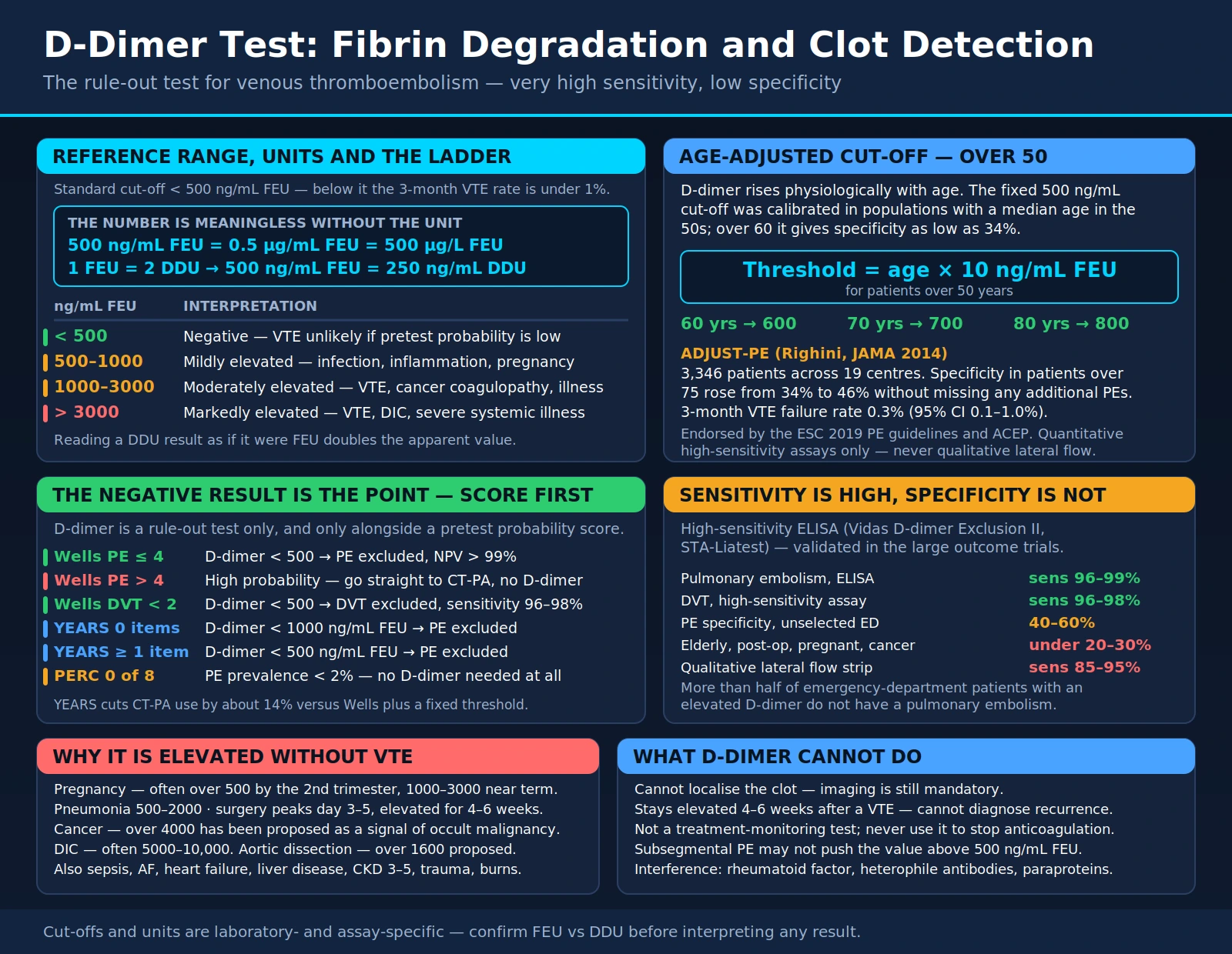
D-dimer is a fibrin degradation product released when cross-linked fibrin clots are broken down by plasmin. It is widely used to help rule out venous thromboembolism (VTE), which includes deep vein thrombosis (DVT) and pulmonary embolism (PE). The StatPearls chapter on the test describes it as highly sensitive but not specific: a normal result in a person with low or intermediate clinical probability makes a clot unlikely, while a raised result has many possible causes besides a clot.
Table of Contents
- Overview — What Is D-Dimer?
- Biochemistry — How D-Dimer Is Formed
- How the Test Works
- Reference Ranges and Units
- Wells Score and Clinical Decision Rules
- Age-Adjusted D-Dimer Threshold
- Causes of Elevated D-Dimer
- D-Dimer in DIC and Critical Illness
- Pregnancy and Other Special Populations
- COVID-19 and D-Dimer
- Quantitative vs. Qualitative Assays
- Limitations and False Positives
- Anticoagulation Duration and the HERDOO2 Rule
- Key Research Papers
- Connections
- Featured Videos
Overview — What Is D-Dimer?
D-dimer is a small protein fragment produced when a blood clot (thrombus) dissolves. The name refers to a specific structural feature of fibrin: after thrombin converts soluble fibrinogen into fibrin monomers, Factor XIIIa (a transglutaminase activated by thrombin) cross-links adjacent fibrin D domains together, forming a stable, reinforced clot. When the fibrinolytic system — driven by plasmin — degrades this cross-linked fibrin network, it releases fragments called D-dimer, named for the two D domains that remain joined after plasmin cleaves the fibrin polymer.
Because D-dimer reflects both clot formation and clot dissolution, it is a marker of clotting activity anywhere in the body, not in one specific blood vessel. A raised D-dimer shows that fibrin has been laid down and is being broken down somewhere; it does not show where. This is the reason for its high sensitivity and low specificity: many processes that activate coagulation produce D-dimer, from wounds and surgery to pulmonary embolism.
Clinically, D-dimer is used as a rule-out test for venous thromboembolism. In the Christopher Study of 3,306 patients with suspected PE, 1,057 (32.0%) had a "PE unlikely" score and a normal D-dimer; of the 1,028 left untreated, 5 (0.5%) had a nonfatal VTE over 3 months (van Belle 2006). In a randomized trial in suspected DVT, 39% of patients in the D-dimer group needed no ultrasound, and 0.4% of those in whom DVT had been ruled out had a VTE over 3 months (Wells 2003). Ruling a clot out this way spares people CT pulmonary angiography (with its radiation and contrast) or compression ultrasound.
The coagulation cascade responsible for D-dimer production has two pathways: the intrinsic pathway (contact activation via Factor XII, XI, IX, VIII) and the extrinsic pathway (tissue factor plus Factor VII). Both converge on a common pathway involving Factor X, Factor V, prothrombin (Factor II), thrombin, and finally fibrinogen. D-dimer appears at the very end of this process — after the clot has formed and after plasmin has acted. The StatPearls chapter notes that results can be affected if blood is sampled too early, during clot formation, and that blood taken after an anticoagulant has been given may test negative; it gives D-dimer a plasma half-life of 6 to 8 hours.
Biochemistry — How D-Dimer Is Formed
D-dimer is the end product of a two-stage sequence: a clot must first be built and chemically reinforced, and then it must be enzymatically taken apart. Neither step alone produces D-dimer. This is why the molecule is a marker of completed clot turnover rather than of coagulation activity in the abstract.
Step 1: Cross-Linked Fibrin Clot Formation
When coagulation is triggered — by tissue factor exposure, endothelial injury, or contact activation — thrombin cleaves fibrinopeptides A and B from circulating fibrinogen, converting it into fibrin monomers. These monomers polymerize into fibrin protofibrils, which associate laterally into fibers. Thrombin also activates Factor XIII into Factor XIIIa, a transglutaminase that forms covalent bonds between the gamma chains of adjacent fibrin monomers and, more slowly, between their alpha chains. The resulting cross-linked polymer is mechanically stronger and more resistant to enzymatic digestion than uncross-linked fibrin.
Step 2: Plasmin-Mediated Fibrinolysis
Tissue plasminogen activator (t-PA) and urokinase plasminogen activator convert plasminogen into plasmin, the principal fibrinolytic enzyme. Plasmin cleaves fibrin at lysine and arginine residues throughout the clot. Because Factor XIIIa has already joined the gamma chains, plasmin cannot fully disassemble the polymer into its original subunits; instead it liberates a characteristic set of fragments. One of them is D-dimer — two D domains from neighboring fibrin monomers still joined through their cross-link. StatPearls notes that most fibrin degradation products are cleared mainly by the liver, while D-dimer itself is cleared primarily by the kidneys and the reticuloendothelial system.
Why the Cross-Link Matters Analytically
D-dimer is not generated from fibrinogen or from uncross-linked fibrin. That biochemical constraint is what allows monoclonal antibody assays to distinguish D-dimer from circulating fibrinogen and from other fibrin(ogen) degradation products. It also explains how the result is read: a raised D-dimer means a clot was built and is being dismantled — whether that clot is a deep vein thrombus, a pulmonary embolus, a surgical wound, a resolving hematoma, or the diffuse microthrombi of disseminated intravascular coagulation. StatPearls adds that people with Factor XIII deficiency have continually low D-dimer levels.
How the Test Works
D-dimer is measured from a plasma sample; StatPearls describes collection into a tube containing 3.2% sodium citrate, which binds calcium so that the sample does not clot in the tube. The plasma is separated by centrifugation and analyzed by immunoassay. Modern D-dimer assays use monoclonal antibodies that recognize an epitope present on cross-linked fibrin degradation products and not on fibrinogen.
Three main assay formats are in common use:
- Enzyme-linked immunosorbent assay (ELISA): In a 2004 systematic review, ELISA and quantitative rapid ELISA ranked highest for sensitivity: about 0.96 for DVT and 0.95 for PE, with negative likelihood ratios of 0.09 to 0.13 (Stein 2004). The 2019 European Society of Cardiology (ESC) guideline gives quantitative ELISA or ELISA-derived assays a diagnostic sensitivity of 95% or more (Konstantinides 2019).
- Latex agglutination / immunoturbidimetric assays: Automated analyzers use latex particles coated with anti-D-dimer antibodies. When D-dimer is present, particle agglutination increases turbidity, which is measured photometrically.
- Whole-blood point-of-care assays: These tests run on a drop of whole blood without centrifugation. The 2019 ESC guideline states that point-of-care D-dimer assays have lower sensitivity than laboratory-based tests and limits their use to patients with a low pre-test probability (Konstantinides 2019).
D-dimer assays differ in the antibody used, the capture method, the instrument and the calibration standard, and these differences change how the assays perform, so evidence for one assay cannot simply be extrapolated to another (Linkins 2017). StatPearls lists hemolysis of the sample, lipemia and heterophile antibodies among the causes of falsely raised results.
Reference Ranges and Units
StatPearls gives a typical normal D-dimer of less than 500 ng/mL (0.50 µg/mL), with 500 ng/mL or more counted as positive, and notes that no critical level has been established. MedlinePlus does not give a numeric range; it explains that a low or normal result means a clotting disorder is probably absent, that blood thinners can cause a false-negative result, and that a high result needs further tests to find where a clot is or what kind of clotting disorder is present.
Units are a common source of confusion. There is no universally accepted international standard or calibrator for the test, so laboratories use different reporting units, thresholds and methods (StatPearls):
- FEU (fibrinogen equivalent units): In an international survey of 409 laboratories, 246 (60%) reported D-dimer in FEU, and at least 28 different combinations of measurement units were in use (Lippi 2015).
- DDU (D-dimer units): A review of COVID-19 papers found that most did not state whether results were in DDU or FEU — a difference of about two-fold — and found at least four reporting errors in 21 papers (Favaloro 2020). The same numeric result therefore means different things depending on the unit.
- Mass units: results are written as ng/mL, µg/L, µg/mL or mg/L. StatPearls writes the usual cut-off as 500 ng/mL or 0.50 µg/mL; ng/mL and µg/L are numerically identical (500 ng/mL = 500 µg/L).
The cut-offs quoted on this page are given in the units the cited studies used (ng/mL or µg/L). A cut-off validated for one assay and unit does not automatically transfer to another (Linkins 2017), and laboratory reports give the value together with the unit and the laboratory's own threshold.
Wells Score and Clinical Decision Rules
D-dimer is validated as a rule-out test when combined with a structured clinical pre-test probability assessment. The 2019 ESC guideline notes that the negative predictive value of D-dimer testing is high and the positive predictive value of a raised result is low (Konstantinides 2019). StatPearls describes clinical decision tools such as the Wells and revised Geneva scores as the first step, and states that D-dimer testing is not recommended for patients with a high pre-test probability, who go directly to imaging. The main validated algorithms are:
Wells PE Score (Pulmonary Embolism)
The Wells PE score was derived from 40 clinical variables in a prospective cohort and assigns points to seven items (Wells 2000):
- Clinical symptoms of DVT: 3 points
- No alternative diagnosis: 3 points
- Heart rate > 100: 1.5 points
- Immobilization or surgery in the previous four weeks: 1.5 points
- Previous DVT or PE: 1.5 points
- Hemoptysis: 1 point
- Malignancy: 1 point
In the two-level version, a score of 4 or less means "PE unlikely" and more than 4 means "PE likely". In the derivation study, 7.8% of patients scoring 4 or less had PE, but when their D-dimer was also negative the rate was 2.2% in the derivation set and 1.7% in the validation set (Wells 2000). In the Christopher Study's algorithm, "PE unlikely" patients had D-dimer testing and PE was excluded if the result was normal, while all others went to CT; the 3-month VTE rate after a normal D-dimer was 0.5% (van Belle 2006).
Wells DVT Score (Deep Vein Thrombosis)
The Wells DVT score uses a leg-focused set of items. StatPearls describes it as considering recent malignancy; recent immobilization, including recent surgery; asymmetric leg swelling; collateral veins; tenderness along the deep veins; and a history of DVT — and as subtracting 2 points if an alternative diagnosis is as likely as, or more likely than, DVT. The modified Wells score adds 1 point for a previously documented DVT; a score of 1 or less means DVT is unlikely and 2 or more means DVT is likely.
In a randomized trial of outpatients with suspected DVT (530 assigned to ultrasound alone, 566 to D-dimer testing), patients judged clinically unlikely to have DVT who had a negative D-dimer did not have ultrasound. Over 3 months, 0.4% of those in whom DVT had been ruled out in the D-dimer group had a VTE, compared with 1.4% in the ultrasound-only group (P=0.16); ultrasound use fell from 1.34 to 0.78 tests per patient, and 39% of the D-dimer group needed no ultrasound at all (Wells 2003). StatPearls states that a high-sensitivity D-dimer below 500 ng/mL in a patient with low or moderate pre-test probability rules out DVT, and that D-dimer cannot reliably exclude DVT at high pre-test probability.
YEARS Algorithm
The YEARS algorithm (van der Hulle 2017) uses three clinical items plus a D-dimer threshold that depends on them:
- Clinical signs of DVT
- Hemoptysis
- PE the most likely diagnosis
If no YEARS items are present and D-dimer is below 1000 ng/mL, PE is considered excluded. If one or more YEARS items are present and D-dimer is below 500 ng/mL, PE is considered excluded. All other patients have CT pulmonary angiography. In the prospective YEARS study of 3,465 patients at 12 hospitals in the Netherlands, 18 of the 2,946 patients in whom PE was ruled out and who were left untreated were diagnosed with VTE over 3 months (0.61%). CT pulmonary angiography was not indicated in 1,651 patients (48%) with YEARS, compared with 1,174 (34%) had the Wells rule with a fixed 500 ng/mL threshold been applied — an absolute difference of 14% (van der Hulle 2017). The 2019 ESC guideline describes the YEARS approach as one way to adapt the D-dimer cut-off to clinical probability (Konstantinides 2019).
PERC Rule (Pulmonary Embolism Rule-Out Criteria)
The PERC rule is a block of eight criteria; if all are met, the rule is used to stop the work-up without a D-dimer: age under 50, pulse under 100, oxygen saturation of 95% or more, no unilateral leg swelling, no hemoptysis, no surgery or trauma requiring hospitalization within 4 weeks, no prior DVT or PE, and no estrogen use (Kline 2008). The rule is meant to prevent ordering a D-dimer in a very-low-probability patient whose slightly raised non-specific result would then lead to imaging. The 2019 ESC guideline describes PERC as selecting patients whose likelihood of PE is so low that a diagnostic work-up need not be started, and notes the low overall prevalence of PE in the studies (Konstantinides 2019).
The criteria were derived by Kline et al. (J Thromb Haemost 2004, PMID 15304025) from 3,148 emergency department patients at 10 US hospitals. The authors estimated a test threshold of 1.8% — the probability below which a D-dimer is not worth ordering. In the two validation groups, 1.4% of rule-negative patients in the low-risk group and 0% in the very-low-risk group had PE. In a 2008 prospective study of 8,138 patients in 13 emergency departments, low clinician suspicion (a gestalt estimate below 15%) together with a negative PERC was present in 1,666 patients (20%), with a false-negative rate of 1.0% for VTE or death within 45 days (Kline 2008).
The PROPER trial, a crossover cluster-randomized noninferiority trial in 14 emergency departments in France, enrolled 1,916 patients with a low gestalt clinical probability of PE. During the PERC period, one PE (0.1%) was diagnosed during the 3-month follow-up, compared with none in the control period, which met the noninferiority test; CT pulmonary angiography was used in 13% versus 23% of patients (Freund 2018).
Age-Adjusted D-Dimer Threshold
D-dimer rises with age, so a fixed cut-off rules out fewer older patients. In a meta-analysis of 13 cohorts including 12,497 patients with a non-high clinical probability, the specificity of the conventional 500 µg/L cut-off fell from 57.6% in patients aged 51–60 to 39.4% at 61–70, 24.5% at 71–80 and 14.7% over 80. The age-adjusted cut-off (age × 10 µg/L) gave higher specificity in every age group — 62.3%, 49.5%, 44.2% and 35.2% — while sensitivity stayed above 97% (Schouten 2013). A lower specificity means more people without a clot are sent to imaging.
The age-adjusted D-dimer threshold was prospectively validated in the ADJUST-PE study (Righini et al., JAMA 2014, PMID 24643601):
- Formula: for patients aged 50 or older, cut-off = age × 10 µg/L; below 50, the conventional 500 µg/L cut-off applies.
- Design: a prospective management outcome study of 3,346 emergency department outpatients with suspected PE at 19 centers in Belgium, France, the Netherlands and Switzerland. Clinical probability was assessed with the simplified revised Geneva score or the two-level Wells score, and D-dimer was measured with highly sensitive assays.
- Safety: patients with a non-high or unlikely clinical probability and a D-dimer between 500 µg/L and their age-adjusted cut-off had no CT and no treatment. Their 3-month failure rate was 1 of 331 patients — 0.3% (95% CI 0.1–1.7%).
- Imaging avoided in the oldest patients: among the 673 patients aged 75 or older with a non-high clinical probability, the age-adjusted cut-off raised the proportion in whom PE could be excluded on D-dimer from 43 (6.4%) to 200 (29.7%), without any additional false-negative findings.
- Independent confirmation: Schouten et al. (BMJ 2013, Search PubMed) concluded in their systematic review and meta-analysis that age-adjusted cut-offs substantially increase specificity without modifying sensitivity in patients aged 50 or more with a non-high clinical probability.
The 2019 ESC guideline states that age-adjusted cut-offs (age × 10 µg/L for patients aged over 50) may improve the performance of D-dimer testing in older people (Konstantinides 2019). StatPearls notes that the approach is supported by both American and European medical colleges, although it is not approved by the United States Food and Drug Administration.
The age-adjusted threshold is part of a rule-out strategy for people with a low or intermediate (non-high) pre-test probability. In people with a high pre-test probability, StatPearls describes going directly to imaging rather than D-dimer testing. Combining the two strategies was tested in a post hoc analysis of the YEARS cohort: fully integrating the age-adjusted cut-off into YEARS would have spared CT in 1,627 patients (47%) instead of 1,651 (48%), at the cost of four additional missed PE diagnoses, and the authors found no added value (van der Pol 2017).
Groups in Which the Age-Adjusted Threshold Has Not Been Established
- Pregnancy. Pregnancy raises D-dimer through a mechanism unrelated to age. The pregnancy-adapted algorithm that has been prospectively tested uses the YEARS thresholds, not an age-adjusted cut-off (van der Pol 2019).
- Point-of-care and qualitative assays. A qualitative test gives no number against which an age-based cut-off can be applied, and the ESC guideline limits point-of-care assays to patients with a low pre-test probability (Konstantinides 2019).
- Other assays. Cut-offs and performance differ between assays, so results for one assay cannot simply be extrapolated to another (Linkins 2017).
Causes of Elevated D-Dimer
Because D-dimer reflects clot formation and dissolution anywhere in the body, any condition that activates coagulation or triggers inflammation with fibrin deposition can raise it. The list is long, which is why D-dimer is a poor rule-in test despite being a useful rule-out test: in the 2004 systematic review, positive likelihood ratios were in the general range of 1.5 to 2.5, which does not greatly increase the certainty of a diagnosis (Stein 2004).
Thrombotic Conditions (Primary Indications)
- Deep vein thrombosis (DVT): The main clinical indication. Pooled sensitivity of ELISA assays for DVT was 0.96 in the 2004 systematic review (Stein 2004).
- Pulmonary embolism (PE): Pooled sensitivity of ELISA assays for PE was 0.95 (Stein 2004). StatPearls describes the size and location of a clot as determining the severity of symptoms.
- Arterial thrombosis: Myocardial infarction, stroke and acute arterial occlusion also activate coagulation and fibrinolysis and can raise D-dimer. StatPearls notes that D-dimer is less specific for arterial events than for venous events, so its usefulness in arterial thrombosis is limited. The rule-out algorithms above were built for venous thromboembolism; a normal D-dimer does not exclude an acute coronary syndrome.
- Cerebral venous thrombosis: D-dimer is often raised. In its stroke section, StatPearls notes that a D-dimer can help suggest a central venous thrombosis but that a negative D-dimer does not exclude the diagnosis.
- Aortic dissection: D-dimer has been studied as part of rule-out strategies for aortic dissection; it is not a standard stand-alone test for it.
- Mesenteric vein thrombosis: As with other venous clots, D-dimer is usually raised.
Physiological Elevation
- Pregnancy: D-dimer rises during pregnancy, including the postpartum period (StatPearls), and the 2019 ESC guideline notes that it is more frequently elevated in pregnancy (Konstantinides 2019). Trimester-specific thresholds have been proposed but are not universally adopted. See Pregnancy and Other Special Populations.
- Older age: As discussed above, D-dimer rises with age, which is what age-adjusted thresholds address.
- Neonates: D-dimer is physiologically higher in the first weeks of life.
- Other listed causes: StatPearls also lists cigarette smoking, short bursts of strenuous exercise, oral contraceptive pills, immobilization, diabetes and elevated triglyceride levels.
Inflammatory and Infectious Conditions
- Sepsis and ARDS: One of the most common causes of markedly raised D-dimer in the ICU. Bacterial products and inflammatory cytokines drive tissue factor expression, activating coagulation broadly; acute respiratory distress syndrome adds lung fibrin deposition. StatPearls estimates that disseminated intravascular coagulation (DIC) occurs in up to 30% to 50% of cases of severe sepsis, the most common cause of DIC.
- Pneumonia and other infections: StatPearls describes D-dimer as a dubious marker of clotting in the setting of inflammation, ischemia, infarction and infection, and lists infections such as sepsis and HIV. Pneumonia can mimic PE clinically and biochemically.
- Inflammatory bowel disease: Active Crohn's disease or ulcerative colitis is associated with a hypercoagulable state and D-dimer elevation.
- Autoimmune diseases: StatPearls lists autoimmune diseases, and MedlinePlus lists rheumatoid arthritis, among the causes of a raised D-dimer.
Malignancy
- Cancer-associated coagulopathy: Many tumors express tissue factor and other procoagulant molecules, creating a chronic hypercoagulable state (Trousseau syndrome). StatPearls lists malignancy among the causes of a raised D-dimer, and notes that up to 20% of patients with metastatic adenocarcinoma or lymphoproliferative disease have DIC.
- Cryptogenic stroke: StatPearls reports that D-dimer levels in cryptogenic stroke caused by cancer-related hypercoagulability tend to be much higher than in cryptogenic stroke without cancer (10,070 ng/mL versus 500 to 700 ng/mL) and states that a raised D-dimer in cryptogenic stroke should prompt screening for an occult malignancy. A raised D-dimer is not by itself a cancer test.
Post-Procedural and Traumatic
- Surgery: Surgery causes fibrin deposition at wound sites, and StatPearls lists recent surgery among the causes of a raised D-dimer, which makes biochemical rule-out of VTE less useful after an operation.
- Trauma: Significant traumatic injury activates coagulation both at wound sites and systemically.
- Burns: Extensive burns trigger systemic inflammatory and coagulation activation.
Cardiovascular
- Atrial fibrillation: Stasis within the left atrial appendage promotes intracardiac thrombus formation and D-dimer elevation.
- Heart failure: StatPearls and MedlinePlus list congestive heart failure and heart disease among the causes of a raised D-dimer.
- Aortic aneurysm: Mural thrombus within an aneurysm produces D-dimer; StatPearls notes that 1% to 5% of patients with chronic conditions such as solid tumors and aortic aneurysms have DIC.
Hematologic
- Disseminated intravascular coagulation (DIC): The condition with the most extreme D-dimer values. DIC involves simultaneous widespread clot formation and fibrinolysis, consuming platelets and clotting factors. A fibrin-related marker such as D-dimer is part of the ISTH DIC score (see D-Dimer in DIC and Critical Illness).
- Sickle cell disease: Chronic vaso-occlusion and vascular inflammation keep baseline D-dimer higher, complicating VTE work-up in this group.
- Thrombophilias and hemolysis: StatPearls lists thrombophilias, and HELLP syndrome (hemolysis, elevated liver enzymes and low platelets), among the causes of a raised D-dimer.
Organ Dysfunction
- Liver disease: StatPearls lists cirrhosis among the causes of a raised D-dimer. The liver synthesizes most coagulation factors and inhibitors, including plasminogen activator inhibitor-1 (PAI-1), so liver failure produces a complex mixed coagulopathy.
- Kidney disease: StatPearls states that D-dimer is cleared primarily by the kidneys and the reticuloendothelial system, so reduced kidney function is one route to a higher baseline value.
- Stroke: StatPearls notes that D-dimer levels typically rise after a stroke, and that higher levels correspond to incomplete recanalization, stroke progression and recurrence.
D-Dimer in DIC and Critical Illness
Disseminated intravascular coagulation is the condition in which D-dimer reaches its most extreme values, and it is also the condition in which the test has a formal, scored diagnostic role rather than a rule-out role. DIC is simultaneous, widespread coagulation activation and fibrinolysis: microthrombi form throughout the circulation while platelets and clotting factors are consumed, so the patient can be clotting and bleeding at the same time.
The ISTH DIC Score
The International Society on Thrombosis and Haemostasis scoring system (Taylor et al., Thromb Haemost 2001, PMID 11816725) gives D-dimer — or another fibrin-related marker — a central weighting. As tabulated in a 2024 scoping review of DIC criteria (Matsuoka 2024):
- No increase: 0 points
- Moderate increase: 2 points
- Strong increase: 3 points
Three other items complete the score: platelet count, prolongation of the prothrombin time, and fibrinogen level. A total of 5 or more is compatible with overt DIC (Matsuoka 2024). In a prospective validation in 217 intensive care patients, the 0–8-point score had a sensitivity of 91% and a specificity of 97% against an expert-opinion standard, and each 1-point increase carried an odds ratio of 1.25 for 28-day mortality (Bakhtiari 2004). StatPearls describes acute DIC as thrombocytopenia and consumption of coagulation factors with a prolonged PT and aPTT and an elevated D-dimer — a pattern that separates it from the isolated D-dimer rise of ordinary VTE.
DIC Always Has a Trigger
DIC is a downstream consequence of another illness, and StatPearls describes treatment as centering on the underlying disorder. Triggers it lists include:
- Sepsis — the most common cause; DIC is estimated to occur in up to 30% to 50% of cases of severe sepsis
- Major trauma
- Obstetric complications — placental abruption, amniotic fluid embolism, HELLP syndrome
- Malignancy, including metastatic adenocarcinoma, lymphoproliferative disease and acute leukemia (acute promyelocytic leukemia is a well-known example)
- Transfusion reactions
- Snake bites — StatPearls adds that, in many cases, a rise in D-dimer is the first indicator of venom-induced consumption coagulopathy
Because both DIC and a large pulmonary embolism can produce a very high D-dimer, the number alone does not separate them. The ISTH score combines D-dimer with platelet count, PT and fibrinogen, and imaging remains the way to find a PE. MedlinePlus notes that D-dimer may also be used to monitor how well DIC treatment is working.
Pregnancy and Other Special Populations
Some groups have a physiologically higher D-dimer baseline, which lowers the share of people in whom the standard cut-off rules a clot out. In several of these groups the baseline is highest in the people at greatest thrombotic risk, so the test is least discriminating where it would be most useful.
Pregnancy: Physiology
Pregnancy is a hypercoagulable state. Fibrinogen and several clotting factors (including Factors VII, VIII and X) rise, Protein S falls, and fibrinolytic activity decreases. The net effect is more fibrin turnover and therefore more D-dimer, with no thrombus anywhere. StatPearls lists pregnancy, including the postpartum period, among the causes of a raised D-dimer.
Pregnancy: How Much the Test Helps by Trimester
The usefulness of D-dimer falls as pregnancy advances. In the Pregnancy-Adapted YEARS study, CT pulmonary angiography was avoided in 65% of women who entered the study in the first trimester and in 32% of those who entered in the third trimester (van der Pol 2019).
Pregnancy: Diagnostic Algorithms That Have Been Studied
The Pregnancy-Adapted YEARS algorithm (van der Pol 2019) assessed the three YEARS items with a D-dimer: PE was ruled out if no item was present and D-dimer was below 1000 ng/mL, or if one or more items were present and D-dimer was below 500 ng/mL. Women with symptoms of DVT had compression ultrasonography first, and if it showed a clot, CT pulmonary angiography was not performed. Of 510 women screened, 12 were excluded, and PE was diagnosed in 20 (4.0%) at baseline; during the 3-month follow-up one woman (0.21%) was diagnosed with a popliteal DVT and none with PE. CT pulmonary angiography was avoided in 195 women (39%). The 2019 ESC guideline also presents a dedicated diagnostic algorithm for suspected PE in pregnancy and notes that D-dimer is more frequently elevated during pregnancy (Konstantinides 2019). There are no universally adopted trimester-specific reference ranges, and the age-adjusted formula has not been established in pregnancy. StatPearls notes that CT pulmonary angiography is now an accepted alternative to ventilation-perfusion scanning in pregnancy.
Active Malignancy
People with active cancer often carry a raised D-dimer (StatPearls), so a positive result adds less information in this group than in the general population. A result below the cut-off keeps its rule-out value only within the algorithms and populations in which it was studied.
COVID-19 and D-Dimer
The COVID-19 pandemic brought D-dimer into wide public awareness, as SARS-CoV-2 infection was recognized to cause a hypercoagulable state often called COVID-19-associated coagulopathy (CAC), typically described as a raised D-dimer and fibrinogen with relatively preserved platelet counts. In one early series of 183 patients in Wuhan, however, 71.4% of non-survivors (and 0.6% of survivors) met the criteria for DIC during their hospital stay (Tang, J Thromb Haemost 2020, PMID 32073213).
Proposed Mechanisms of COVID-19 Hypercoagulability
- Endothelial injury: SARS-CoV-2 uses the ACE2 receptor, and endothelial damage exposes tissue factor, triggering the extrinsic coagulation pathway.
- Inflammatory cytokines: IL-6, IL-1β and TNF-α can suppress natural anticoagulants (Protein C, Protein S, antithrombin) and raise plasminogen activator inhibitor-1 (PAI-1), which slows fibrinolysis.
- Complement activation: SARS-CoV-2 has been reported to activate the complement system, contributing to platelet activation and microthrombi.
- Neutrophil extracellular traps (NETs): DNA-histone complexes released by neutrophils can act as a scaffold for fibrin deposition and activate Factor XII.
D-Dimer as a COVID-19 Severity and Mortality Predictor
Observational cohorts during the pandemic found that a raised D-dimer on hospital admission was associated with worse outcomes. StatPearls notes that a D-dimer of 1000 ng/mL in a patient with COVID-19 corresponds to severe disease, and that management decisions are based mainly on the clinical picture rather than on changing D-dimer levels.
- In a retrospective cohort of 191 adult inpatients in Wuhan, a D-dimer greater than 1 µg/mL on admission was associated with higher odds of in-hospital death (odds ratio 18.42, 95% CI 2.64–128.55), alongside older age and a higher SOFA score (Zhou 2020).
- In a retrospective study of 449 patients with severe COVID-19 (Tang et al., J Thromb Haemost 2020, PMID 32220112), D-dimer, prothrombin time and age were positively correlated with 28-day mortality. Overall 28-day mortality did not differ between heparin users and non-users (30.3% vs 29.7%); among patients with a D-dimer more than 6-fold above the upper limit of normal, mortality was lower in heparin users (32.8% vs 52.4%), as it was in those with a sepsis-induced coagulopathy score of 4 or more (40.0% vs 64.2%). The design was observational, so the association does not show cause and effect.
- In an earlier report from the same group — Tang et al., J Thromb Haemost 2020, PMID 32073213 — non-survivors of COVID-19 pneumonia had significantly higher D-dimer and fibrin degradation product levels on admission, and longer prothrombin and activated partial thromboplastin times, than survivors; overall mortality was 11.5%. This was one of the first signals that the illness carried a coagulopathic component.
- A review of COVID-19 papers found that most did not state whether D-dimer was reported in DDU or FEU, units that differ about two-fold, so cut-offs quoted from different studies are not always comparable (Favaloro 2020).
COVID-Associated Pulmonary Embolism
Early pandemic series reported a high rate of thrombosis in hospitalized COVID-19 patients:
- Klok et al. (Thromb Res 2020, PMID 32291094) studied 184 ICU patients with COVID-19 pneumonia, all receiving at least standard-dose thromboprophylaxis. The cumulative incidence of thrombotic complications was 31%; VTE confirmed by CT pulmonary angiography and/or ultrasonography accounted for 27% and arterial thrombotic events for 3.7%. PE was the most frequent thrombotic complication (25 events, 81%).
Long COVID and D-Dimer
In some people with Long COVID (post-acute sequelae of SARS-CoV-2 infection), persistent symptoms including fatigue, shortness of breath and brain fog have been linked to ongoing microclot formation. Fibrin amyloid microclots — identified by fluorescence microscopy — have been reported in blood samples from Long COVID patients, and some studies have found modestly raised D-dimer months after acute infection; this remains an area of active research rather than established clinical practice.
Quantitative vs. Qualitative Assays
The distinction between quantitative and qualitative D-dimer assays determines which clinical algorithms can be applied and how results are interpreted.
Quantitative Assays
Quantitative assays give a numeric result (in FEU or DDU). They:
- Allow age-adjusted thresholds (age × 10 µg/L from age 50, as in ADJUST-PE)
- Allow the YEARS algorithm (a threshold of 1000 or 500 ng/mL depending on the items present)
- Carry prognostic information through the degree of elevation, as in DIC scoring and COVID-19 studies
- Can be followed over time, for example in DIC (MedlinePlus)
- Include the laboratory ELISA assays that ranked highest for sensitivity in the 2004 systematic review — about 0.95 for PE and 0.96 for DVT (Stein 2004)
Qualitative Assays
Qualitative assays give a positive or negative result against a fixed cut-off. The 2019 ESC guideline describes point-of-care D-dimer assays as less sensitive than laboratory-based tests and limits their use to patients with a low pre-test probability (Konstantinides 2019). Because they give no number, they cannot be combined with age-adjusted or YEARS thresholds.
High-Sensitivity vs. Standard Assays
Not all quantitative assays are equivalent. D-dimer assays vary in antibody, capture method, instrument and calibration standard, and the evidence available for one assay cannot simply be extrapolated to another (Linkins 2017). The major outcome studies — the Christopher Study, ADJUST-PE and YEARS — each used their own assays, and ADJUST-PE specified highly sensitive D-dimer measurement (Righini 2014). The 2019 ESC guideline gives quantitative ELISA or ELISA-derived assays a sensitivity of 95% or more (Konstantinides 2019). Whether a given laboratory's assay matches those used in the studies is a property of that assay, documented by its manufacturer and the laboratory.
Limitations and False Positives
The limitations of D-dimer testing matter as much as its strengths. Where specificity is low, a raised result leads to imaging in many people who have no clot, with the contrast and radiation that imaging brings.
Low Specificity
The fundamental limitation of D-dimer is that it is raised by many conditions other than VTE. Positive likelihood ratios of the assays were in the general range of 1.5 to 2.5 in a 2004 systematic review (Stein 2004). Specificity falls with age: with the conventional 500 µg/L cut-off it was 14.7% in patients over 80 (Schouten 2013). Pregnancy, malignancy and recent surgery are other causes with frequent non-specific elevations (StatPearls). Used without a clinical probability assessment, D-dimer leads to a great deal of extra testing.
Cannot Localize the Clot
A raised D-dimer shows that a clot has formed and is being dissolved somewhere in the body. It cannot show whether the source is a leg vein, pulmonary artery, portal vein, cerebral sinus or coronary artery. Imaging is needed to confirm and locate a clot (MedlinePlus).
Cannot Distinguish Old from New Clot Activity
D-dimer can stay raised for a time after an acute VTE event, even during treatment. This makes it less reliable for diagnosing a recurrence soon after a prior VTE, because a new elevation cannot be told apart from persistent elevation from the earlier event. StatPearls notes that patients with recurrent symptoms in the same leg can go directly to imaging, because of their higher risk and the lower specificity of D-dimer in this group.
Not a Treatment-Monitoring Test for VTE
D-dimer is not a routine marker of anticoagulation response: levels often fall during treatment, but the pace varies and depends on the underlying condition, and MedlinePlus notes that blood thinners can cause a false-negative result. One narrowly validated use is in deciding the length of treatment after a first unprovoked VTE in women (see the HERDOO2 rule below).
Post-Surgical and Post-Trauma Populations
Surgery and trauma reliably raise D-dimer for some time, due to wound healing and fibrinolysis at the injured sites. In these groups a raised result says little about whether a VTE is present.
Analytical Interference
StatPearls lists heterophile antibodies, hemolysis of the sample and lipemia among causes of falsely raised results, and a case report describes heterophile-antibody interference in a D-dimer assay (Lippi 2014). Because assays differ in their antibodies (Linkins 2017), an interference that affects one platform may not affect another.
Subsegmental PE Controversy
Some subsegmental PEs (small clots in peripheral pulmonary arteries) may not raise D-dimer above the usual cut-off, particularly in ambulatory patients. The clinical significance of small subsegmental PEs — increasingly detected on high-resolution CT — is debated, and their management remains controversial.
Anticoagulation Duration and the HERDOO2 Rule
D-dimer is not used to titrate day-to-day anticoagulation. There is one narrowly validated use in deciding treatment length, and it is easy to over-generalize.
After a first unprovoked venous thromboembolism, the question is how long to continue anticoagulation: stopping carries a risk of recurrence, and continuing carries a risk of bleeding. The HERDOO2 rule (Rodger et al., BMJ 2017, PMID 28314711) was developed to identify women at low risk of recurrence. The acronym enumerates four risk markers, each worth one point:
- Hyperpigmentation, Edema, or Redness in either leg — post-thrombotic signs
- D-dimer of 250 µg/L or higher, measured while the patient is still taking anticoagulation
- Obesity, defined as BMI of 30 or above
- Older age, defined as 65 years or above
In the validation study (2,785 participants at 44 centres in seven countries, after 5 to 12 months of initial treatment), women with 0 or 1 of these markers were classified as low risk and stopped anticoagulation. Their recurrence rate was 3.0% per patient-year (95% CI 1.8% to 4.8%), and the authors concluded that such women can safely discontinue anticoagulants after short-term treatment. Among high-risk women and men who stopped, the rate was 8.1% per patient-year. The rule does not apply to men: the study report states that no subgroup of men could be identified as low risk.
Two constraints limit the rule. First, HERDOO2 applies only to unprovoked events; a VTE that followed surgery, immobilization, or another transient trigger is a different question. Second, the D-dimer in this rule is drawn on treatment, was measured with one specific assay in the study, and is interpreted against a 250 µg/L threshold, not the 500 µg/L diagnostic cut-off — the two numbers answer different questions and are not interchangeable.
Key Research Papers
- Righini M et al. (2014). Age-adjusted D-dimer cutoff levels to rule out pulmonary embolism: the ADJUST-PE study. JAMA 311(11):1117–1124. — PubMed PMID: 24643601
- Wells PS et al. (2003). Evaluation of D-dimer in the diagnosis of suspected deep-vein thrombosis. N Engl J Med 349(13):1227–1235. — PubMed PMID: 14507948
- van Belle A et al. (2006). Effectiveness of managing suspected pulmonary embolism using an algorithm combining clinical probability, D-dimer testing, and computed tomography. JAMA 295(2):172–179. — PubMed PMID: 16403929
- Tang N et al. (2020). Anticoagulant treatment is associated with decreased mortality in severe coronavirus disease 2019 patients with coagulopathy. J Thromb Haemost 18(5):1094–1099. — PubMed PMID: 32220112
- Klok FA et al. (2020). Incidence of thrombotic complications in critically ill ICU patients with COVID-19. Thromb Res 191:145–147. — PubMed PMID: 32291094
- Schouten HJ et al. (2013). Diagnostic accuracy of conventional or age adjusted D-dimer cut-off values in older patients with suspected venous thromboembolism: systematic review and meta-analysis. BMJ 346:f2492. — Search PubMed
- Kline JA et al. (2008). Emergency clinician-performed compression ultrasonography for deep venous thrombosis of the lower extremity. Ann Emerg Med 52(4):437–445. — PubMed PMID: 18562044
- Le Gal G et al. (2006). Prediction of pulmonary embolism in the emergency department: the revised Geneva score. Ann Intern Med 144(3):165–171. — PubMed PMID: 16461960
- Freund Y et al. (2018). Effect of the Pulmonary Embolism Rule-Out Criteria on subsequent thromboembolic events among low-risk emergency department patients: the PROPER randomized clinical trial. JAMA 319(6):559–566. — PubMed PMID: 29450523
- Di Nisio M, van Es N, Büller HR (2016). Deep vein thrombosis and pulmonary embolism. Lancet 388(10063):3060–3073. — PubMed PMID: 27375038
- Tritschler T et al. (2018). Venous thromboembolism: advances in diagnosis and treatment. JAMA 320(15):1583–1594. — PubMed PMID: 30326130
- Marcus AJ (1994). Thrombosis and inflammation as multicellular processes: significance of cell-cell interactions. Semin Hematol 31(4):261–269. — PubMed PMID: 7831571 (Editorial note: this entry was previously credited to "Weitz JI et al. (2021), Semin Thromb Hemost"; PubMed indexes this title only under Marcus AJ, 1994 and 1995.)
- Kline JA et al. (2004). Clinical criteria to prevent unnecessary diagnostic testing in emergency department patients with suspected pulmonary embolism. J Thromb Haemost 2(8):1247–1255. — PubMed PMID: 15304025 (Derivation and validation of the PERC rule.)
- Stein PD et al. (2004). D-dimer for the exclusion of acute venous thrombosis and pulmonary embolism: a systematic review. Ann Intern Med 140(8):589–602. — PubMed PMID: 15096330
- Taylor FB Jr et al. (2001). Towards definition, clinical and laboratory criteria, and a scoring system for disseminated intravascular coagulation. Thromb Haemost 86(5):1327–1330. — PubMed PMID: 11816725
- Tang N et al. (2020). Abnormal coagulation parameters are associated with poor prognosis in patients with novel coronavirus pneumonia. J Thromb Haemost 18(4):844–847. — PubMed PMID: 32073213
- Linkins LA, Takach Lapner S (2017). Review of D-dimer testing: good, bad, and ugly. Int J Lab Hematol 39(Suppl 1):98–103. — PubMed PMID: 28447414
- Rodger MA et al. (2017). Validating the HERDOO2 rule to guide treatment duration for women with unprovoked venous thrombosis: multinational prospective cohort management study. BMJ 356:j1065. — PubMed PMID: 28314711
- Wells PS et al. (2000). Derivation of a simple clinical model to categorize patients probability of pulmonary embolism: increasing the models utility with the SimpliRED D-dimer. Thromb Haemost 83(3):416–420. — PubMed PMID: 10744147
- Kline JA et al. (2008). Prospective multicenter evaluation of the pulmonary embolism rule-out criteria. J Thromb Haemost 6(5):772–780. — PubMed PMID: 18318689
- van der Hulle T et al. (2017). Simplified diagnostic management of suspected pulmonary embolism (the YEARS study): a prospective, multicentre, cohort study. Lancet 390(10091):289–297. — PubMed PMID: 28549662
- van der Pol LM et al. (2017). No added value of the age-adjusted D-dimer cut-off to the YEARS algorithm in patients with suspected pulmonary embolism. J Thromb Haemost 15(12):2317–2324. — PubMed PMID: 28941051
- van der Pol LM et al. (2019). Pregnancy-adapted YEARS algorithm for diagnosis of suspected pulmonary embolism. N Engl J Med 380(12):1139–1149. — PubMed PMID: 30893534
- Konstantinides SV et al. (2020). 2019 ESC Guidelines for the diagnosis and management of acute pulmonary embolism developed in collaboration with the European Respiratory Society (ERS). Eur Heart J 41(4):543–603. — PubMed PMID: 31504429
- Zhou F et al. (2020). Clinical course and risk factors for mortality of adult inpatients with COVID-19 in Wuhan, China: a retrospective cohort study. Lancet 395(10229):1054–1062. — PubMed PMID: 32171076
- Favaloro EJ, Thachil J (2020). Reporting of D-dimer data in COVID-19: some confusion and potential for misinformation. Clin Chem Lab Med 58(8):1191–1199. — PubMed PMID: 32432563
- Lippi G, Tripodi A, Simundic AM, Favaloro EJ (2015). International survey on D-dimer test reporting: a call for standardization. Semin Thromb Hemost 41(3):287–293. — PubMed PMID: 25839870
- Lippi G, Ippolito L, Tondelli MT, Favaloro EJ (2014). Interference from heterophilic antibodies in D-dimer assessment. A case report. Blood Coagul Fibrinolysis 25(3):277–279. — PubMed PMID: 24253242
- Bakhtiari K, Meijers JC, de Jonge E, Levi M (2004). Prospective validation of the International Society of Thrombosis and Haemostasis scoring system for disseminated intravascular coagulation. Crit Care Med 32(12):2416–2421. — PubMed PMID: 15599145
- Matsuoka T et al. (2024). The transition of the criteria for disseminated intravascular coagulation and the targeted patients in randomized controlled trials over the decades: a scoping review. Thromb J 22(1):112. — PubMed PMID: 39716150
External Authoritative Resources
Connections
- All Lab Tests
- Coagulation Panel
- PT/INR
- aPTT
- Cardiovascular Disease
- Complete Blood Count
- Inflammatory Markers
- BNP / NT-proBNP
- Cardiac Troponin
- High-Sensitivity CRP (hs-CRP)
- Kidney Function Tests
- Pulmonary Embolism — the diagnosis this test is used to rule out.
- Fibrinogen
- D-Dimer and Snake Venom — Dr. Bryan Ardis’s argument about raised D-dimer after vaccination, as he states it.