ANA Test: Antinuclear Antibody for Autoimmune Disease
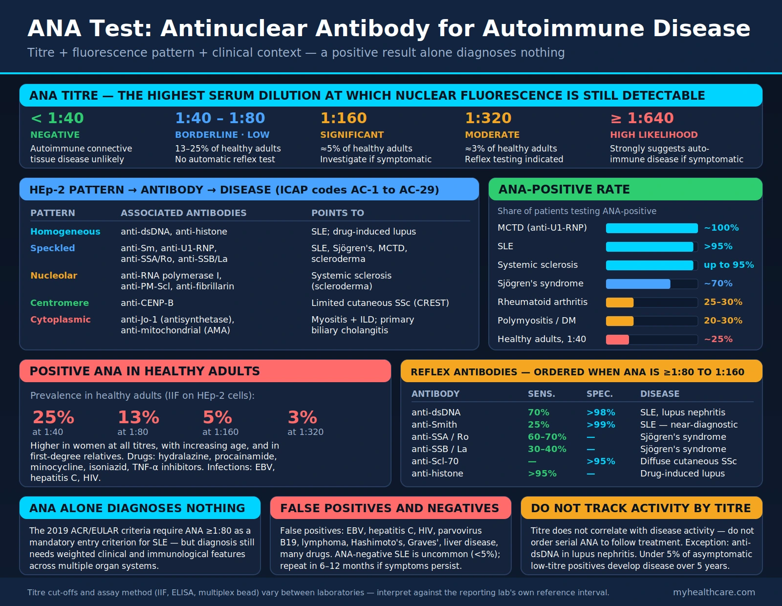
The antinuclear antibody (ANA) test is the main screening test for systemic autoimmune rheumatic diseases. It detects autoantibodies directed against components of the cell nucleus; across the published studies pooled in a 2018 meta-analysis, 95.9% of 13,080 people with systemic lupus erythematosus (SLE) were reported ANA-positive. Understanding the ANA result requires interpreting titer, fluorescence pattern, and clinical context together — a positive result alone does not diagnose any disease.
Table of Contents
Overview
The ANA (antinuclear antibody) test detects autoantibodies that target components of the cell nucleus — including DNA, histones, RNA-binding proteins, and nuclear enzymes. These autoantibodies are a hallmark of systemic autoimmune connective tissue diseases, where the immune system mistakenly attacks the body's own nuclear material.
ANA is used as a screening test for systemic autoimmune diseases, particularly:
- Systemic lupus erythematosus (SLE) — 95.9% of 13,080 patients reported ANA-positive across published studies (Leuchten 2018)
- Sjögren's syndrome — ANA-positive in 79.3% of 10,500 patients at diagnosis in an international registry (Brito-Zerón 2018)
- Systemic sclerosis (scleroderma) — autoantibody-negative disease is rare: fewer than 2% of 874 patients in a Canadian cohort were negative on every method tested (Hudson 2014)
- Polymyositis and dermatomyositis — ANA results vary; myositis-associated antibodies such as anti-Jo-1 are the more characteristic finding
- Mixed connective tissue disease (MCTD) — anti-U1-RNP positivity is mandatory for the diagnosis (Dima 2018)
The test is performed by indirect immunofluorescence (IIF) on HEp-2 cells, which are a human epithelial cell line whose large nuclei make nuclear fluorescence patterns easy to visualize. Many laboratories also use ELISA or multiplex bead-based assays for initial screening; international recommendations emphasise IIF as the reference method and note that alternative platforms differ in their antigen profiles, sensitivity and specificity (Agmon-Levin 2014). For SLE, the 2018 meta-analysis gave a sensitivity of 97.8% but a specificity of 74.7% at a titer of 1:80 (Leuchten 2018) — so a positive result is interpreted in clinical context.
When Ordered
An ANA test is typically ordered when symptoms could indicate a systemic autoimmune connective tissue disease. Features that commonly prompt it include:
- Persistent fatigue with joint pain — especially polyarthralgia or inflammatory arthritis in multiple joints
- Butterfly (malar) rash — fixed erythema across the cheeks and nasal bridge, sparing the nasolabial folds; hallmark of SLE
- Raynaud's phenomenon — episodic vasospasm of fingers/toes triggered by cold or stress; associated with multiple connective tissue diseases
- Unexplained serositis — pleuritis (pleurisy), pericarditis, or peritonitis without infectious cause
- Dry eyes and dry mouth (sicca symptoms) — cardinal features of Sjögren's syndrome
- Unexplained cytopenias — immune-mediated low platelet count (thrombocytopenia) or low white blood cell count (leukopenia)
- Photosensitivity — unusual skin reactions to sun exposure
- Recurrent pregnancy loss — a feature associated with antiphospholipid syndrome and lupus
- Skin thickening — especially progressive hardening of the fingers, hands, or face, suggesting systemic sclerosis
- Unexplained renal disease — lupus nephritis can present with hematuria, proteinuria, or declining kidney function
- Known autoimmune disease — repeat ANA testing is common in practice: over a quarter of ANA tests in Ontario in 2008–2015 were repeats (Lake 2020)
Because low-titer positives are common in healthy people (see below), testing people without symptoms turns up many positives unrelated to disease; reviews describe the resulting unnecessary referrals and examinations of ANA-positive persons (Conrad 2017), and Tan 1997 noted that ANA assays “can create misunderstanding when the limitations are not fully appreciated.”
HEp-2 Cell Patterns
When ANA is performed by indirect immunofluorescence (IIF) on HEp-2 cells, the pattern of nuclear fluorescence provides important diagnostic guidance. Different patterns reflect which nuclear antigens the autoantibodies are targeting, and each pattern directs which specific antibody tests to order next.
- Homogeneous (diffuse) pattern (AC-1): The entire nucleus stains uniformly, including the chromosomes in dividing cells. ICAP lists dsDNA, nucleosomes and histones as the associated antigens, and SLE, drug-induced lupus and juvenile idiopathic arthritis as the associated conditions. ICAP stresses that it must be told apart from the dense fine speckled pattern (below), whose clinical significance is quite different (Chan 2015).
- Speckled patterns: Fine or coarse discrete dots scattered throughout the nucleus, sparing the nucleolus. The nuclear fine speckled pattern was the most frequent pattern in both healthy people and patients in a study of 918 healthy individuals and 153 patients (Mariz 2011). Associated with anti-Sm, anti-U1-RNP, anti-SSA/Ro, and anti-SSB/La antibodies; ICAP lists MCTD, SLE, Sjögren's syndrome, dermatomyositis and SSc/polymyositis overlap. Fine speckled (AC-4) is linked to SS-A/Ro and SS-B/La among other antigens; large/coarse speckled (AC-5) to U1-RNP, Sm and RNA polymerase III (Chan 2015).
- Dense fine speckled (DFS) pattern (AC-2): Produced mainly by antibodies to DFS70/LEDGF. In the Mariz 2011 study this pattern occurred only in healthy individuals. A 2017 review describes it as the most frequent pattern in high-titer ANA-positive healthy persons and reports isolated anti-DFS70 antibodies in less than 1% of patients with ANA-associated rheumatic diseases but in 2–22% of healthy persons (Conrad 2017); a multi-cohort study likewise found anti-DFS70 more prevalent in healthy individuals than in patients with systemic autoimmune rheumatic diseases (Mahler 2012).
- Nucleolar pattern: Staining concentrated in the nucleolus (the RNA-synthesis center of the nucleus). Associated mainly with systemic sclerosis (scleroderma) and SSc/polymyositis overlap. Specific antibodies include anti-RNA polymerase I, anti-PM-Scl (overlap myositis/scleroderma), and anti-fibrillarin (anti-U3-RNP) (Chan 2015).
- Centromere pattern (AC-3): About 30–50 discrete dots corresponding to the centromere regions of chromosomes, aligned in dividing cells. Produced by antibodies to kinetochore proteins (CENP-A and CENP-B, sometimes CENP-C). ICAP lists limited cutaneous systemic sclerosis (lcSSc), formerly called CREST syndrome (Calcinosis, Raynaud's, Esophageal dysmotility, Sclerodactyly, Telangiectasia), and primary biliary cholangitis as the associated conditions (Chan 2015).
- Cytoplasmic pattern: Staining in the cytoplasm rather than the nucleus. Not strictly antinuclear, but seen on the same HEp-2 test; international recommendations emphasise defining nuclear and cytoplasmic staining (Agmon-Levin 2014). The cytoplasmic fine speckled pattern (AC-20) is associated with anti-Jo-1 (anti-histidyl-tRNA synthetase) and the antisynthetase syndrome (myositis, interstitial lung disease, mechanic's hands, arthritis, Raynaud's phenomenon). Anti-mitochondrial antibodies give a reticular cytoplasmic pattern (AC-21), common in primary biliary cholangitis (Chan 2015).
Standardized pattern nomenclature is provided by the International Consensus on ANA Patterns (ICAP). Its 2015 report grouped patterns into nuclear, cytoplasmic and mitotic classes and coded them AC-1 to AC-28 (Chan 2015); AC-29, a pattern associated with antibodies to DNA topoisomerase I (Scl-70), was defined in 2018 (Andrade 2018). The codes aim to make reporting consistent across laboratories.
Titer Interpretation
The ANA titer is reported as a dilution ratio, representing the highest dilution of the patient's serum at which antinuclear fluorescence is still detectable. Higher titers indicate greater quantities of autoantibody.
ANA Titer — Typical Reporting Bands (cut-offs vary by laboratory)
ANA Titer — General Disease Likelihood (cut-offs vary by laboratory)
- <1:40 (Negative): Negative at the usual screening dilution. Autoimmune connective tissue disease is less likely, though ANA-negative SLE occurs, and anti-Ro/SSA antibodies can be missed on standard HEp-2 cells because those cells contain little of the Ro60 antigen (Keech 1996).
- 1:40–1:80 (Borderline): Common in healthy adults: in a 15-laboratory international study, 31.7% of healthy individuals were positive at 1:40 and 13.3% at 1:80 (Tan 1997). The 2019 EULAR/ACR SLE classification criteria use ≥1:80 as their entry threshold (Aringer 2019).
- 1:160 (Commonly used higher cut-off): 5.0% of healthy individuals were positive at 1:160; the authors noted that this cut-off would exclude 95% of normal individuals but would confirm disease in only a portion of patients (Tan 1997).
- 1:320 or higher: 3.3% of healthy individuals remained positive at 1:320 (Tan 1997); at this titer the meta-analytic sensitivity for SLE fell to 86.0% (Leuchten 2018).
- 1:640 or higher: Higher titers are found more often in patients with autoimmune rheumatic diseases than in ANA-positive healthy people (Mariz 2011), but titer is still read together with pattern and symptoms.
Important caveat: a 2004 review found no serologic test that reliably measures SLE disease activity. Anti-dsDNA, used as an activity marker for decades, performs best in lupus nephritis, though the review judged its overall predictive value for activity to be small (Reveille 2004).
Positive ANA in Healthy Adults
One of the most clinically important facts about the ANA test is its high background positivity in the general healthy population. ANA-positive healthy people can be drawn into unnecessary referrals and examinations (Conrad 2017).
ANA positivity in sera from healthy adults aged 20–60, tested by IIF on HEp-2 cells in 15 international laboratories (Tan 1997):
- At 1:40 dilution: 31.7%
- At 1:80 dilution: 13.3%
- At 1:160 dilution: 5.0%
- At 1:320 dilution: 3.3%
In a US national sample of 4,754 people aged 12 and older (NHANES 1999–2004), ANA prevalence by IIF was 13.8% (Satoh 2012).
Several factors increase the background rate of ANA positivity in otherwise healthy individuals:
- Sex: ANA was more prevalent in females than males (17.8% versus 9.6%) in the US sample (Satoh 2012)
- Age: ANA prevalence increased with age in the US sample (Satoh 2012), although Tan 1997 found no significant difference across age groups from 20 to 60
- First-degree relatives of people with lupus: These relatives have a higher ANA prevalence than healthy controls (Langkilde 2017)
- Medications: Drugs linked to drug-induced lupus — most often hydralazine, procainamide, isoniazid and minocycline, and increasingly TNF-alpha inhibitors — are associated with ANA positivity (Marzano 2009; Vaglio 2018)
- Infections: Low-titer ANA are found in chronic bacterial and viral infections (Grygiel-Górniak 2018)
Follow-up data on healthy ANA-positive people are limited but reassuring. In the Mariz 2011 study, none of 40 ANA-positive healthy individuals re-evaluated after 3.6–5.0 years had developed an autoimmune rheumatic disease, and 29 (72.5%) remained ANA-positive. Healthy people with anti-DFS70 antibodies did not develop such disease during a 4-year observation period (Conrad 2017). This is why an ANA result is interpreted alongside symptoms, physical examination and other laboratory findings.
Associated Autoimmune Diseases
Different autoimmune diseases have characteristic ANA positivity rates and associated specific antibody profiles. These associations shape which follow-up antibody tests are relevant when ANA is positive.
- Systemic lupus erythematosus (SLE): 95.9% of 13,080 patients reported ANA-positive across published studies (Leuchten 2018). The SLE-specific antibodies anti-dsDNA and anti-Smith (anti-Sm) form one immunologic domain of the 2019 EULAR/ACR classification criteria, which count anti-dsDNA only from an immunoassay with at least 90% specificity for SLE against relevant disease controls (Aringer 2019). Anti-Sm is detectable in 5–30% of SLE patients and is valued for its high specificity (Migliorini 2005).
- Sjögren's syndrome: In an international registry of 10,500 patients, 79.3% were ANA-positive at diagnosis, 73.2% anti-Ro (anti-SSA) positive and 45.1% anti-La (anti-SSB) positive (Brito-Zerón 2018). Anti-Ro can be missed on standard HEp-2 cells, which contain little Ro60: in one laboratory study, 3 of 14 anti-Ro-positive sera were ANA-negative on regular HEp-2 cells (Keech 1996), which is why anti-Ro/SSA is sometimes measured directly. Maternal anti-Ro/SSA and anti-La/SSB cross the placenta; autoimmune congenital heart block occurs in about 2% of anti-Ro/SSA-exposed pregnancies (De Carolis 2020).
- Systemic sclerosis (scleroderma): Autoantibody-negative SSc is rare: fewer than 2% of 874 patients in a Canadian cohort were negative on every method tested (Hudson 2014). In a cohort of 1,325 patients, anti-topoisomerase I (anti-Scl-70) occurred in both limited and diffuse disease and carried the highest incidence of clinically significant pulmonary fibrosis (86.1% and 84% at 15 years, respectively); anti-centromere-positive limited cutaneous SSc had the highest 20-year survival and the lowest incidence of pulmonary fibrosis, while its rate of pulmonary hypertension was similar to the cohort average (Nihtyanova 2020).
- Drug-induced lupus (DIL): Positive ANA with anti-histone antibodies is the typical laboratory profile of systemic drug-induced lupus, and anti-histone antibodies are regarded as its serum marker (Marzano 2009); a 2022 review notes they are probably less prevalent than once thought, because modern biologic agents do not appear to elicit them (Lee 2022). The drugs most frequently implicated in systemic DIL are hydralazine (antihypertensive), procainamide (antiarrhythmic), isoniazid (tuberculosis treatment) and minocycline (antibiotic) (Marzano 2009); TNF-alpha inhibitors are increasingly recognised (Vaglio 2018). Stopping the drug usually results in improvement (He 2018).
- Mixed connective tissue disease (MCTD): Anti-U1-RNP positivity is mandatory for the diagnosis (Dima 2018), alongside an overlap of SLE, scleroderma, and polymyositis features; high titers of anti-RNP are regarded as diagnostic of MCTD (Migliorini 2005). ICAP links the large/coarse speckled pattern (AC-5) to U1-RNP (Chan 2015).
- Polymyositis and dermatomyositis: The antisynthetase syndrome is characterized by antibodies against aminoacyl-tRNA synthetases, including anti-Jo-1; it presents most often with interstitial lung disease, myositis and arthritis, with Raynaud's phenomenon, fever and rashes also common (Marco 2020), and ICAP adds mechanic's hands (Chan 2015).
- Rheumatoid arthritis (RA): ANA positivity in RA varies widely between studies; in a registry of 814 Japanese patients without coexisting systemic autoimmune disease, 41.5% were ANA-positive, and the authors concluded ANA should be interpreted alongside other serological markers rather than as a standalone tool (Nakano 2025). Anti-cyclic citrullinated peptide (anti-CCP) and rheumatoid factor (RF) are the antibodies used for RA.
- Primary biliary cholangitis (PBC): Anti-mitochondrial antibodies (AMA) are the hallmark finding, present in 90–95% of patients (Lleo 2017); a 2023 meta-analysis gave AMA a pooled sensitivity of 84% and specificity of 98% (Xu 2023). On HEp-2 cells AMA give a reticular cytoplasmic pattern, and ICAP also lists the multiple nuclear dots and centromere patterns among those associated with PBC (Chan 2015).
Reflex Testing
When ANA is positive at a clinically significant titer (≥1:80 to 1:160, depending on laboratory and clinical context), specific antinuclear antibodies are commonly measured next to narrow down which disease is most likely. This is called "reflex testing." The ICAP pattern associations above indicate which antigens are likely.
- Anti-double-stranded DNA (anti-dsDNA): Used when SLE is suspected, especially with a homogeneous pattern. The 2019 EULAR/ACR criteria count anti-dsDNA only from an immunoassay with at least 90% specificity for SLE against relevant disease controls (Aringer 2019). It has long been used to track SLE activity and performs best in lupus nephritis, though a 2004 review judged its overall predictive value for activity small (Reveille 2004).
- Anti-Smith (anti-Sm): Highly specific for SLE but detectable in only 5–30% of SLE patients, more often in Black patients (Migliorini 2005). It sits in the same SLE-specific-antibody domain of the 2019 EULAR/ACR classification criteria as anti-dsDNA (Aringer 2019).
- Anti-SSA (anti-Ro) and anti-SSB (anti-La): Characteristic of Sjögren's syndrome. Anti-Ro/SSA is associated with neonatal lupus and congenital heart block; a 2020 review describes weekly fetal echocardiography from week 16 to at least week 24 of pregnancy as the strongly recommended way to detect heart block before birth (De Carolis 2020). Both antibodies can be present in SLE as well.
- Anti-topoisomerase I (anti-Scl-70): Found in both diffuse and limited cutaneous systemic sclerosis; in the 1,325-patient cohort these groups had the highest incidence of clinically significant pulmonary fibrosis, and anti-Scl-70-positive diffuse disease had the lowest 20-year survival (Nihtyanova 2020). ICAP defined a distinct HEp-2 pattern associated with these antibodies, AC-29 (Andrade 2018).
- Anti-centromere (anti-CENP-B): Associated with limited cutaneous systemic sclerosis/CREST syndrome, and also with primary biliary cholangitis (Chan 2015). In the 1,325-patient cohort, anti-centromere-positive limited disease had the lowest incidence of pulmonary fibrosis, renal crisis and cardiac involvement, while pulmonary hypertension occurred at about the cohort-average rate (Nihtyanova 2020).
- Anti-Jo-1 (anti-histidyl-tRNA synthetase): One of the aminoacyl-tRNA synthetase antibodies that characterise antisynthetase syndrome, whose most frequent features are interstitial lung disease, myositis and arthritis (Marco 2020). On HEp-2 cells it is associated with the cytoplasmic fine speckled pattern (AC-20) (Chan 2015).
- Anti-U1-RNP: Anti-U1-RNP positivity is mandatory for the diagnosis of mixed connective tissue disease (Dima 2018). Anti-RNP is also detectable in 25–47% of SLE patients (Migliorini 2005). ICAP links the large/coarse speckled pattern (AC-5) to U1-RNP (Chan 2015).
- Anti-histone: Regarded as the serum marker of systemic drug-induced lupus (Marzano 2009), but also found in idiopathic SLE and probably less common in lupus linked to modern biologic drugs (Lee 2022). Drug-induced lupus is defined by its temporal relation to continuous drug exposure and its resolution after the drug is stopped (Marzano 2009), so the antibody alone does not establish it.
Many laboratories offer an "ANA reflex panel" that automatically tests for multiple specific antibodies when ANA is positive, streamlining the diagnostic workup.
Clinical Context and Limitations
The ANA test is a screening tool, not a standalone diagnostic test. Several important clinical principles govern its interpretation:
- ANA alone does not diagnose SLE or any autoimmune disease. The 2019 EULAR/ACR classification criteria for SLE use ANA at a titer of ≥1:80 on HEp-2 cells (or an equivalent positive test), at least once, as an obligatory entry criterion; a patient is then classified only on reaching at least 10 points from weighted criteria in 7 clinical and 3 immunologic domains. In the validation cohort the criteria had a sensitivity of 96.1% and a specificity of 93.4% (Aringer 2019). These are classification criteria, developed largely for research; within them a positive ANA is necessary but far from sufficient.
- False positives are common. ANA are found in healthy people and in non-rheumatic conditions, including chronic bacterial or viral infections, hematological cancers, atopic dermatitis and other immune disorders, and with some drugs; the testing method itself can also produce false positives, and a 2018 review concludes that the ANA result by itself has no diagnostic value (Grygiel-Górniak 2018).
- False negatives are rare but occur. In the 2018 meta-analysis, 95.9% of SLE patients were reported ANA-positive, so a small minority tested negative (Leuchten 2018). Some antibodies are under-detected on standard HEp-2 cells — anti-Ro/SSA is the best-documented example (Keech 1996) — which is why specific antibodies are sometimes measured directly when clinical features are strong.
- Repeat testing is common. The 2019 EULAR/ACR SLE criteria accept a positive ANA found at any time (“at least once”) (Aringer 2019). Over a quarter of ANA tests in Ontario in 2008–2015 were repeat tests, which the study's authors described as potentially unnecessary, harmful and costly (Lake 2020).
- Titer changes are not a reliable activity measure. A 2004 review found no serologic test that reliably measures SLE disease activity; anti-dsDNA performs best in lupus nephritis (Reveille 2004).
- Laboratory methodology matters. IIF on HEp-2 cells is the reference method (Agmon-Levin 2014). Newer methods screen larger volumes more quickly and at less cost, but an American College of Rheumatology committee noted they do not detect all ANAs and that false positives and negatives have been reported (Meroni and Schur 2010). A result from one method may not be directly comparable to another.
- Interpretation is a specialist task. Reading a positive ANA together with systemic symptoms, pattern, titer, and specific reflex antibodies is a specialized clinical skill, usually that of rheumatology; decisions about testing and its meaning belong with a clinician.
Key Research Papers
- Tan EM, Feltkamp TE, Smolen JS, et al. (1997). Range of antinuclear antibodies in "healthy" individuals. Arthritis and Rheumatism — PubMed PMID: 9324014
- Satoh M, Chan EK, Ho LA, et al. (2012). Prevalence and sociodemographic correlates of antinuclear antibodies in the United States. Arthritis and Rheumatism — PubMed PMID: 22237992
- Leuchten N, Hoyer A, Brinks R, et al. (2018). Performance of Antinuclear Antibodies for Classifying Systemic Lupus Erythematosus: A Systematic Literature Review and Meta-Regression of Diagnostic Data. Arthritis Care & Research — PubMed PMID: 28544593
- Aringer M, Costenbader K, Daikh D, et al. (2019). 2019 European League Against Rheumatism/American College of Rheumatology Classification Criteria for Systemic Lupus Erythematosus. Arthritis & Rheumatology — PubMed PMID: 31385462
- Chan EK, Damoiseaux J, Carballo OG, et al. (2015). Report of the First International Consensus on Standardized Nomenclature of Antinuclear Antibody HEp-2 Cell Patterns 2014-2015. Frontiers in Immunology — PubMed PMID: 26347739
- Andrade LEC, Klotz W, Herold M, et al. (2018). International consensus on antinuclear antibody patterns: definition of the AC-29 pattern associated with antibodies to DNA topoisomerase I. Clinical Chemistry and Laboratory Medicine — PubMed PMID: 29813025
- Agmon-Levin N, Damoiseaux J, Kallenberg C, et al. (2014). International recommendations for the assessment of autoantibodies to cellular antigens referred to as anti-nuclear antibodies. Annals of the Rheumatic Diseases — PubMed PMID: 24126457
- Meroni PL, Schur PH (2010). ANA screening: an old test with new recommendations. Annals of the Rheumatic Diseases — PubMed PMID: 20511607
- Mariz HA, Sato EI, Barbosa SH, et al. (2011). Pattern on the antinuclear antibody-HEp-2 test is a critical parameter for discriminating antinuclear antibody-positive healthy individuals and patients with autoimmune rheumatic diseases. Arthritis and Rheumatism — PubMed PMID: 20954189
- Mahler M, Parker T, Peebles CL, et al. (2012). Anti-DFS70/LEDGF antibodies are more prevalent in healthy individuals compared to patients with systemic autoimmune rheumatic diseases. The Journal of Rheumatology — PubMed PMID: 22942262
- Conrad K, Röber N, Andrade LE, et al. (2017). The Clinical Relevance of Anti-DFS70 Autoantibodies. Clinical Reviews in Allergy & Immunology — PubMed PMID: 27350273
- Grygiel-Górniak B, Rogacka N, Puszczewicz M (2018). Antinuclear antibodies in healthy people and non-rheumatic diseases - diagnostic and clinical implications. Reumatologia — PubMed PMID: 30237629
- Langkilde H, Voss A, Heegaard N, et al. (2017). Autoantibodies persist in relatives to systemic lupus erythematosus patients during 12 years follow-up. Lupus — PubMed PMID: 27831535
- Brito-Zerón P, Acar-Denizli N, Ng WF, et al. (2018). How immunological profile drives clinical phenotype of primary Sjögren's syndrome at diagnosis: analysis of 10,500 patients (Sjögren Big Data Project). Clinical and Experimental Rheumatology — PubMed PMID: 30156539
- Keech CL, Howarth S, Coates T, et al. (1996). Rapid and sensitive detection of anti-Ro (SS-A) antibodies by indirect immunofluorescence of 60kDa Ro HEp-2 transfectants. Pathology — PubMed PMID: 8714273
- De Carolis S, Garufi C, Garufi E, et al. (2020). Autoimmune Congenital Heart Block: A Review of Biomarkers and Management of Pregnancy. Frontiers in Pediatrics — PubMed PMID: 33415090
- Hudson M, Satoh M, Chan JY, et al. (2014). Prevalence and clinical profiles of 'autoantibody-negative' systemic sclerosis subjects. Clinical and Experimental Rheumatology — PubMed PMID: 24144389
- Nihtyanova SI, Sari A, Harvey JC, et al. (2020). Using Autoantibodies and Cutaneous Subset to Develop Outcome-Based Disease Classification in Systemic Sclerosis. Arthritis & Rheumatology — PubMed PMID: 31682743
- Marzano AV, Vezzoli P, Crosti C (2009). Drug-induced lupus: an update on its dermatologic aspects. Lupus — PubMed PMID: 19762393
- Vaglio A, Grayson PC, Fenaroli P, et al. (2018). Drug-induced lupus: Traditional and new concepts. Autoimmunity Reviews — PubMed PMID: 30005854
- He Y, Sawalha AH (2018). Drug-induced lupus erythematosus: an update on drugs and mechanisms. Current Opinion in Rheumatology — PubMed PMID: 29870500
- Lee AYS (2022). Clinical use of anti-histone antibodies in idiopathic and drug-induced lupus. Immunological Medicine — PubMed PMID: 35383534
- Migliorini P, Baldini C, Rocchi V, et al. (2005). Anti-Sm and anti-RNP antibodies. Autoimmunity — PubMed PMID: 15804705
- Dima A, Jurcut C, Baicus C (2018). The impact of anti-U1-RNP positivity: systemic lupus erythematosus versus mixed connective tissue disease. Rheumatology International — PubMed PMID: 29796907
- Marco JL, Collins BF (2020). Clinical manifestations and treatment of antisynthetase syndrome. Best Practice & Research Clinical Rheumatology — PubMed PMID: 32284267
- Nakano K, Fujita S, Hiramatsu-Asano S, et al. (2025). Clinical Significance of Antinuclear Antibodies in Patients with Rheumatoid Arthritis: From SETOUCHI-RA Registry. Journal of Clinical Medicine — PubMed PMID: 40095466
- Lleo A, Marzorati S, Anaya JM, et al. (2017). Primary biliary cholangitis: a comprehensive overview. Hepatology International — PubMed PMID: 29164395
- Xu Q, Zhu W, Yin Y (2023). Diagnostic value of anti-mitochondrial antibody in patients with primary biliary cholangitis: A systemic review and meta-analysis. Medicine — PubMed PMID: 37960792
- Reveille JD (2004). Predictive value of autoantibodies for activity of systemic lupus erythematosus. Lupus — PubMed PMID: 15230281
- Lake S, Yao Z, Gakhal N, et al. (2020). Frequency of repeat antinuclear antibody testing in Ontario: a population-based descriptive study. CMAJ Open — PubMed PMID: 32184282
PubMed Topic Searches
- Antinuclear antibody ANA test interpretation — PubMed literature search
- ANA HEp-2 immunofluorescence patterns — PubMed literature search
- ANA positive healthy adults prevalence — PubMed literature search
- Anti-dsDNA antibody lupus diagnosis — PubMed literature search
- Anti-Smith antibody SLE specificity — PubMed literature search
- Anti-SSA Sjögren's syndrome diagnosis — PubMed literature search
- Systemic sclerosis antinuclear antibody — PubMed literature search
- Drug-induced lupus anti-histone antibody — PubMed literature search
- ANA reflex testing autoimmune disease — PubMed literature search
- Mixed connective tissue disease anti-RNP — PubMed literature search
- Antinuclear antibody screening guidelines — PubMed literature search
- 2019 ACR/EULAR lupus classification criteria — PubMed literature search
External Authoritative Resources
Connections
- All Lab Tests
- Lupus (SLE)
- Sjögren's Syndrome
- Rheumatoid Arthritis
- Scleroderma
- Polymyositis
- Dermatomyositis
- Inflammatory Markers
- ESR Test
- Ferritin Test
- Complete Blood Count
- Comprehensive Metabolic Panel
- Vitamin D Test
- Antiphospholipid Syndrome
- Lupus Nephritis
- Pericarditis
- Hashimoto's Thyroiditis
- Primary Biliary Cholangitis
- Vitamin D3