Syncope (Fainting)
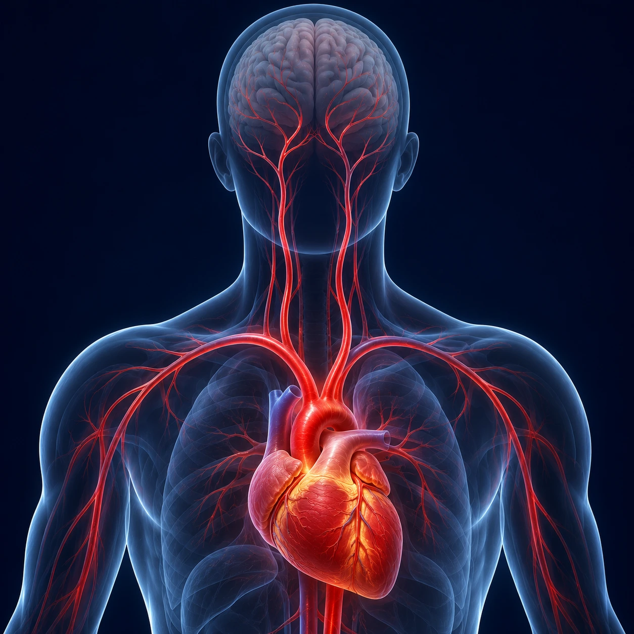
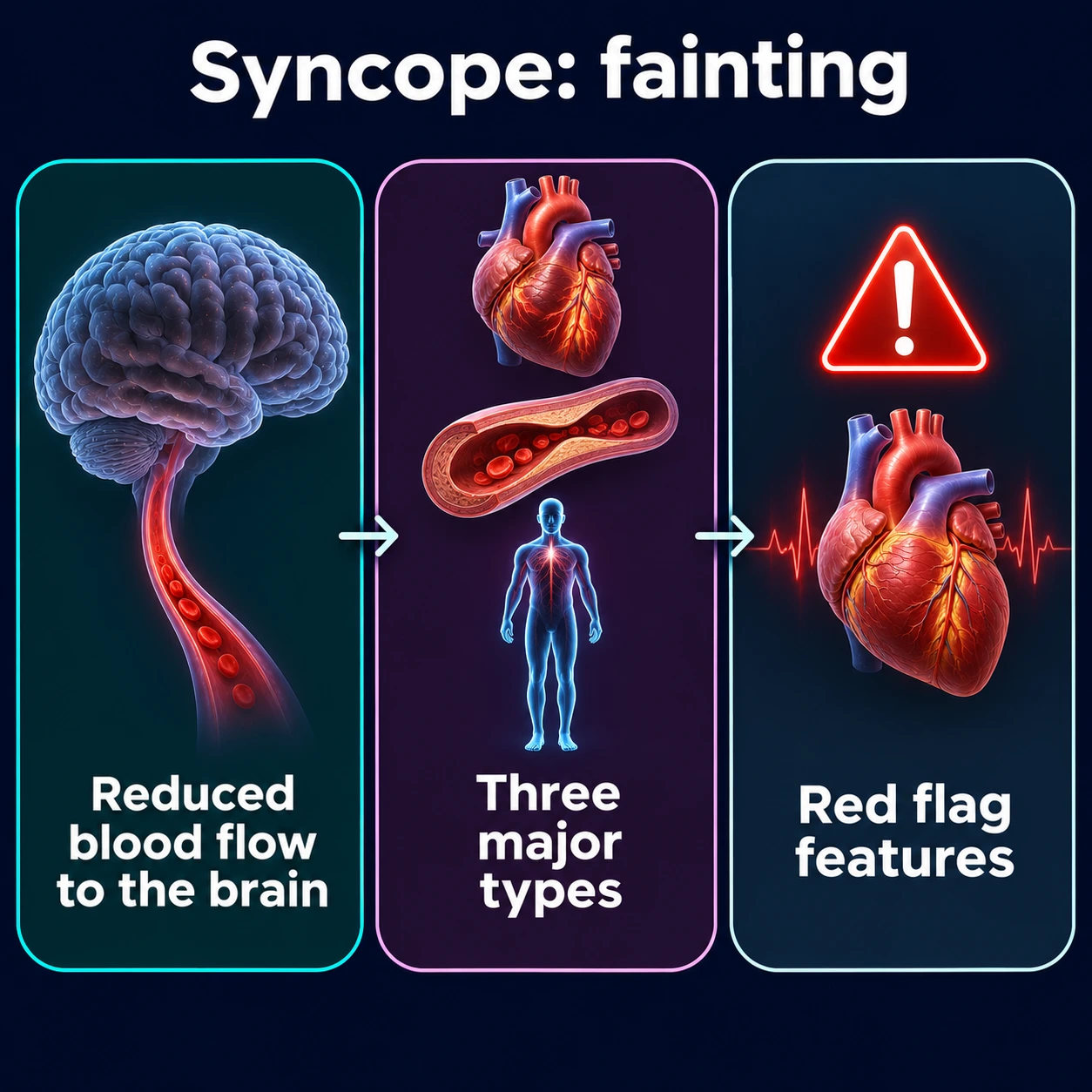
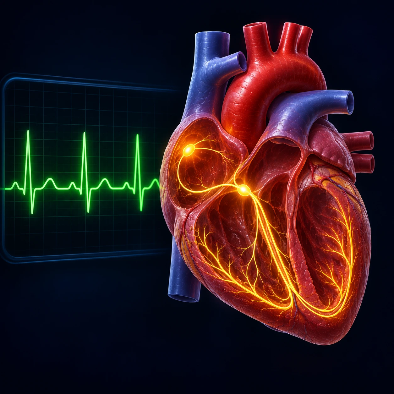
Table of Contents
- What Syncope Is
- Pathophysiology — Why the Brain Shuts Off
- Classification: Three Major Types
- Red Flags: High-Risk Features
- Initial Evaluation: History and Electrocardiogram
- Risk Stratification
- Cardiac Testing
- Treatment by Type
- When to Call 911
- Key Research Papers
- PubMed Topic Searches
- Connections
- Featured Videos
What Syncope Is
Syncope is a transient, self-terminating loss of consciousness caused by global cerebral hypoperfusion, with rapid onset, short duration, and spontaneous complete recovery. The key elements that define it are all four of those features together: it comes on quickly, it resolves on its own, the person returns to full awareness, and the underlying cause is a temporary interruption of blood flow to the entire brain. This distinguishes syncope from closely related conditions that are frequently confused with it in both clinical and lay settings. Seizure involves tonic-clonic motor activity, a postictal period of prolonged confusion lasting many minutes, and tongue biting along the lateral surface — none of which are characteristic of syncope. Coma is sustained unresponsiveness that does not self-terminate. Drop attacks are sudden falls without any loss of consciousness at all. Each of these distinctions carries serious diagnostic and legal consequences.
Syncope is far more common than most people appreciate. Studies estimate that approximately 40% of people will experience at least one episode in their lifetime, and a nationally representative US analysis estimated syncope-related hospital stays cost about $2.4 billion a year, about $5,400 per stay. Despite its frequency, misdiagnosis remains a persistent problem. One clinic study of 74 patients with a diagnosis of epilepsy found another explanation in 41.9%, including 25.7% with vasovagal syncope confirmed on a tilt test; it was a small, selected group, and its authors note that as many as 20–30% of epilepsy diagnoses may be wrong. That is a misclassification with enormous real-world consequences, because an epilepsy diagnosis triggers mandatory restrictions on driving and, in some occupations, on employment. Conversely, some patients with actual seizure disorder go undiagnosed because their episodes are attributed to fainting. The history, particularly an eyewitness account of the episode, remains the single most powerful tool for distinguishing between the two.
Pathophysiology — Why the Brain Shuts Off
The brain is exquisitely sensitive to reductions in blood flow. Under normal conditions, cerebral blood flow is maintained at approximately 50 mL per 100 grams of brain tissue per minute across a wide range of systemic blood pressures — a process called cerebrovascular autoregulation. When systolic blood pressure drops to roughly 50–60 mmHg, cerebral blood flow falls below the autoregulatory threshold to around 25 mL per 100 g per minute, and consciousness is lost within approximately 10 seconds. The brain does not require a prolonged insult; a very brief interruption suffices. Recovery occurs because the horizontal position achieved during a fall immediately restores gravitational drainage from the legs back toward the heart, increases venous return, and normalizes cardiac output and cerebral perfusion. This is why a person who has fainted nearly always recovers rapidly — and why propping someone upright before they have fully recovered can actually prolong the episode or cause a second one.
The vasovagal mechanism, the most common cause of syncope, operates through the Bezold-Jarisch reflex. A triggering stimulus — emotional distress, pain, the sight of blood, prolonged standing in a warm environment, or sudden fear — causes venous pooling in the lower extremities. This reduces preload, the volume of blood returning to the right heart. The left ventricle responds by contracting vigorously against a near-empty chamber. Mechanoreceptors in the ventricular wall sense this unusual hypercontractile state and transmit signals to the brainstem that are paradoxically interpreted as systemic hypertension. The brainstem then orders sympathetic withdrawal and increased vagal tone simultaneously, producing a combination of hypotension and bradycardia that together crash cerebral perfusion. The prodrome — pallor, profuse sweating, nausea, yawning, blurred or tunnel vision, and a sense of warmth — reflects this autonomic storm and typically lasts 30 seconds to a few minutes before consciousness is lost. The presence of a recognizable prodrome is reassuring, because it gives the patient time to lie down and abort the episode, and it strongly suggests a benign mechanism.
Orthostatic hypotension syncope arises from a failure of the autonomic nervous system to compensate for the shift in blood distribution that occurs when a person stands. Normally, standing causes blood to pool transiently in the capacitance vessels of the legs and abdomen, reducing venous return to the heart. Baroreceptors in the aortic arch and carotid sinus detect the resulting fall in blood pressure and within seconds trigger sympathetic vasoconstriction and a modest increase in heart rate to restore perfusion. When this reflex is impaired — by volume depletion, autonomic neuropathy from diabetes or Parkinson's disease, multiple system atrophy, or medications including alpha-1 blockers, diuretics, vasodilators, and certain antidepressants — systolic blood pressure drops by 20 mmHg or more (or diastolic by 10 mmHg or more) within three minutes of standing, and syncope or presyncope results. Unlike vasovagal syncope, orthostatic syncope is directly and reproducibly tied to positional change and typically lacks the autonomic prodrome of pallor and sweating.
Classification: Three Major Types
Reflex or neurally mediated syncope is often quoted at roughly half of all cases, though shares vary by setting: in the community-based Framingham Heart Study, vasovagal syncope was 21.2%, cardiac 9.5%, orthostatic 9.4%, and 36.6% had no known cause. It carries an excellent prognosis in most patients. This broad category is further subdivided by trigger. Vasovagal syncope — sometimes called "common fainting" or neurocardiogenic syncope — is precipitated by prolonged standing, emotional triggers, pain, or heat, and is the type most people encounter at least once in their lives. Situational syncope occurs reflexively in response to specific physiological maneuvers: cough syncope (common in patients with obstructive lung disease), micturition syncope (typically in older men rising at night to urinate), defecation syncope, and swallowing syncope in the setting of esophageal disease. Carotid sinus syndrome is triggered by mechanical pressure on the carotid sinus — from turning the head sharply, wearing a tight collar, or shaving — and is most common in older men; carotid sinus massage during evaluation reproduces it. In the absence of structural heart disease, the long-term mortality associated with reflex syncope is no higher than in the general population, and the primary burden is recurrence, injury from falls, and quality-of-life impairment.
Orthostatic hypotension syncope was 9.4% of cases in the Framingham study. Its defining feature is that symptoms occur predictably after positional change from lying or sitting to standing and resolve promptly when the patient returns to a horizontal position. The diagnostic step — measuring blood pressure in both lying and standing positions after one and three minutes — is simple and inexpensive, yet is frequently omitted in the initial evaluation. Drug review is the single most effective intervention, because a large proportion of cases are iatrogenic: antihypertensives, diuretics, alpha-blockers used for benign prostatic hyperplasia, tricyclic antidepressants, and dopaminergic agents for Parkinson's disease are among the most common culprits. When no reversible medication cause is found, neurodegenerative autonomic failure — including Parkinson's disease, multiple system atrophy, and pure autonomic failure — is the next group doctors look at.
Cardiac syncope is quoted at 10 to 15% of cases (9.5% in Framingham) but carries the highest mortality of the three types, so hospitals treat it as the most urgent to evaluate. It is subdivided into arrhythmic and structural causes. Arrhythmic cardiac syncope can arise from abnormally slow rhythms — sick sinus syndrome, sinoatrial block, high-degree atrioventricular block — or from fast rhythms including ventricular tachycardia, ventricular fibrillation, and supraventricular tachycardias that compromise cardiac output. Structural cardiac syncope results from a fixed or dynamic obstruction to blood flow: severe aortic stenosis, hypertrophic cardiomyopathy (HCM) with outflow obstruction, massive pulmonary embolism, and cardiac tamponade are the most important examples. Syncope that occurs during physical exertion — rather than afterward — is particularly worrying for outflow tract obstruction or exercise-induced arrhythmia, and hospitals respond with urgent echocardiography and cardiac monitoring regardless of how benign the patient appears when examined at rest.
Red Flags — High-Risk Features
Recognizing high-risk features is the central clinical task when evaluating a patient with syncope, because missing them can be fatal. Cardiac red flags include syncope that occurs during exertion (not after), syncope while supine (which eliminates orthostatic and most vasovagal mechanisms immediately), a family history of sudden cardiac death or an inherited arrhythmia syndrome such as Brugada syndrome, long QT syndrome, arrhythmogenic right ventricular cardiomyopathy (ARVC), or hypertrophic cardiomyopathy, and syncope with no warning prodrome at all. Palpitations immediately before loss of consciousness suggest an arrhythmic trigger. Any trauma sustained during the fall — lacerations, fractures, head injury — indicates that the fall was sudden and unprotected, consistent with an abrupt loss of postural tone before the patient could react. An abnormal electrocardiogram (ECG) is perhaps the single most important red flag identified in clinical risk scores: QT prolongation, a Brugada coved-type ST-elevation pattern in leads V1–V3, a delta wave consistent with Wolff-Parkinson-White syndrome, left bundle branch block, complete AV block, or electrocardiogram changes consistent with a prior myocardial infarction all represent high-risk findings requiring immediate admission and monitoring.
Distinguishing syncope from seizure is a diagnostic challenge that has genuine consequences. A seizure typically begins with an aura specific to the anatomical focus of the discharge — an olfactory hallucination, a rising epigastric sensation, visual phenomena — and is followed by tonic-clonic motor activity, sustained rhythmic jerking, and a postictal period of confusion, drowsiness, and disorientation lasting more than five minutes. Syncope, by contrast, recovers to full alertness within seconds to two or three minutes. Biting the lateral surface of the tongue is strongly associated with seizure; biting the tongue tip is more common with vasovagal syncope. Brief, low-amplitude clonic twitches of the limbs can occur during syncope itself — so-called convulsive syncope — but prolonged, coordinated tonic-clonic shaking lasting more than 30 seconds is virtually never syncope. Urinary incontinence, often cited as a seizure marker, occurs in both conditions and is not reliably discriminating.
Hospitals rule out several high-risk diagnoses rapidly because they are immediately life-threatening. Pulmonary embolism is considered when syncope is accompanied by pleuritic chest pain, dyspnea, calf swelling, or in the setting of recent immobilization or hypercoagulable state. Aortic dissection presents with syncope alongside tearing or ripping chest or back pain radiating between the shoulder blades and a pulse or blood pressure differential between arms. Ruptured ectopic pregnancy is considered in any woman of reproductive age presenting with syncope and abdominal pain, with or without vaginal bleeding, as hemoperitoneum can progress to hemorrhagic shock rapidly.
Initial Evaluation — History and Electrocardiogram
The clinical history is the single most powerful diagnostic tool in syncope evaluation and correctly identifies the underlying mechanism in approximately 50% of cases when taken carefully. A careful history characterizes the triggering context (was the patient standing in a warm environment, rising from a seated position, engaged in vigorous exercise, emotionally upset, or experiencing pain?), the nature and duration of the prodrome (did they feel dizzy, nauseated, sweaty, or warm before losing consciousness, and if so for how long?), an eyewitness account if available (what did the patient look like during the episode — pale or cyanotic? Did limbs jerk, and if so for how long? Did the eyes deviate?), the speed of recovery (seconds vs. minutes), and the state after recovery (fully alert and oriented, or confused and fatigued?). The medication list deserves close scrutiny for antihypertensives, diuretics, QT-prolonging drugs (antipsychotics, certain antibiotics, methadone), and sympathomimetics. Family history of sudden cardiac death, inherited arrhythmia syndromes, or structural heart disease is specifically asked about. Comorbidities — known coronary artery disease, reduced ejection fraction, diabetes with autonomic neuropathy, Parkinson's disease — significantly alter pretest probability for different syncope types.
The syncope guidelines list a 12-lead electrocardiogram for virtually every patient presenting with syncope. It is inexpensive, immediately available, non-invasive, and capable of identifying the high-risk findings that triage the patient directly to hospital admission. Clinicians look for QT prolongation (corrected QT, or QTc, greater than 450 milliseconds in men or 470 milliseconds in women, consistent with long QT syndrome), the Brugada type-1 coved pattern of ST elevation in leads V1 through V3, a short PR interval with a delta wave at the onset of the QRS consistent with Wolff-Parkinson-White pre-excitation, left or right bundle branch block, degrees of atrioventricular block, ST-segment or T-wave changes suggesting acute or prior myocardial infarction, and the epsilon wave in V1–V3 characteristic of ARVC. Any one of these findings on the initial electrocardiogram defines a high-risk electrocardiogram and is treated as grounds for hospitalization with continuous cardiac monitoring regardless of how well the patient looks in the emergency department at the time of evaluation.
Risk Stratification
Multiple validated clinical decision tools exist to help clinicians determine which patients with syncope require hospitalization and which can safely be discharged for outpatient evaluation. The Canadian Syncope Risk Score uses nine items: predisposition to vasovagal syncope, heart disease, any systolic blood pressure reading below 90 or above 180 mmHg in the emergency department, troponin above the 99th percentile, an abnormal QRS axis (below −30 or above 100 degrees), a QRS longer than 130 milliseconds, a QTc longer than 480 milliseconds, an emergency department diagnosis of cardiac syncope, and an emergency department diagnosis of vasovagal syncope. Scores range from −3 to +11. The risk of a serious adverse event within 30 days, including arrhythmia, myocardial infarction (MI), procedures, or death, ran from 0.4% at −3 to 83.6% at +11. The reported cut-offs are −2 (sensitivity 99.2%) and −1 (sensitivity 97.7%). A validation in 3,819 patients at 9 Canadian emergency departments found 97.8% sensitivity and 44.3% specificity at −1.
The San Francisco Syncope Rule offers a simpler five-item checklist: a history of congestive heart failure, an abnormal electrocardiogram, complaint of dyspnea, a hematocrit below 30%, or a systolic blood pressure below 90 mmHg at triage. The presence of any single one of these factors identifies a patient as high risk. In the original study it had 96% sensitivity and 62% specificity for a serious outcome within seven days. Its simplicity makes it easy to apply at bedside, but later testing was less favorable: a systematic review of 12 studies and 5,316 patients found a pooled sensitivity of 87% (95% confidence interval 79–93%) and specificity of 52% (43–62%). The most common missed serious outcome was cardiac arrhythmia.
The EGSYS Score, developed from the Evaluation of Guidelines in Syncope Study, incorporates findings that point toward or away from a cardiac etiology. Points are added for the presence of electrocardiogram abnormality, palpitations before the episode, exertional or supine syncope, and signs of structural heart disease, while points are subtracted for predisposing factors (crowding, hot environment, prolonged standing) and a prodrome of autonomic symptoms (nausea, vomiting, flushing). A score of three or more identifies a cardiac mechanism with a sensitivity near 92%. Used together, these three tools provide complementary perspectives on risk and help standardize discharge decisions in busy emergency departments where resource constraints and clinical gestalt vary widely between providers.
Cardiac Testing
Echocardiography is indicated for all patients in whom a structural cardiac etiology is suspected or cannot be excluded. This includes anyone with an abnormal electrocardiogram, known or suspected heart disease, a murmur audible on examination, exertional syncope, or syncope in the context of dyspnea. Echocardiography assesses left ventricular systolic and diastolic function, valvular pathology (severe aortic stenosis is the most important structural cause of syncope), the presence of HCM and its degree of outflow obstruction, pericardial effusion causing tamponade physiology, and right ventricular dysfunction consistent with pulmonary hypertension or massive pulmonary embolism. It is non-invasive and provides information that directly changes management.
Ambulatory cardiac monitoring is the cornerstone of the arrhythmia diagnosis strategy. The choice of monitor depends on the frequency of episodes. A 24–48 hour Holter monitor is appropriate when episodes are very frequent (daily or near-daily). A 2-week external event recorder is better suited to weekly episodes. A 30-day loop recorder that stores a continuous rhythm strip and allows patient-triggered event marking is used for monthly episodes. For patients whose episodes are infrequent — once every several months or less often — the implantable cardiac monitor (ICM), also called an implantable loop recorder (ILR), provides up to three years of continuous rhythm monitoring from a small device inserted subcutaneously near the sternum under local anesthesia. Multiple randomized controlled trials have established the ICM as the gold standard for infrequent unexplained syncope, with arrhythmia diagnoses achieved in 35 to 50% of patients at one to two years, compared to 20% or less with conventional monitoring strategies. Earlier insertion — at the first evaluation rather than after exhausting all non-invasive options — has been shown to be more cost-effective and diagnostically efficient.
Exercise testing is recommended when syncope occurs during or immediately after exertion to exclude exercise-induced arrhythmia and evaluate hemodynamic response to increased cardiac demand. Tilt table testing tilts the patient from supine to 60–80 degrees head-up for a standardized duration, sometimes with pharmacological provocation using sublingual nitroglycerin or intravenous isoproterenol, to reproduce vasovagal syncope under controlled conditions. A positive tilt test confirms the diagnosis and classifies the response as cardioinhibitory (predominant bradycardia or asystole), vasodepressor (predominant hypotension without bradycardia), or mixed. This classification directly guides whether a pacemaker is appropriate therapy for patients with recurrent vasovagal syncope — the cardioinhibitory subtype with documented asystole is the one in which pacemaker therapy has demonstrated clinical benefit.
Treatment by Type
For vasovagal syncope, education and reassurance are not merely adjuncts to therapy — they are the most important intervention in most patients. Understanding that fainting is a benign autonomic reflex, that it will not kill them, and that they can learn to recognize and abort their prodrome substantially reduces fear, catastrophizing, and health-care utilization. The guidelines describe identifying and, where possible, avoiding personal triggers: prolonged standing in warm crowded spaces, extreme heat, dehydration, alcohol, and blood-draw situations. When avoidance is not possible, physical counterpressure maneuvers — isometric hand-grip (crossing the fingers of both hands and pulling outward), leg crossing with leg muscle tensing, or squatting — applied at the very first symptom of a prodrome were tested in the Physical Counterpressure Maneuvers Trial (PC-Trial). Among 223 patients followed for about 14 months, 31.6% of those trained in the maneuvers fainted again, against 50.9% with usual care, a relative risk reduction of 39% with no adverse events reported. Increased dietary salt intake (2–4 grams per day above usual) and liberal fluid intake (2–2.5 liters per day) increase circulating volume and raise the threshold for vasovagal episodes. Fludrocortisone, a synthetic mineralocorticoid that promotes sodium and water retention, has been used for volume expansion, though randomized trial evidence for benefit is mixed. In the POST 2 trial of 210 patients the result just missed significance (hazard ratio 0.69, 95% confidence interval 0.46–1.03), and it was 0.51 when counted only after 2 weeks of dose stabilization. Midodrine, an alpha-1 adrenergic agonist that increases peripheral vascular resistance, has demonstrated modest benefit in several trials. In the POST 4 trial (2021) of 133 young, otherwise healthy people, 42% on midodrine fainted again against 61% on placebo (relative risk 0.69), with side effects similar in both groups. It carries caution because it causes supine hypertension and is contraindicated in urinary retention and severe heart disease. Beta-blockers, historically prescribed for vasovagal syncope on theoretical grounds, are not routinely recommended — the Prevention of Syncope Trial (POST) of metoprolol in 208 patients and a placebo-controlled trial of atenolol in 50 patients both failed to demonstrate a significant reduction in syncope recurrence. An earlier hint that beta-blockers might help older adults did not hold up in POST 5 (2026): among 51 patients aged 40 and over, 31% on metoprolol and 40% on placebo fainted (P=0.69), and pooled data from 4 trials gave 34% in both groups. Pacemaker implantation has been tested in cardioinhibitory vasovagal syncope in patients aged 40 and over. In ISSUE-3, patients had a recorded pause in the heartbeat (asystole) of at least 3 seconds with fainting or at least 6 seconds without, and 2-year recurrence was 57% with the pacemaker off against 25% with it on. In the SPAIN trial of 46 patients, whose slow heartbeat was brought on by a tilt test, 72% with pacing against 28% with sham pacing had at least a 50% drop in episodes. A newer procedure, cardioneuroablation, uses a catheter to quiet the nerves that slow the heart. In a 48-patient randomized trial, 8% fainted again against 54% of controls over 2 years; a 2025 pooled analysis of 37 mostly non-randomized studies reported 8.9% recurrence, with results that varied widely between studies. Selective serotonin reuptake inhibitors (SSRIs), particularly paroxetine, have some evidence for benefit in refractory frequent vasovagal syncope, likely through central modulation of the autonomic nervous system.
Orthostatic hypotension syncope is frequently treatable with straightforward measures. The first and most effective step is a comprehensive review of the medication list, with discontinuation or dose reduction of any agent that contributes to vasodilation, volume depletion, or sympathetic blunting — this single intervention resolves or substantially improves orthostatic syncope in a large proportion of patients. Non-pharmacological measures include increasing salt and fluid intake, wearing graduated compression stockings to reduce venous pooling in the legs, elevating the head of the bed by 30 degrees (which activates the renin-angiotensin system overnight), and rising slowly from supine to seated to standing with pauses at each position. When these measures are insufficient, midodrine (taken during waking hours, avoided within four hours of bedtime to prevent supine hypertension) is a first-choice pharmacological agent. Pyridostigmine, an acetylcholinesterase inhibitor, selectively amplifies sympathetic ganglionic transmission without causing supine hypertension and is particularly useful in patients who require a medication that can be dosed continuously. Droxidopa, a norepinephrine prodrug, is approved specifically for neurogenic orthostatic hypotension — the form caused by autonomic failure from Parkinson's disease, multiple system atrophy, and pure autonomic failure — and has demonstrated efficacy in randomized trials in those populations.
Cardiac arrhythmia syncope requires targeted therapy matched to the specific rhythm disorder identified. Sinus node dysfunction and high-degree atrioventricular block causing syncope are treated with permanent pacemaker implantation, which is highly effective and durable. Ventricular tachycardia or fibrillation causing syncope, particularly in the setting of structural heart disease or reduced ejection fraction, is treated with an implantable cardioverter-defibrillator (ICD), which also provides backup pacing. Catheter ablation has become a first-line treatment for many supraventricular tachycardias (SVT) including atrioventricular nodal reentrant tachycardia (AVNRT) and accessory pathway-mediated tachycardia (Wolff-Parkinson-White syndrome, WPW), with cure rates exceeding 95% for AVNRT and 90% for WPW ablation. Selected forms of ventricular tachycardia in structurally normal hearts — right ventricular outflow tract tachycardia, idiopathic left ventricular tachycardia — are also highly amenable to catheter ablation. Antiarrhythmic medications are guided by electrophysiology study findings and are generally reserved for patients who are not candidates for or who have declined ablation. Structural syncope from severe aortic stenosis or HCM with outflow obstruction requires structural intervention — valve replacement (surgical or transcatheter) for aortic stenosis, septal myectomy or alcohol septal ablation for HCM — rather than medication alone.
When to Call 911
Not all syncope is an emergency, but several circumstances require immediate emergency evaluation rather than a scheduled office visit. Call 911 for syncope that occurs during physical exercise — even mild exertion like walking — as this raises the possibility of structural outflow obstruction or exercise-induced arrhythmia, both of which can be immediately life-threatening. Syncope in a person with a known pacemaker, ICD, or established heart disease, particularly when it occurs without the usual prodrome, warrants emergency evaluation because it may represent device malfunction or a new ventricular arrhythmia that the device failed to detect or treat. Syncope accompanied by chest pain, severe shortness of breath, or a sensation of pounding palpitations immediately before the event should be treated as a potential cardiac emergency. Any episode in which loss of consciousness persists longer than five minutes, or in which the person does not return to their normal cognitive state promptly after regaining consciousness, is not simple syncope and requires immediate evaluation for seizure, stroke, or metabolic derangement. Syncope with focal neurological deficits afterward — arm or leg weakness, speech difficulty, facial drooping, vision loss — may represent a transient ischemic attack or stroke and is an emergency. Syncope resulting in significant trauma such as a head injury, facial laceration, or suspected fracture requires emergency care both for the injury itself and because traumatic syncope without prodrome suggests a mechanism that put the patient at risk before they could protect themselves. Finally, two or more syncopal episodes in rapid succession within a short time frame suggest an ongoing physiological instability that requires monitoring rather than outpatient reassurance.
Key Research Papers
- Shen W-K, Sheldon RS, Benditt DG, et al. 2017 ACC/AHA/HRS Guideline for the Evaluation and Management of Patients With Syncope. J Am Coll Cardiol. 2017. — PubMed PMID: 28286221
- Brignole M, Moya A, de Lange FJ, et al. 2018 ESC Guidelines for the diagnosis and management of syncope. Eur Heart J. 2018. — PubMed PMID: 29562304
- Thiruganasambandamoorthy V, Kwong K, Wells GA, et al. Development of the Canadian Syncope Risk Score to predict serious adverse events after emergency department assessment of syncope. CMAJ. 2016. — PubMed PMID: 27378464
- Martin TP, Hanusa BH, Kapoor WN. Risk stratification of patients with syncope. Ann Emerg Med. 1997;29(4):459–466. — PubMed PMID: 9095005
- Quinn JV, Stiell IG, McDermott DA, Sellers KL, Kohn MA, Wells GA. Derivation of the San Francisco Syncope Rule to predict patients with short-term serious outcomes. Ann Emerg Med. 2004;43(2):224–232. — PubMed PMID: 14747812
- Saccilotto RT, Nickel CH, Bucher HC, Steyerberg EW, Bingisser R, Koller MT. San Francisco Syncope Rule to predict short-term serious outcomes: a systematic review. CMAJ. 2011;183(15):E1116–E1126. — PubMed PMID: 21948723
- Thiruganasambandamoorthy V, Sivilotti MLA, Le Sage N, et al. Multicenter Emergency Department Validation of the Canadian Syncope Risk Score. JAMA Intern Med. 2020;180(5):737–744. — PubMed PMID: 32202605
- Brignole M, Menozzi C, Moya A, et al. Pacemaker therapy in patients with neurally mediated syncope and documented asystole: Third International Study on Syncope of Uncertain Etiology (ISSUE-3). Circulation. 2012. — PubMed PMID: 22565936
- Baron-Esquivias G, Morillo CA, Moya-Mitjans A, et al. Dual-chamber pacing with closed loop stimulation in recurrent reflex vasovagal syncope: the SPAIN Study. J Am Coll Cardiol. 2017. — PubMed PMID: 28958328
- van Dijk N, Quartieri F, Blanc JJ, et al. Effectiveness of physical counterpressure manoeuvres in preventing vasovagal syncope: The Physical Counterpressure Manoeuvres Trial (PC-Trial). J Am Coll Cardiol. 2006. — PubMed PMID: 17045903
- Moya A, García-Civera R, Croci F, et al. Diagnosis, management, and outcomes of patients with syncope and bundle branch block. Eur Heart J. 2011;32(12):1535–1541. — PubMed PMID: 21444367
- Task Force for the Diagnosis and Management of Syncope; European Society of Cardiology (ESC). Guidelines for the diagnosis and management of syncope (version 2009). Eur Heart J. 2009. — PubMed PMID: 19713422
- Sheldon R, Connolly S, Rose S, et al. Prevention of Syncope Trial (POST): a randomized, placebo-controlled study of metoprolol in the prevention of vasovagal syncope. Circulation. 2006. — PubMed PMID: 16505178
- Madrid AH, Ortega J, Rebollo JG, et al. Lack of efficacy of atenolol for the prevention of neurally mediated syncope in a highly symptomatic population: a prospective, double-blind, randomized and placebo-controlled study. J Am Coll Cardiol. 2001;37(2):554–559. — PubMed PMID: 11216978
- Sheldon R, Raj SR, Rose MS, et al. Fludrocortisone for the Prevention of Vasovagal Syncope: A Randomized, Placebo-Controlled Trial. J Am Coll Cardiol. 2016;68(1):1–9. — PubMed PMID: 27364043
- Sheldon R, Faris P, Tang A, et al. Midodrine for the Prevention of Vasovagal Syncope: A Randomized Clinical Trial. Ann Intern Med. 2021;174(10):1349–1356. — PubMed PMID: 34339231
- Sepehrvand N, Hatami S, Raj SR, et al. Beta-Blockers for Vasovagal Syncope in Aging Subjects: A Randomized Controlled Trial and Individual Patient Data Meta-Analysis of Clinical Trials. CJC Open. 2026;8(8):939–947. — PubMed PMID: 42609753
- Piotrowski R, Baran J, Sikorska A, Krynski T, Kulakowski P. Cardioneuroablation for Reflex Syncope: Efficacy and Effects on Autonomic Cardiac Regulation-A Prospective Randomized Trial. JACC Clin Electrophysiol. 2023;9(1):85–95. — PubMed PMID: 36114133
- Ababei A, Ursu CG, Bistriceanu MIA, et al. Cardioneuroablation for Vasovagal Syncope: An Updated Systematic Review and Single-Arm Meta-Analysis. Biomedicines. 2025;13(7). — PubMed PMID: 40722829
- Zaidi A, Clough P, Cooper P, Scheepers B, Fitzpatrick AP. Misdiagnosis of epilepsy: many seizure-like attacks have a cardiovascular cause. J Am Coll Cardiol. 2000;36(1):181–184. — PubMed PMID: 10898432
- Sun BC, Emond JA, Camargo CA. Direct medical costs of syncope-related hospitalizations in the United States. Am J Cardiol. 2005;95(5):668–671. — PubMed PMID: 15721118
- Puppala VK, Dickinson O, Benditt DG. Syncope: classification and risk stratification. J Cardiol. 2014;63(3):171–177. — PubMed PMID: 24405895
- Soteriades ES, Evans JC, Larson MG, et al. Incidence and prognosis of syncope. N Engl J Med. 2002;347(12):878–885. — PubMed PMID: 12239256
PubMed Topic Searches
Connections
- All Symptoms
- Related symptoms: Dizziness and Vertigo · Lightheadedness · Heart Palpitations · Chest Pain
- Cardiac conditions: Arrhythmia · Atrial Fibrillation · Heart Failure · Aortic Stenosis
- Neurological: Postural Orthostatic Tachycardia Syndrome · Multiple System Atrophy · Epilepsy
- Lab Tests index