Kawasaki Disease
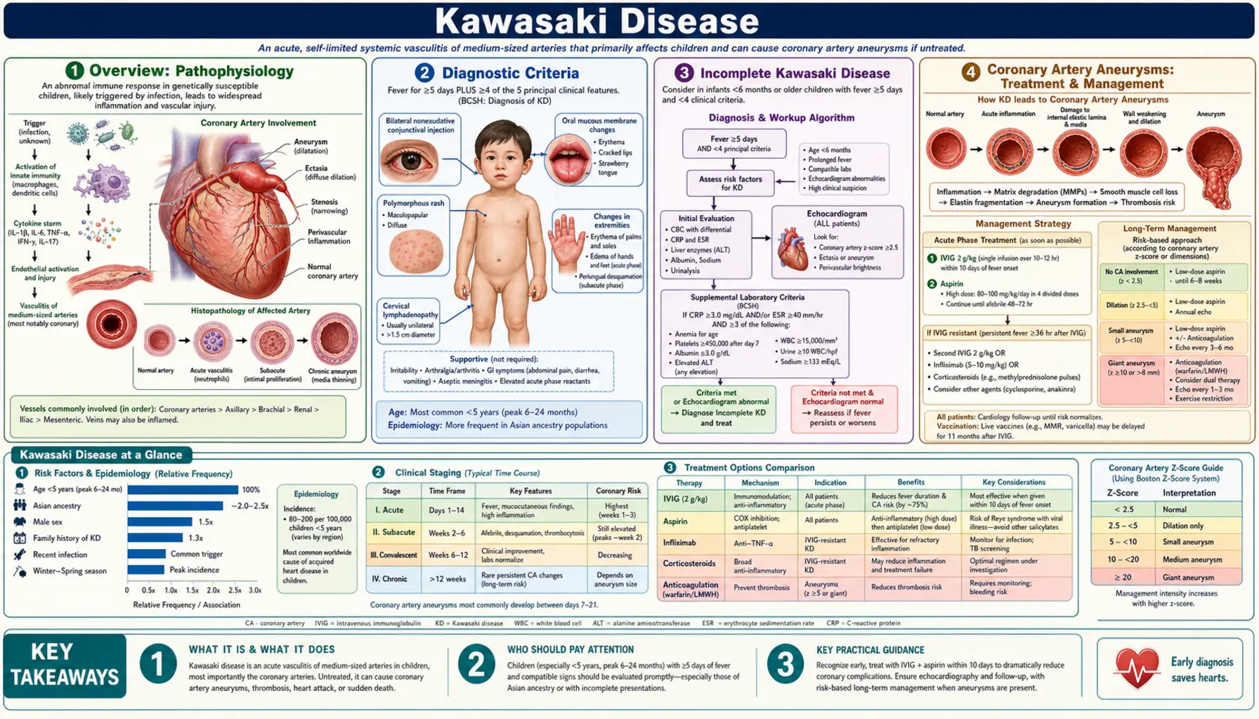
- Overview
- Diagnostic Criteria
- Incomplete Kawasaki Disease
- Coronary Artery Aneurysms
- Treatment
- Echocardiographic Monitoring
- Complications
- Long-Term Outcomes
- Key Research Papers
- Connections
Overview
Kawasaki disease (KD) is an acute, self-limited vasculitis of childhood whose cause is unknown. The 2017 American Heart Association (AHA) scientific statement describes it as the leading cause of acquired heart disease in children in developed countries, with coronary artery aneurysms in about 25% of untreated cases (McCrindle et al. 2017, PubMed PMID: 28356445). The condition was first reported in Japan in 1967 by Kawasaki, in a paper titled "Infantile acute febrile mucocutaneous lymph node syndrome with specific desquamation of the fingers and toes. Clinical observation of 50 cases" (Kawasaki and Naoe 2014, PubMed PMID: 24595558).
KD is a vasculitis of medium-sized arteries in many organs (Zhu and Ang 2021, PubMed PMID: 35194409), but the coronary arteries are where lasting damage matters most. Vasculitis destroys the normal arterial architecture and is followed by aneurysmal dilatation, particularly of the proximal coronary arteries (Brogan et al. 2020, PubMed PMID: 31843876). These coronary artery aneurysms (CAA) shape the long-term outlook.
A 2021 review reports that about 80% of cases occur in children under 5 years and 50% in children under 2, with a male-to-female ratio of about 1.5:1 in all countries. Recorded incidence in children under 5 is highest in Japan (369 per 100,000 in 2018), followed by Korea (195 per 100,000 in 2014) and Taiwan (75 per 100,000 in 2011); in Western countries it is 4–25 per 100,000, and 17.5–20.8 per 100,000 in the USA. In the USA, incidence is about 2.5 times higher in children of Asian/Pacific Islander descent and 1.5 times higher in African American children than in white children (Zhu and Ang 2021, PubMed PMID: 35194409).
No causative agent has been identified. Epidemiological features suggest an inciting infection or agent, and genetic factors raise susceptibility, as shown by the high incidence in children of Asian ethnicity and in first-degree relatives (Shulman and Rowley 2015, PubMed PMID: 25907703). Many bacteria and viruses have been isolated from patients, but their role is unclear; seasonality varies by region (winter peaks in Japan, winter-spring peaks in the USA) (Zhu and Ang 2021, PubMed PMID: 35194409).
Diagnostic Criteria
Kawasaki disease is a clinical diagnosis. The classic criteria in the 2017 AHA statement, as summarized in a 2021 review, require fever for at least 5 days plus at least 4 of 5 principal features (Zhu and Ang 2021, PubMed PMID: 35194409):
- Bilateral non-purulent conjunctival injection — bulbar redness that spares the limbus, typically beginning shortly after fever onset.
- Polymorphous rash — usually appears within 5 days of fever onset; diffuse maculopapular is the most common pattern, scarlatiniform or erythema multiforme-like patterns are common, and urticarial or fine micropustular eruptions are less common; bullous, vesicular and petechial rashes are not associated with KD.
- Changes in the lips and oral cavity — redness of the lips and oropharyngeal mucosa, cracked or peeling lips, and "strawberry tongue" (also seen in streptococcal scarlet fever); oral ulcers and pharyngeal exudates are not seen in KD.
- Changes of the extremities — redness, induration and swelling of the hands and feet in the acute phase; peeling of the fingers and toes 2–3 weeks after onset, and deep transverse nail grooves (Beau's lines) at 1–2 months.
- Cervical lymphadenopathy — at least one node over 1.5 cm in diameter, usually unilateral and in the anterior cervical triangle; it is the least commonly observed principal feature.
According to the same review, KD can also be diagnosed with only 4 days of fever when 4 or more principal features are present (particularly redness and swelling of the hands and feet), and some experienced clinicians have treated KD after 3 days of fever. The fever is typically high (above 39–40 °C) and remittent; untreated, it can last 1–3 weeks, and it typically resolves within 36 hours of IVIG treatment (Zhu and Ang 2021, PubMed PMID: 35194409).
The principal features may not be present at the same time, and they overlap with many common childhood febrile illnesses, which makes diagnosis difficult (Zhu and Ang 2021, PubMed PMID: 35194409). The AHA statement describes prompt diagnosis as essential (McCrindle et al. 2017, PubMed PMID: 28356445).
Incomplete Kawasaki Disease
Incomplete Kawasaki disease describes children with prolonged fever who do not meet the classic criteria. "Atypical" KD — a presentation with features not usually seen in KD, such as facial nerve palsy or hearing loss — is regarded in a 2021 review as a separate entity from incomplete KD (Zhu and Ang 2021, PubMed PMID: 35194409).
Infants are a particular concern. Children under 6 months may have prolonged fever as their only finding and are at substantial risk of coronary aneurysms, so the 2017 AHA statement added evaluation for incomplete KD in infants with 7 or more days of unexplained fever, as well as in children with 5 or more days of fever and 2–3 principal features (Zhu and Ang 2021, PubMed PMID: 35194409). British surveillance data show coronary involvement despite IVIG in 19% of children overall and 39% of those under 1 year, partly related to delayed diagnosis and treatment (Brogan et al. 2020, PubMed PMID: 31843876).
The AHA algorithm for incomplete KD, as described in a 2022 review, uses laboratory and echocardiographic criteria when clinical features are insufficient (Kim 2022, PubMed PMID: 34922423):
- CRP ≥3 mg/dL and/or ESR ≥40 mm/hr in a child with prolonged unexplained fever
- Supplemental laboratory criteria (3 or more count as a positive evaluation) (Zhu and Ang 2021, PubMed PMID: 35194409):
- Low serum albumin (hypoalbuminemia)
- Anemia for age
- Elevated alanine aminotransferase (ALT)
- Platelet count ≥450,000/mm³ after day 7 of fever
- White blood cell count ≥15,000/mm³
- Urinalysis with ≥10 white blood cells per high-power field (sterile pyuria)
- Echocardiographic criteria, positive if any one is met: a Z-score ≥2.5 for the left anterior descending (LAD) or right coronary artery (RCA); a coronary artery aneurysm; or 3 or more other suggestive features, including decreased left ventricular function, mitral regurgitation, pericardial effusion, or LAD or RCA Z-scores of 2–2.5 (Kim 2022, PubMed PMID: 34922423)
In this algorithm, elevated inflammatory markers together with compatible supplemental laboratory or echocardiographic findings allow a diagnosis of incomplete KD, and the algorithm calls for IVIG treatment (Kim 2022, PubMed PMID: 34922423). The evaluation also includes serial clinical and laboratory re-examination (Zhu and Ang 2021, PubMed PMID: 35194409).
Coronary Artery Aneurysms
The defining complication of Kawasaki disease is damage to the coronary arteries. Coronary artery aneurysms develop in about 25% of untreated cases (McCrindle et al. 2017, PubMed PMID: 28356445). A 2021 review reports that treatment with high-dose IVIG within the first 10 days of illness lowers this to about 5% (Zhu and Ang 2021, PubMed PMID: 35194409).
The 2017 AHA statement classifies coronary arteries by the Z-score — the internal diameter expressed as standard deviations from the mean for body surface area — as set out in a 2022 review (Kim 2022, PubMed PMID: 34922423):
- No involvement: Z-score always below 2
- Dilation only: Z-score 2 to below 2.5 (or, if initially below 2, a fall of 1 or more during follow-up)
- Small aneurysm: Z-score 2.5 to below 5
- Medium aneurysm: Z-score 5 to below 10, with an absolute dimension below 8 mm
- Large or giant aneurysm: Z-score 10 or more, or an absolute dimension of 8 mm or more
Different published Z-score formulas can give noticeably different values for the same artery, and the gap widens for larger aneurysms; the review notes that this can change an aneurysm's classification (Kim 2022, PubMed PMID: 34922423). The most common aneurysm locations are the bifurcation of the left main coronary artery and the proximal right coronary artery (Brogan et al. 2020, PubMed PMID: 31843876).
Many aneurysms regress. In a US cohort of 2,860 patients, 500 (17%) had coronary aneurysms and 75% of these regressed; a larger Z-score at diagnosis predicted worse outcomes (Friedman et al. 2016, PubMed PMID: 27633390). In a 34-institution international registry of 1,651 patients with aneurysms, luminal diameter had normalized at 10 years in 99% of small, 92% of medium and 57% of large aneurysms (McCrindle et al. 2020, PubMed PMID: 32750313). Regression in size is not the same as a normal artery: pathology studies describe disruption of the intima and media, intimal myofibroblastic proliferation, and arterial remodelling that can progress over months to years, with stenoses forming particularly at the junction between the aneurysm and normal artery (Brogan et al. 2020, PubMed PMID: 31843876).
Large and giant aneurysms carry the highest risk. In the international registry, among patients with Z-scores of 10 or more, the 10-year cumulative incidence was 20% for luminal narrowing, 18% for coronary thrombosis and 14% for major adverse cardiovascular events; no complications were observed in patients with Z-scores below 10 (McCrindle et al. 2020, PubMed PMID: 32750313). A 2020 review reports that within 30 years of the illness, large or giant aneurysms are associated with up to a 50% risk of thrombotic coronary occlusion, progressive stenoses requiring revascularisation, or acute coronary syndrome (Brogan et al. 2020, PubMed PMID: 31843876).
Treatment
The mainstay of initial treatment is high-dose intravenous immunoglobulin (IVIG) together with aspirin, ideally within the first 10 days of illness; according to a 2021 review, the AHA statement also supports treatment after day 10 when signs of ongoing inflammation persist (Zhu and Ang 2021, PubMed PMID: 35194409).
Primary Treatment
- IVIG as a single infusion. A 1991 multicenter randomized trial in 549 children compared a single infusion of 2 g per kg over 10 hours with 400 mg per kg daily for four days; the four-day regimen had a higher adjusted relative prevalence of coronary abnormalities at two weeks (1.94; 95% CI 1.01–3.71), and the single infusion shortened fever and normalized inflammatory markers faster, with similar adverse-effect rates (Newburger et al. 1991, PubMed PMID: 1709446).
- Aspirin. A 2021 review reports acute-phase doses of 80–100 mg/kg/day divided every 6 hours in the USA and 30–50 mg/kg/day in Japan and Western Europe, then low-dose aspirin (3–5 mg/kg/day) until there is no evidence of coronary aneurysms by 6–8 weeks after onset; the switch to low dose can follow 48–72 hours without fever, although some clinicians continue the higher dose until the 14th day of illness (Zhu and Ang 2021, PubMed PMID: 35194409). The 2017 AHA statement lists both acute-phase dose ranges (Zhu and Ang 2021, PubMed PMID: 35194409). In a retrospective multicentre cohort of 1,213 children treated with aspirin and IVIG, low-dose aspirin (3–5 mg/kg per day) was not inferior to high-dose aspirin (80 mg/kg per day) for coronary artery abnormalities (22.2% vs 20.5%) (Dallaire et al. 2017, PubMed PMID: 28562282).
IVIG Resistance
About 10–20% of patients do not respond to the initial IVIG infusion (McCrindle et al. 2017, PubMed PMID: 28356445). Refractory KD is defined as recurrent or persistent fever 36 hours after the initial infusion is completed (Zhu and Ang 2021, PubMed PMID: 35194409), and these children are at increased risk of coronary aneurysms (Brogan et al. 2020, PubMed PMID: 31843876). Options described in a 2021 review of the AHA statement (Zhu and Ang 2021, PubMed PMID: 35194409):
- A second IVIG infusion at the same dose — retrospective series suggest efficacy.
- Corticosteroids — pulse intravenous methylprednisolone (20–30 mg/kg for 3 days, with or without an oral prednisolone taper), or a longer prednisolone course with a 2–3-week taper. For children predicted to be at high risk of IVIG resistance, Japanese trials (including the RAISE study, which used the Kobayashi score) added corticosteroids to first-line IVIG, and a meta-analysis found fewer coronary aneurysms; the AHA statement says a longer tapered steroid course may be considered for such patients.
- Infliximab (a TNF-alpha blocking antibody) — may be considered as an alternative to a second IVIG infusion or corticosteroids in refractory KD. As primary therapy it was tested in a 2014 phase 3 trial of 196 children: adding infliximab 5 mg/kg to IVIG did not reduce treatment resistance (11.2% vs 11.3%), although it shortened fever and lowered some inflammatory markers (Tremoulet et al. 2014, PubMed PMID: 24572997).
- Cyclosporine — may be considered when a second IVIG infusion, infliximab or corticosteroids have failed; clinical data are limited.
- Anakinra (an IL-1 receptor antagonist) — used in highly refractory cases, with data limited to case reports at the time of the review and trials under way.
Risk Prediction Scores
Three Japanese scoring systems were developed to predict IVIG resistance before treatment:
- Kobayashi score (546 development and 204 validation patients): 7 variables — day of illness at initial treatment, age in months, percentage of neutrophils, platelet count, serum aspartate aminotransferase (AST), sodium and C-reactive protein; a cut-off of 4 points gave 86% sensitivity and 68% specificity (Kobayashi et al. 2006, PubMed PMID: 16735679).
- Egami score (320 patients): 1 point each for age under 6 months, treatment before day 4 of illness, platelet count ≤30 × 1010/L and CRP ≥8 mg/dL, and 2 points for ALT ≥80 IU/L; a cut-off of 3 points gave 78% sensitivity and 76% specificity (Egami et al. 2006, PubMed PMID: 16887442).
- Sano score (112 patients): C-reactive protein ≥7.0, total bilirubin ≥0.9 and AST ≥200 IU/L (thresholds as the abstract gives them); patients with at least two of the three were classed as likely non-responders (Sano et al. 2007, PubMed PMID: 16896641).
The sensitivity of these Japanese scores has been found to be low in other ethnic groups, most notably in North America (Zhu and Ang 2021, PubMed PMID: 35194409), and identifying high-risk patients has proven difficult in ethnically diverse populations (Shulman and Rowley 2015, PubMed PMID: 25907703).
Echocardiographic Monitoring
The AHA guidelines call for echocardiography at three time points to detect coronary artery abnormalities (de et al. 2018, PubMed PMID: 30285057):
- At baseline — the AHA statement asks for the initial study as soon as possible (Kim 2022, PubMed PMID: 34922423).
- At 1–2 weeks after treatment.
- At 4–6 weeks after treatment.
In a review of 464 patients whose coronary arteries were normal at baseline and at 2 weeks, 98.3% still had normal Z-scores at 6 weeks; the authors suggest the 6-week study may be unnecessary in uncomplicated KD with Z-scores below 2.0 in the first 2 weeks (de et al. 2018, PubMed PMID: 30285057). Coronary dimensions are expressed as Z-scores normalized for body surface area (Kim 2022, PubMed PMID: 34922423). A 2020 review notes that the bifurcation of the left main coronary artery and the proximal right coronary artery are the most common aneurysm sites and lists them as the minimum to be clearly imaged (Brogan et al. 2020, PubMed PMID: 31843876).
Long-term follow-up in the 2017 AHA statement is set by risk level, based on the largest coronary Z-score, as summarized in a 2022 review (Kim 2022, PubMed PMID: 34922423):
- No involvement: may be discharged from cardiology between 4 weeks and 12 months; low-dose aspirin for 4–6 weeks, then stopped.
- Dilation only: discharge between 4 weeks and 12 months if the artery returns to normal, reassessment every 2–5 years if dilation persists; low-dose aspirin until regression.
- Small aneurysm, current or persistent: assessment at 6 months, then yearly; testing for inducible myocardial ischemia every 2–3 years; low-dose aspirin.
- Medium aneurysm, current or persistent: assessment at 3, 6 and 12 months, then every 6–12 months; ischemia testing every 1–3 years; low-dose aspirin, with dual antiplatelet therapy "may be considered".
- Large or giant aneurysm, current or persistent: assessment at 3, 6, 9 and 12 months, then every 3–6 months; ischemia testing every 6–12 months; low-dose aspirin plus anticoagulation (warfarin or low-molecular-weight heparin) listed as "reasonably indicated".
Aneurysms that regress move to less frequent follow-up in the same scheme (Kim 2022, PubMed PMID: 34922423).
Complications
KD affects medium-sized arteries in many organs, so problems can appear outside the coronary arteries. A 2021 review lists the following (Zhu and Ang 2021, PubMed PMID: 35194409):
- Heart — besides coronary aneurysms: pericarditis, myocarditis, valvulitis, new valve regurgitation and cardiogenic shock; arrhythmia, a prolonged PR interval and nonspecific ST and T wave changes may appear on the ECG.
- Kawasaki shock syndrome — a rare presentation with low systolic blood pressure or signs of poor perfusion.
- Liver and gallbladder — hepatitis; gallbladder hydrops or biliary sludging; 40–60% of patients have mild transaminase or GGT elevation and about 10% mild hyperbilirubinemia.
- Pancreas and lungs — pancreatitis; interstitial pneumonitis.
- Urinary tract — sterile pyuria in up to 80% of children, a finding that lacks specificity.
- Nervous system — about 30% of children who undergo a lumbar puncture have CSF pleocytosis with mononuclear predominance and normal glucose and protein; transient peripheral facial nerve palsy and transient high-frequency sensorineural hearing loss are among the atypical features.
- Joints — arthritis in KD is mild and self-limited, unlike the persistent arthritis of systemic juvenile idiopathic arthritis.
- Hemophagocytic syndromes — reported but uncommon. Macrophage activation syndrome occurred in five of ten children with Kawasaki-like disease during the 2020 SARS-CoV-2 epidemic in Bergamo, Italy, compared with none of 19 earlier patients (Verdoni et al. 2020, PubMed PMID: 32410760).
Long-Term Outcomes
Outcome depends mainly on the coronary arteries. In the international registry of patients with aneurysms, the medium-term risk of complications was confined to those with maximum Z-scores of 10 or more (McCrindle et al. 2020, PubMed PMID: 32750313). The AHA statement bases long-term risk stratification on maximal coronary luminal dimensions normalized as Z-scores, and states that patients with aneurysms need life-long, uninterrupted cardiology follow-up (McCrindle et al. 2017, PubMed PMID: 28356445).
For children with persistent large or giant aneurysms, KD becomes a lifelong cardiovascular condition. A 2020 review reports (Brogan et al. 2020, PubMed PMID: 31843876):
- The risk of thrombosis within an aneurysm is greatest in the first 2 years after the illness but persists for life.
- Coronary stenoses can develop as the artery remodels, particularly where the aneurysm joins normal artery; calcification of the aneurysm wall is common.
- Heart failure and serious arrhythmias may also occur later in life, and aneurysms of non-coronary arteries can occur when coronary involvement is extensive.
The same review describes long-term management as prevention and early detection of thrombosis or stenosis, plus general measures to lower cardiovascular risk (such as lipid lowering, blood pressure control and not smoking). For giant aneurysms it describes low-dose aspirin (or clopidogrel) with an added anticoagulant, and for very young children it describes low-molecular-weight heparin as preferable to warfarin. It also covers stress imaging for ischemia, coronary CT or MRI, catheter-based treatment of thrombosis, and coronary bypass, noting that arterial grafts stay open much longer than vein grafts (Brogan et al. 2020, PubMed PMID: 31843876). Statins have been studied in children with medium to giant aneurysms (13 children in one pravastatin study), with trials under way (Zhu and Ang 2021, PubMed PMID: 35194409).
Multisystem Inflammatory Syndrome in Children (MIS-C), identified during the COVID-19 pandemic, shares many features with KD, including coronary aneurysms, and a 2021 review describes it as a clinically distinct entity (Zhu and Ang 2021, PubMed PMID: 35194409). In a UK series, MIS-C patients were older than a historical KD cohort (median age 9 vs 2.7 years), and in US series the peak of SARS-CoV-2 infections preceded the peak of MIS-C cases by 25–31 days (Zhu and Ang 2021, PubMed PMID: 35194409). In Bergamo, 10 children with Kawasaki-like disease seen between February and April 2020 were older than 19 earlier KD patients (mean 7.5 vs 3.0 years) and more often had cardiac involvement, shock syndrome and macrophage activation syndrome; the monthly incidence was 30 times higher (Verdoni et al. 2020, PubMed PMID: 32410760). Whether SARS-CoV-2 triggers KD features, causes a separate entity, or produces an overlap syndrome remains unknown (Rife and Gedalia 2020, PubMed PMID: 32924089).
Key Research Papers
- Kawasaki T. [Acute febrile mucocutaneous syndrome with lymphoid involvement with specific desquamation of the fingers and toes in children]. Arerugi. 1967;16(3):178-222. (In Japanese.) — PubMed PMID: 6062087
- Kawasaki T, Naoe S. History of Kawasaki disease. Clin Exp Nephrol. 2014;18(2):301-304. — PubMed PMID: 24595558
- Newburger JW, Takahashi M, Gerber MA, et al. Diagnosis, treatment, and long-term management of Kawasaki disease: a statement for health professionals from the Committee on Rheumatic Fever, Endocarditis, and Kawasaki Disease, American Heart Association. Circulation. 2004;110(17):2747-2771. — PubMed PMID: 15505111
- Kobayashi T, Inoue Y, Takeuchi K, et al. Prediction of intravenous immunoglobulin unresponsiveness in patients with Kawasaki disease. Circulation. 2006;113(22):2606-2612. — PubMed PMID: 16735679
- Newburger JW, Takahashi M, Beiser AS, et al. A single intravenous infusion of gamma globulin as compared with four infusions in the treatment of acute Kawasaki syndrome. N Engl J Med. 1991;324(23):1633-1639. — PubMed PMID: 1709446
- Tremoulet AH, Jain S, Jaggi P, et al. Infliximab for intensification of primary therapy for Kawasaki disease: a phase 3 randomised, double-blind, placebo-controlled trial. Lancet. 2014;383(9930):1731-1738. — PubMed PMID: 24572997
- Son MB, Newburger JW. Kawasaki disease. Pediatr Rev. 2013;34(4):151-162. — PubMed PMID: 23547061
- Friedman KG, Gauvreau K, Hamaoka-Okamoto A, et al. Coronary artery aneurysms in Kawasaki disease: risk factors for progressive disease and adverse cardiac events in the US population. J Am Heart Assoc. 2016;5(9):e003289. — PubMed PMID: 27633390
- Egami K, Muta H, Ishii M, et al. Prediction of resistance to intravenous immunoglobulin treatment in patients with Kawasaki disease. J Pediatr. 2006;149(2):237-240. — PubMed PMID: 16887442
- Sano T, Kurotobi S, Matsuzaki K, et al. Prediction of non-responsiveness to standard high-dose gamma-globulin therapy in patients with acute Kawasaki disease before starting initial treatment. Eur J Pediatr. 2007;166(2):131-137. — PubMed PMID: 16896641
- Shulman ST, Rowley AH. Kawasaki disease: insights into pathogenesis and approaches to treatment. Nat Rev Rheumatol. 2015;11(8):475-482. — PubMed PMID: 25907703
- Verdoni L, Mazza A, Gervasoni A, et al. An outbreak of severe Kawasaki-like disease at the Italian epicentre of the SARS-CoV-2 epidemic: an observational cohort study. Lancet. 2020;395(10239):1771-1778. — PubMed PMID: 32410760
- McCrindle BW, Rowley AH, Newburger JW, et al. Diagnosis, treatment, and long-term management of Kawasaki disease: a scientific statement for health professionals from the American Heart Association. Circulation. 2017;135(17):e927-e999. (Erratum: Circulation. 2019;140(5):e181-e184.) — PubMed PMID: 28356445
- Dallaire F, Fortier-Morissette Z, Blais S, et al. Aspirin dose and prevention of coronary abnormalities in Kawasaki disease. Pediatrics. 2017;139(6):e20170098. — PubMed PMID: 28562282
- McCrindle BW, Manlhiot C, Newburger JW, et al. Medium-term complications associated with coronary artery aneurysms after Kawasaki disease: a study from the International Kawasaki Disease Registry. J Am Heart Assoc. 2020;9(15):e016440. — PubMed PMID: 32750313
- de Ferranti SD, Gauvreau K, Friedman KG, et al. Association of initially normal coronary arteries with normal findings on follow-up echocardiography in patients with Kawasaki disease. JAMA Pediatr. 2018;172(12):e183310. — PubMed PMID: 30285057
- Zhu F, Ang JY. 2021 update on the clinical management and diagnosis of Kawasaki disease. Curr Infect Dis Rep. 2021;23(3):3. — PubMed PMID: 35194409
- Kim SH. Diagnosis of coronary artery abnormalities in Kawasaki disease: recent guidelines and z score systems. Clin Exp Pediatr. 2022;65(9):430-438. — PubMed PMID: 34922423
- Brogan P, Burns JC, Cornish J, et al. Lifetime cardiovascular management of patients with previous Kawasaki disease. Heart. 2020;106(6):411-420. — PubMed PMID: 31843876
- Rife E, Gedalia A. Kawasaki disease: an update. Curr Rheumatol Rep. 2020;22(10):75. — PubMed PMID: 32924089
PubMed Topic Searches
Connections
- Pediatrics
- Intussusception
- Croup
- Juvenile Idiopathic Arthritis
- Cardiology — Acquired Heart Disease
- Rheumatology — Vasculitis
- Infectious Disease — Fever of Unknown Origin