Retinitis Pigmentosa
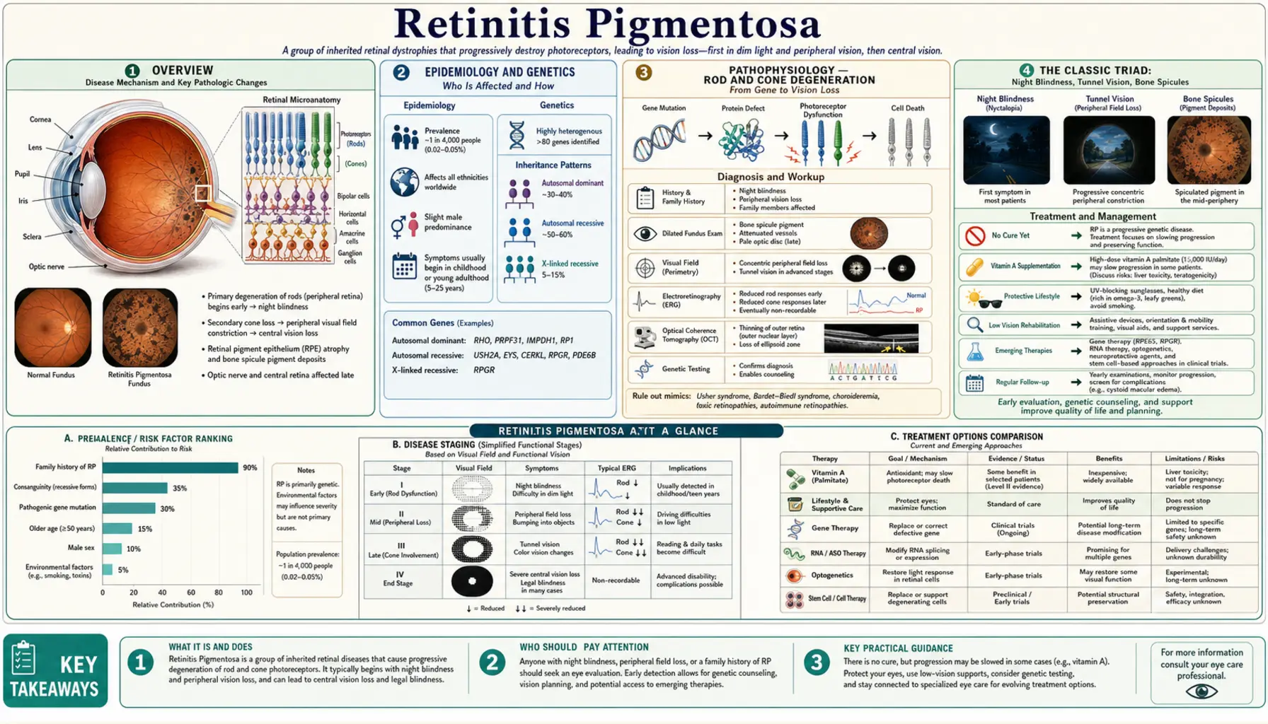
Table of Contents
- Overview
- Epidemiology and Genetics
- Pathophysiology — Rod and Cone Degeneration
- The Classic Triad: Night Blindness, Tunnel Vision, Bone Spicules
- Inheritance Patterns
- Syndromic Forms — Usher and Bardet-Biedl
- Diagnosis — ERG, Visual Field, Imaging
- Vitamin A Palmitate — A Controversial but Studied Treatment
- Lutein, Zeaxanthin, and DHA
- Gene Therapy — Voretigene Neparvovec for RPE65 Mutations
- Retinal Prostheses — Epiretinal and Subretinal Implants
- Living with Retinitis Pigmentosa
- Key Research Papers
- Connections
- Featured Videos
Overview
Retinitis pigmentosa (RP) is a group of inherited retinal dystrophies characterized by the primary degeneration of rod and cone photoreceptors, the light-sensing cells of the retina (Verbakel 2018). In the typical course, people lose night vision in adolescence, side vision in young adulthood, and central vision in later life as rods and then cones are lost (Hartong 2006). The clinical hallmarks are an abnormal fundus with bone-spicule pigment deposits and narrowed (attenuated) retinal vessels, a diminished or absent electroretinogram (ERG), and a reduced visual field (Ferrari 2011).
RP is not one disease but many: variants in more than 80 genes have been associated with non-syndromic RP alone (Verbakel 2018). Course and severity differ between genetic subtypes, and Ferrari and colleagues note that even patients of the same age who carry the same RP1 mutation can differ notably in visual field and ERG (Ferrari 2011).
The 2020 Cochrane review gives a worldwide prevalence of about 1 in 4,000, with about 100,000 people affected in the United States (Schwartz 2020). Estimates that include syndromic and systemic forms range from 1 in 3,000 to 1 in 7,000 (Ferrari 2011), and a 2023 systematic review puts the number affected worldwide at more than 1 million (Khojasteh 2023).
The Cochrane review states that there is no proven therapy for RP (Schwartz 2020). One gene therapy, voretigene neparvovec, is FDA-approved, but only for retinal dystrophy caused by confirmed biallelic RPE65 mutations (FDA prescribing information). Other gene-specific approaches — gene replacement, gene editing, antisense oligonucleotides and small-molecule drugs — are in development or in clinical trials (Toms 2020). Because these therapies are gene-specific, a molecular diagnosis decides eligibility.
Epidemiology and Genetics
Verbakel and colleagues describe RP as a leading cause of visual disability (Verbakel 2018). In 2006, Hartong, Berson and Dryja counted well over 100 genes implicated in hereditary retinal degenerations and more than 45 genes for RP, which together accounted for only about 60% of patients (Hartong 2006); by 2018 more than 80 genes had been associated with non-syndromic RP (Verbakel 2018). The genes encode proteins with many different jobs — phototransduction, retinal (visual-cycle) metabolism, outer-segment structure, transport along the photoreceptor cilium, and pre-mRNA splicing — yet all lead to photoreceptor loss (Ferrari 2011).
Genes that account for a notable share of cases, as reported in the 2011 review by Ferrari and colleagues unless stated otherwise:
- RHO (rhodopsin): mutations account for 30 to 40% of autosomal dominant RP, with more than 100 different mutations described. Rhodopsin is the rod visual pigment: the protein opsin bound to the chromophore 11-cis retinaldehyde, which is derived from vitamin A.
- RPGR (retinitis pigmentosa GTPase regulator): mutated in around 70% of patients with X-linked RP, with the ORF15 region a mutational hotspot; RPGR works in intraflagellar transport. RP2 accounts for about 10–15% of X-linked RP, and the two genes together for more than 80% of X-linked cases.
- USH2A: responsible for approximately 80% of Usher syndrome type 2, and variants in Usher genes can also cause non-syndromic RP (Toms 2020).
- EYS (eyes shut homolog): encodes an extracellular-matrix protein; the RP25 locus, where EYS lies, was identified as the cause of 10–20% of autosomal recessive RP in Spain, though it is rare elsewhere.
- PRPH2 (peripherin-2, also called RDS): a structural protein of the photoreceptor outer segment; mutations account for 5–9.5% of autosomal dominant RP, and digenic RP occurs with ROM1.
- PDE6A, PDE6B: subunits of rod phosphodiesterase, part of phototransduction; defects in the PDE6 complex are estimated to account for about 8% of autosomal recessive RP.
- RPE65: more than 60 mutations, accounting for approximately 2% of recessive RP and 16% of Leber congenital amaurosis — the gene targeted by the approved gene therapy.
Hartong and colleagues reported in 2006 that known genes explained about 60% of patients, and Ferrari and colleagues anticipated that molecular testing would become routine and identify the cause in at least 95% of cases. A molecular diagnosis informs genetic counseling of relatives (Verbakel 2018) and eligibility for gene-specific therapy: the approved RPE65 gene therapy is indicated only for confirmed biallelic RPE65 mutations (FDA prescribing information).
Pathophysiology — Rod and Cone Degeneration
The average human retina contains about 92 million rods and 4.6 million cones, according to measurements of eight whole-mounted retinas (Curcio 1990). Cone density peaks at the fovea and falls steeply within 1 mm of its center; the very center is rod-free, and rod density is highest in an elliptical ring at about the eccentricity of the optic disc (Curcio 1990). Rods serve vision in dim light; cones are essential for daylight and high-acuity vision (Léveillard 2004).
In RP the initial defect usually lies in the rods or the supporting retinal pigment epithelium (RPE); most RP genes are not expressed by cones, so cone loss is indirect (Léveillard 2004). The mechanism varies with the gene — a faulty phototransduction protein, a blocked visual cycle, a structurally weak outer segment, failed ciliary transport, or defective RNA splicing (Ferrari 2011).
The visible results are bone-spicule pigment deposits and attenuated retinal vessels on examination (Ferrari 2011) and, on optical coherence tomography (OCT) and fundus autofluorescence imaging, a progressive loss of the outer retinal layers with an altered lipofuscin distribution in a characteristic pattern (Verbakel 2018).
Why cones die after rods is not settled. Léveillard, Sahel and colleagues had shown that factors secreted by rods are essential for cone viability, and in 2004 they identified one: rod-derived cone viability factor (RdCVF), a truncated thioredoxin-like protein expressed by photoreceptors (Léveillard 2004). A second hypothesis is oxidative damage. Rods are a major user of oxygen in the retina, and after rods die the oxygen level in the outer retina rises; in the rd1 mouse model of RP, a mixture of antioxidants reduced markers of oxidative damage in cones and was accompanied by a twofold increase in cone density (Komeima 2006 — an animal study).
The Classic Triad: Night Blindness, Tunnel Vision, Bone Spicules
Ferrari and colleagues list three clinical hallmarks of RP: an abnormal fundus with bone-spicule deposits and attenuated retinal vessels, an abnormal ERG, and a reduced visual field (Ferrari 2011).
Nyctalopia (Night Blindness)
Night blindness is typically the first symptom, appearing in adolescence in the typical course (Hartong 2006; Verbakel 2018). People describe difficulty seeing in dim light and slow adjustment when moving from bright to dark surroundings. Measures of retinal function such as the ERG show diminished photoreceptor function generally many years before symptomatic night blindness, visual-field scotomas or reduced acuity appear (Hartong 2006).
Night blindness also has non-progressive genetic causes: congenital stationary night blindness is listed among the conditions that some RHO and PDE6B variants cause (Ferrari 2011).
Progressive Concentric Visual Field Constriction (Tunnel Vision)
Night blindness is followed by concentric visual field loss (Verbakel 2018). The loss is progressive and reaches central vision later in the disease course (Schwartz 2020). Trials have tracked it with automated Humphrey perimetry (Berson 2004; Berson 2010) and with Goldmann kinetic perimetry (Maguire 2021).
Bone Spicule Pigmentation and Other Fundus Features
The fundus of established RP shows dark bone-spicule pigment deposits and attenuated retinal vessels (Ferrari 2011).
Cystoid macular edema (CME) — fluid-filled spaces in the macula, seen on OCT — is a common complication. In a 2025 cohort of 580 patients with genetically confirmed non-syndromic RP, 30.9% developed CME in at least one eye over the disease course: 51.4% of those with autosomal dominant forms, 28.1% with autosomal recessive forms and 7.5% with X-linked RP (Testa 2025). A 2018 systematic review of 23 studies found oral carbonic anhydrase inhibitors (acetazolamide, methazolamide) and topical ones (dorzolamide, brinzolamide) effective as first-line treatments, with oral acetazolamide having the strongest clinical basis and outperforming topical dorzolamide; rebound of CME was common in the long term (Bakthavatchalam 2018).
Posterior subcapsular cataract is the most prevalent cataract type in younger people with RP, and a 2023 systematic review concluded that cataract surgery appears able to play an important role in restoring vision in RP, while noting the concerns surgeons raise about operating on these eyes (Khojasteh 2023).
Inheritance Patterns
RP can be inherited as autosomal dominant, autosomal recessive or X-linked; rarer forms include X-linked dominant, mitochondrial and digenic RP (Ferrari 2011). Ferrari and colleagues estimate that non-syndromic RP is autosomal dominant in 20–25% of cases, autosomal recessive in 15–20%, X-linked in 10–15% and sporadic or simplex in 30%, with about 5% early-onset and grouped with Leber congenital amaurosis.
Autosomal Dominant RP (AD-RP)
One altered copy of the gene is enough to cause disease, so each child of an affected parent has a 50% chance of inheriting it. The genes accounting for the largest shares are RHO (30–40% of AD-RP), PRPH2 (5–9.5%), RP1 (about 5–10%) and PRPF31 (1–8% in different cohorts); PRPF8 and PRPF3 account for about 3% and 1% (Ferrari 2011).
Autosomal Recessive RP (AR-RP)
Two altered copies are needed, one from each parent; the parents are usually unaffected carriers. When both parents are carriers, each brother or sister of an affected person has a 25% chance of being affected and a 50% chance of being an unaffected carrier, as GeneReviews sets out for recessive Refsum disease (Waterham, GeneReviews). Reported gene shares include USH2A (10%), CRB1 (6.5%), PDE6A and PDE6B (4% each), CNGA1 (2.3%) and RPE65 (2%) (Ferrari 2011).
X-Linked RP (XL-RP)
X-linked RP makes up about 10–15% of RP; around 70% of patients carry RPGR mutations and 10–15% carry RP2 mutations (Ferrari 2011). The DHAX trial investigators describe X-linked RP as a severe inherited retinal degeneration with a frequency of 1 in 100,000 persons (Hoffman 2014). Because males have a single X chromosome, one altered copy causes disease in males.
Simplex RP and Digenic RP
About 30% of non-syndromic cases are sporadic or simplex, with no other affected family member (Ferrari 2011). Digenic RP has been reported in families carrying a PRPH2 (RDS) mutation together with a ROM1 mutation; in all cases reported, the PRPH2 mutation was the same (L185P), while three different ROM1 mutations were found (Ferrari 2011).
Syndromic Forms — Usher and Bardet-Biedl
Most RP affects only the eye, but 20–30% of patients also have an associated non-ocular condition (Verbakel 2018; Ferrari 2011).
Usher Syndrome
Usher syndrome combines sensorineural hearing loss with RP, with or without vestibular (balance) dysfunction. It is the most common cause of deaf-blindness worldwide, with a prevalence of 4 to 17 per 100,000, and has been estimated to represent 18% of all RP cases (Toms 2020). It has three clinical types (Toms 2020):
- Usher type I: congenital, severe to profound hearing loss; vestibular hypofunction (infants typically do not walk before 18 months); RP with prepubertal onset and legal blindness in the fourth decade. Genes: MYO7A (more than 50% of type 1), USH1C, CDH23, PCDH15, USH1G and CIB2. The most severe subtype, about 25–44% of Usher cases.
- Usher type II: congenital, moderate to severe hearing loss with the high frequencies most affected; normal vestibular function; RP with onset in the second decade and legal blindness in the sixth decade. Genes: USH2A (about 80%), ADGRV1 and WHRN. The most common form, over half of all cases.
- Usher type III: hearing loss beginning after speech develops and progressing; RP with variable onset, typically in the second decade; vestibular abnormalities in about 50%. Gene: CLRN1. About 2–4% of cases, but particularly prevalent in Finland and among Ashkenazi Jewish people.
Infants with Usher type I are detected through newborn hearing screening, and Toms and colleagues describe bilateral cochlear implants within the first 2 years of life as the standard approach for type 1; hearing loss across the types is managed with hearing aids and cochlear implants. There is no approved treatment for the retinal disease in Usher syndrome; gene replacement, gene editing, antisense oligonucleotides and small-molecule drugs are in development or in clinical trials (Toms 2020).
Bardet-Biedl Syndrome (BBS)
Bardet-Biedl syndrome is an autosomal recessive ciliopathy with more than 20 implicated genes; receptor trafficking in and out of primary cilia is compromised, affecting many organ systems, and hyperphagia is a prominent symptom alongside early-onset obesity (Shoemaker 2024). Ferrari and colleagues list rod-cone dystrophy (more than 90% of patients), truncal obesity (72%), postaxial polydactyly, cognitive impairment, male hypogonadotrophic hypogonadism, genitourinary malformations and renal abnormalities, and report a frequency of 1 in 120,000 among Caucasians, with higher rates in Newfoundland (1 in 13,000) and among Bedouin populations (1 in 13,500, and up to 1 in 6,900 in the Jahra district) (Ferrari 2011).
Other Syndromic Forms
Kearns-Sayre syndrome: a mitochondrial disorder defined by onset before age 20, progressive external ophthalmoplegia and pigmentary retinopathy, accompanied by cardiac conduction defects, raised cerebrospinal-fluid protein or cerebellar ataxia; half of patients develop cardiac complications, most often conduction disease that can progress to complete heart block (Kabunga 2015). Its molecular basis is a rearrangement (deletion) of mitochondrial DNA (Maceluch 2006). The fundus shows a salt-and-pepper pigmentary retinopathy, and vision remains good in most patients (Tsang 2018).
Refsum disease: adult Refsum disease is caused by biallelic variants in PHYH or PEX7 and marked by raised plasma phytanic acid, RP beginning in late childhood or later, and variable anosmia, polyneuropathy, deafness, ataxia and ichthyosis; cardiac arrhythmia and cardiomyopathy can develop later in life (Waterham, GeneReviews). Unlike most forms of RP, it is accessible to therapy (Rüether 2010). Phytanic acid comes mostly from foods of ruminant animals (cow, sheep, goat), some fish and walnuts, and dietary restriction helps resolve ichthyosis, sensory neuropathy and ataxia; GeneReviews also lists fasting, sudden weight loss, ibuprofen and amiodarone among the circumstances and agents to be avoided (Waterham, GeneReviews). Rüether and colleagues report that a diet low in phytanic acid ameliorates polyneuropathy and ataxia and slows or even stops the other manifestations, and describe the ophthalmologist's role in early identification as crucial because the retinal degeneration appears early (Rüether 2010).
Diagnosis — ERG, Visual Field, Imaging
Diagnosis combines history, fundus examination, electrophysiology and imaging. In RP, photoreceptor function measured with an electroretinogram is markedly reduced or even absent, and OCT and fundus autofluorescence show progressive loss of the outer retinal layers and an altered lipofuscin distribution (Verbakel 2018).
Fundoscopy and Slit-Lamp Exam
Bone-spicule pigment deposits and attenuated retinal vessels are the fundus hallmarks (Ferrari 2011). A slit-lamp examination can show posterior subcapsular cataract, the most prevalent cataract type in younger people with RP (Khojasteh 2023).
Electroretinogram (ERG)
The full-field electroretinogram is a mass electrical response of the retina to diffuse flashes of light, used to assess generalized retinal function. The International Society for Clinical Electrophysiology of Vision (ISCEV) publishes minimum protocols for the basic stimuli, recording and reporting, most recently updated in 2022 (Robson 2022). In RP the ERG shows diminished, or absent, a- and b-waves (Ferrari 2011), and the loss is measurable generally many years before symptoms (Hartong 2006). Cone ERG amplitude, including the 30-Hz flicker response, has served as an outcome in the vitamin A and DHA trials (Berson 1993; Berson 2004; Hoffman 2014).
Visual Field Testing
Kinetic (Goldmann) perimetry maps the outer boundary of the field with moving targets; automated static (Humphrey) perimetry tests a grid of fixed points. The supplement trials used Humphrey programs covering the central 30 degrees (30-2) and the mid-periphery (60-4) (Berson 2010), and the RPE65 gene therapy trial reported Goldmann III4e field area (Maguire 2021).
Optical Coherence Tomography (OCT)
OCT gives cross-sectional images of the retinal layers and shows the progressive loss of the outer retina in RP (Verbakel 2018). It also detects cystoid macular edema; the 2025 cohort study cited above used spectral-domain OCT for this purpose (Testa 2025). The width of the ellipsoid zone (the band at the inner/outer segment border) shrank by a mean 0.86° (7%) a year in X-linked RP, a change larger than the test-retest variability, which the authors present as support for ellipsoid-zone width as an outcome measure in clinical trials (Birch 2013).
Fundus Autofluorescence (FAF)
FAF imaging records the natural fluorescence of lipofuscin in the RPE; in RP it shows an altered lipofuscin distribution in a characteristic pattern (Verbakel 2018).
Genetic Testing
A molecular diagnosis supports genetic counseling of patients and relatives (Verbakel 2018) and determines eligibility for gene-specific treatment: the approved gene therapy is indicated only for confirmed biallelic RPE65 mutation-associated retinal dystrophy (FDA prescribing information), and gene-targeted approaches for Usher genes are in development or trials (Toms 2020).
Vitamin A Palmitate — A Controversial but Studied Treatment
Vitamin A palmitate is the most studied supplement in RP, and the evidence is contested. A 2006 review by Hartong, Berson and Dryja states that controlled trials indicate nutritional interventions, including vitamin A palmitate and omega-3-rich fish, slow progression in many patients (Hartong 2006). The 2020 Cochrane review of four trials (944 participants) concluded that it is uncertain whether vitamin A, DHA or both benefit people with RP, rated the evidence as very low certainty, and found no trial showing a benefit on the progression of visual acuity loss (Schwartz 2020).
The pivotal trial, published by Berson and colleagues in Archives of Ophthalmology in 1993 (PubMed PMID: 8512476), randomized 601 patients aged 18 through 49 in a 2 × 2 factorial design for 4 to 6 years to 15,000 IU/day vitamin A, 15,000 IU/day vitamin A plus 400 IU/day vitamin E, trace amounts of both vitamins, or 400 IU/day vitamin E. The two groups receiving 15,000 IU/day vitamin A had on average a slower decline in cone ERG amplitude than the two groups not receiving it (P = .01); the Cochrane review, analysing this trial, reports a slight difference in yearly ERG decline (6.1% versus 7.1%) favoring vitamin A. Among 354 patients with higher starting amplitudes, those on vitamin A were 32% less likely (P = .01), and those on vitamin E 400 IU/day 42% more likely (P = .03), to lose half or more of their amplitude in a given year, which the authors read as a possible adverse effect of vitamin E. Trends in visual field area were not statistically significant, and visual acuity declined about 1 letter per year in all groups (Berson 1993).
Points the research raises:
- ABCA4-related disease: in a mouse model of ABCA4 retinal dystrophy, vitamin A supplementation increased toxic lipofuscin pigments in the RPE, and the authors concluded that vitamin A supplementation should be avoided in patients with ABCA4 mutations or other dystrophies with lipofuscin accumulation (Radu 2008 — an animal study).
- Who was studied: the vitamin A trials enrolled adults (aged 18–49 in 1993, 18–60 in the 2010 lutein trial); the two Cochrane-reviewed trials restricted to X-linked RP tested DHA, not vitamin A (Schwartz 2020).
- Children: none of the vitamin A trials enrolled children, so there are no trial data on this dose in children.
- Pregnancy: pregnancy was an exclusion criterion in the 2010 trial, and its authors state that high-dose vitamin A supplements carry an increased risk of birth defects for women who are pregnant or planning a pregnancy (Berson 2010).
- Liver and bone safety: among 146 adults taking 15,000 IU/day for up to 12 years, no clinical signs of liver toxicity from vitamin A were detected and no serum retinol value exceeded the upper normal limit; long-term doses of 25,000 IU/day or more are considered potentially toxic (Sibulesky 1999). The 2010 trial authors describe annual fasting serum vitamin A and liver function tests as a precaution and mention a slight (0.5–1.0%) increased risk of hip fracture with long-term high-dose vitamin A in patients aged 49 and over (Berson 2010).
- Form: the trials gave vitamin A as retinyl palmitate (Berson 2004; Berson 2010).
Decisions about supplements belong with a clinician.
Lutein, Zeaxanthin, and DHA
Two further supplements have been tested in people with RP, mostly alongside vitamin A: the carotenoid lutein and the omega-3 fatty acid DHA (docosahexaenoic acid).
Lutein and Zeaxanthin
Lutein is concentrated in and around the fovea as yellow macular pigment and has also been found in rod outer segments; it is thought to screen foveal cones from short-wavelength light and may act there as an antioxidant (Berson 2010). In 176 patients with RP, macular pigment optical density rose with serum lutein but was unrelated to serum zeaxanthin (Sandberg 2010).
In the 2010 trial by Berson and colleagues, 225 non-smoking adults aged 18–60, all taking 15,000 IU/day vitamin A palmitate, were randomized to 12 mg/day lutein or a control tablet for 4 years. The primary outcome — Humphrey 30-2 central field sensitivity — showed no significant difference. A prespecified secondary outcome, the Humphrey 60-4 mid-peripheral field, declined more slowly with lutein (P = .05), and decline was slower among those with the highest serum lutein or the largest rise in macular pigment. No significant toxic effects were observed. The authors concluded that lutein slowed mid-peripheral field loss on average among non-smoking adults taking vitamin A; they limited that conclusion to non-smokers because of a concern raised about long-term lutein and lung cancer in smokers over 50, and stated that the long-term safety of lutein had not been established (Berson 2010).
DHA and Omega-3 Fatty Acids
Low blood DHA has been reported in X-linked RP (Hoffman 2004). Three randomized trials tested DHA:
- Berson and colleagues (2004) gave 221 adults aged 18–55, all on 15,000 IU/day vitamin A, 1,200 mg/day DHA or control capsules for 4 years; there was no significant difference in decline of ocular function (Humphrey 30-2, P = .88), including within genetic types (Berson 2004).
- Hoffman and colleagues (2004) gave 44 males with X-linked RP (aged 4–38) 400 mg/day DHA or placebo for 4 years; the rate of cone ERG loss did not differ significantly between groups (P = .16), though subset analyses suggested benefit in some age groups (Hoffman 2004).
- The DHAX trial (Hoffman 2014) randomized 78 males with X-linked RP to 30 mg/kg/day DHA or placebo for 4 years; there was no difference in the rate of cone ERG loss (P = .30) or in rod or maximal ERG, and no severe treatment-emergent adverse events. The Cochrane review notes that one trial reported a slower annual rate of visual field sensitivity loss with DHA, with very low-certainty evidence (Schwartz 2020).
Overall, the 2020 Cochrane review judged the benefit of vitamin A, DHA or both to be uncertain (Schwartz 2020).
Gene Therapy — Voretigene Neparvovec for RPE65 Mutations
The FDA approved voretigene neparvovec-rzyl on December 19, 2017 (FDA approval letter, listed on the agency's product page) for patients with confirmed biallelic RPE65 mutation-associated retinal dystrophy who have viable retinal cells as determined by the treating physician (FDA prescribing information).
The Biology of RPE65-Mediated Disease
RPE65 is an isomerohydrolase expressed in the retinal pigment epithelium and is critical for regenerating the visual pigment needed for both rod and cone vision (Ferrari 2011); the gene encodes the all-trans retinyl ester isomerase of the retinoid (visual) cycle (Russell 2017). Without it, the RPE cannot supply enough 11-cis retinal to the photoreceptors; the photoreceptors degenerate slowly, which is why restoring the enzyme can still recover function (Russell 2017). RPE65 mutations account for about 16% of Leber congenital amaurosis and about 2% of recessive RP (Ferrari 2011).
How the Gene Therapy Is Given
It is an adeno-associated virus (AAV) vector-based gene therapy given by subretinal injection only; each eye is treated on a separate day, no fewer than 6 days apart, and the prescribing information recommends a course of oral corticosteroids around each injection (FDA prescribing information). In the phase 3 trial each eye received 1.5 × 10¹¹ vector genomes in 0.3 mL (Russell 2017).
Clinical Trial Results
The phase 3 trial (Russell S et al., Lancet 2017; PubMed PMID: 28712537) enrolled 31 participants aged 4 to 44 years with confirmed biallelic RPE65 mutations and sufficient viable retina at two US sites, randomized 2:1 to treatment or control; it was open-label, with masked graders for the primary outcome. The primary endpoint was the 1-year change in the multi-luminance mobility test (MLMT), navigating a course at specified light levels. Mean bilateral MLMT change was 1.8 light levels with treatment versus 0.2 in controls (difference 1.6, p = 0.0013), and 13 of 20 treated participants (65%), but no controls, passed the test at the lowest light level tested (1 lux). Full-field light sensitivity improved by more than 2 log units in the treated group (difference versus control −2.11 log units, p = 0.0004), while the change in best-corrected visual acuity was not significant in the primary analysis (p = 0.17). No product-related serious adverse events or harmful immune responses occurred. The trial was funded by the manufacturer (Russell 2017).
Follow-up of the same trial reported that improvements in mobility, light sensitivity and visual field were maintained for 3 to 4 years; 71% of patients with a year-3 visit passed the mobility test at the lowest light level. One patient had a retinal detachment at about year 4, and no product-related serious adverse events were reported (Maguire 2021).
Limitations and Practical Considerations
The therapy is limited to people with confirmed biallelic RPE65 mutations and viable retinal cells, and the prescribing information does not recommend it for infants under 12 months because active retinal cell proliferation may dilute or lose the vector (FDA prescribing information). In follow-up, its safety profile was consistent with vitrectomy and the subretinal injection procedure (Maguire 2021).
The Broader Gene Therapy Pipeline
For other genes, gene replacement, gene editing, antisense oligonucleotides and small-molecule drugs are in development or in clinical trials — for example for the Usher syndrome genes (Toms 2020). Optogenetic therapy, which does not depend on the mutated gene, has been tested in a person with RP (see the next section).
Retinal Prostheses — Epiretinal and Subretinal Implants
Retinal prostheses aim to restore some vision to people blind from end-stage RP or other outer retinal degeneration by electrically stimulating the surviving inner retina: a camera on a pair of glasses feeds a video processor, which converts images into stimulation patterns sent to an electrode array on the retina (da Cruz 2016).
The Epiretinal Implant Approved in 2013
On February 13, 2013, the FDA approved an epiretinal prosthesis under a Humanitarian Device Exemption (HDE H110002) for adults aged 25 or older with severe to profound RP, bare or no light perception in both eyes, and a previous history of useful form vision; if a patient has no light perception, intact inner-retina function must be confirmed first, and the implant goes in one eye, typically the worse-seeing one (FDA approval order). FDA's summary describes the implanted part as:
- A small sealed electronics package that receives power and drives the stimulation
- A coil that receives power and exchanges data with the external coil on the glasses
- A thin-film cable and electrode array tacked to the retinal surface (epiretinal)
The array carries 60 exposed platinum electrodes in a 6 × 10 pattern, 55 of them enabled for use (FDA Summary of Safety and Probable Benefit). In the international trial of 30 blind participants, published by Humayun MS et al. in Ophthalmology in 2012 (PubMed PMID: 22244176), subjects performed significantly better with the system on than off at object localization (96% of subjects), motion discrimination (57%) and discrimination of oriented gratings (23%); the best recorded visual acuity was 20/1260, and mean performance on orientation and mobility tasks was significantly better with the system on. At 5 years, 24 of the 30 remained implanted with functioning systems, and patients still performed significantly better with the device on (da Cruz 2016).
In the trial, 70% of patients had no serious adverse events; the most common serious event was conjunctival erosion or dehiscence over the external part of the implant, and one device was removed (Humayun 2012). The 5-year report recorded only one additional serious adverse event after year 3 (da Cruz 2016).
Other Approaches
A subretinal implant was tested in 29 blind participants with outer retinal degeneration: within up to 12 months, 21 (72%) reached the primary endpoints in daily-living, recognition or mobility tasks, 13 (45%) reported restored visual function they used in daily life, and the best measured acuities were 20/606 and 20/546 (Stingl 2015).
Optogenetics makes surviving retinal cells light-sensitive. In 2021 Sahel and colleagues reported a blind patient with RP who received an intraocular injection of an AAV vector encoding the light-sensitive protein ChrimsonR, combined with engineered goggles that project light pulses onto the retina to activate the transduced retinal ganglion cells. With the goggles, the patient perceived, located, counted and touched objects using the treated eye, which was not possible before the injection or without the goggles. The authors describe it as the first reported case of partial functional recovery in a neurodegenerative disease after optogenetic therapy (Sahel 2021 — a single case).
Living with Retinitis Pigmentosa
A diagnosis of RP brings practical and emotional challenges over many years. Verbakel and colleagues describe management as counseling, visual rehabilitation, and current and emerging therapies (Verbakel 2018).
Low Vision Aids and Assistive Technology
Assistive technology includes smartphone accessibility features (screen readers, magnification, high-contrast settings), apps that read text aloud, describe scenes or connect users with sighted volunteers, and screen-reading and magnification software for computers and tablets.
Optical low vision aids include handheld and stand magnifiers, telescopes for distance viewing, and video magnifiers that show an enlarged image on a screen. Large-print books, audiobooks and e-readers with adjustable font and contrast are other formats.
Orientation and Mobility Training
Orientation and mobility (O&M) specialists teach independent travel skills such as long-cane technique, sighted-guide technique and use of public transport. Guide dogs are another mobility aid.
Driving
Night blindness and a constricted visual field affect driving, first at night. Driving licenses depend on visual field and acuity standards that each licensing authority sets.
Treating Treatable Complications
Two complications of RP can be treated even though the degeneration itself cannot:
- Cystoid macular edema (CME): found over the disease course in 30.9% of a 580-patient genotyped cohort (Testa 2025). In a 2018 systematic review, oral carbonic anhydrase inhibitors had the strongest clinical basis, with topical dorzolamide described as an alternative for patients who do not tolerate oral acetazolamide, and rebound of CME was common in the long term (Bakthavatchalam 2018).
- Posterior subcapsular cataract: the most prevalent cataract type in younger people with RP; a 2023 systematic review concluded that cataract surgery appears able to play an important role in visual restoration, while noting surgeons' concerns about these eyes. Surgery does not change the underlying retinal degeneration (Khojasteh 2023).
Mental Health and Peer Support
Progressive vision loss can bring anxiety, low mood and grief, along with uncertainty about the timeline and the loss of driving and independence. Individual therapy, group therapy and peer support networks are forms of psychological support.
Patient organizations and research foundations fund inherited retinal disease research and run support networks, and disease-specific communities exist for Usher syndrome, Bardet-Biedl syndrome and specific genetic subtypes.
Research and Clinical Trials
ClinicalTrials.gov, the U.S. registry of clinical studies, lists registered RP trials; the RPE65 gene therapy trial (NCT00999609) and the lutein trial (NCT00346333) described above are registered there (Russell 2017; Berson 2010). Many current trials target specific genes, so eligibility often depends on knowing the causative mutation.
Key Research Papers
- Berson EL, Rosner B, Sandberg MA, et al. A randomized trial of vitamin A and vitamin E supplementation for retinitis pigmentosa. Arch Ophthalmol. 1993;111(6):761–772. — Randomized 2 × 2 trial in 601 patients aged 18–49: the groups given 15,000 IU/day vitamin A had a slower average decline in cone ERG amplitude (P = .01), and 400 IU/day vitamin E suggested an adverse effect. (Corrected 2026-10-14: this entry previously named the wrong journal and linked PMID 8479463, an unrelated study of vitamin E and coronary disease.) — PubMed PMID: 8512476
- Berson EL, Rosner B, Sandberg MA, et al. Clinical trial of lutein in patients with retinitis pigmentosa receiving vitamin A. Arch Ophthalmol. 2010;128(4):403–411. — 225 non-smoking adults on vitamin A: no difference on the primary Humphrey 30-2 outcome; slower mid-peripheral (60-4) field loss with 12 mg/day lutein (P = .05). — PubMed PMID: 20385935
- Russell S, Bennett J, Wellman JA, et al. Efficacy and safety of voretigene neparvovec (AAV2-hRPE65v2) in patients with RPE65-mediated inherited retinal dystrophy: a randomised, controlled, open-label, phase 3 trial. Lancet. 2017;390(10097):849–860. — Phase 3 trial in 31 participants with biallelic RPE65 mutations: mean mobility-test change 1.8 versus 0.2 light levels at 1 year (p = 0.0013). — PubMed PMID: 28712537
- Hartong DT, Berson EL, Dryja TP. Retinitis pigmentosa. Lancet. 2006;368(9549):1795–1809. — Seminar review of RP genetics, clinical course, electroretinography and nutritional trials. — PubMed PMID: 17113430
- Wang X, Bai Y, Zuo X, et al. Pathogenic mechanisms of RPGR mutations in X-linked retinitis pigmentosa: integrating clinical pedigree and single-cell transcriptomics. Front Genet. 2026;17:1814462. — Describes a rare RPGR ORF15 frameshift variant in one family; the share of X-linked RP due to RPGR and the ORF15 hotspot are from Ferrari 2011 (below). — PubMed PMID: 42306097
- Usher syndrome: combined retinitis pigmentosa and sensorineural hearing loss. PubMed search: Usher syndrome retinitis pigmentosa hearing loss review — Topic search; the Usher syndrome figures on this page are from Toms 2020 (below). — Search PubMed
- Humayun MS, Dorn JD, da Cruz L, et al. Interim results from the international trial of Second Sight’s visual prosthesis. Ophthalmology. 2012;119(4):779–788. — Single-arm trial of an epiretinal prosthesis in 30 blind subjects: object localization, motion discrimination and grating discrimination were better with the system on than off. — PubMed PMID: 22244176
- Electroretinogram (ERG) in the diagnosis and monitoring of retinitis pigmentosa. PubMed search: electroretinogram retinitis pigmentosa diagnosis monitoring — Topic search; the ERG statements on this page come from Hartong 2006, Ferrari 2011, Verbakel 2018 and the ISCEV standard (Robson 2022). — Search PubMed
- Bardet-Biedl syndrome: ciliopathy with RP, obesity, and polydactyly. PubMed search: Bardet-Biedl syndrome retinitis pigmentosa genetics — Topic search; see Ferrari 2011 and Shoemaker 2024 (below). — Search PubMed
- Carbonic anhydrase inhibitors for cystoid macular edema in retinitis pigmentosa. PubMed search: carbonic anhydrase inhibitor cystoid macular edema retinitis pigmentosa — Topic search; see the 2018 systematic review (Bakthavatchalam 2018) and the 2025 cohort (Testa 2025) below. — Search PubMed
- RHO rhodopsin mutations and autosomal dominant retinitis pigmentosa. PubMed search: rhodopsin RHO mutation autosomal dominant retinitis pigmentosa — Topic search; Ferrari 2011 (below) reports RHO mutations in 30–40% of autosomal dominant RP, with more than 100 mutations described. — Search PubMed
- Refsum disease: phytanic acid accumulation, RP, and dietary treatment. PubMed search: Refsum disease phytanic acid retinitis pigmentosa treatment — Topic search; see the GeneReviews chapter (Waterham) and Rüether 2010 (below). — Search PubMed
- Schwartz SG, Wang X, Chavis P, et al. Vitamin A and fish oils for preventing the progression of retinitis pigmentosa. Cochrane Database Syst Rev. 2020;6(6):CD008428. — Cochrane review of four trials (944 participants): uncertain whether vitamin A, DHA or both benefit people with RP; very low-certainty evidence. — PubMed PMID: 32573764
- Berson EL, Rosner B, Sandberg MA, et al. Clinical trial of docosahexaenoic acid in patients with retinitis pigmentosa receiving vitamin A treatment. Arch Ophthalmol. 2004;122(9):1297–305. — 221 adults on vitamin A: 1,200 mg/day DHA did not slow field loss over 4 years. — PubMed PMID: 15364708
- Hoffman DR, Locke KG, Wheaton DH, et al. A randomized, placebo-controlled clinical trial of docosahexaenoic acid supplementation for X-linked retinitis pigmentosa. Am J Ophthalmol. 2004;137(4):704–18. — 44 males with X-linked RP: 400 mg/day DHA did not significantly change the rate of cone ERG loss. — PubMed PMID: 15059710
- Hoffman DR, Hughbanks-Wheaton DK, Pearson NS, et al. Four-year placebo-controlled trial of docosahexaenoic acid in X-linked retinitis pigmentosa (DHAX trial): a randomized clinical trial. JAMA Ophthalmol. 2014;132(7):866–73. — DHAX trial, 78 males with X-linked RP: 30 mg/kg/day DHA did not slow cone or rod ERG loss. — PubMed PMID: 24805262
- Sandberg MA, Johnson EJ, Berson EL. The relationship of macular pigment optical density to serum lutein in retinitis pigmentosa. Invest Ophthalmol Vis Sci. 2010;51(2):1086–91. — In 176 patients with RP, macular pigment optical density rose with serum lutein. — PubMed PMID: 19797209
- Sibulesky L, Hayes KC, Pronczuk A, et al. Safety of <7500 RE (<25000 IU) vitamin A daily in adults with retinitis pigmentosa. Am J Clin Nutr. 1999;69(4):656–63. — 15,000 IU/day vitamin A for up to 12 years in 146 adults: no clinical signs of liver toxicity detected. — PubMed PMID: 10197566
- Radu RA, Yuan Q, Hu J, et al. Accelerated accumulation of lipofuscin pigments in the RPE of a mouse model for ABCA4-mediated retinal dystrophies following Vitamin A supplementation. Invest Ophthalmol Vis Sci. 2008;49(9):3821–9. — Mouse study: vitamin A supplementation increased RPE lipofuscin in an ABCA4 knockout model. — PubMed PMID: 18515570
- Maguire AM, Russell S, Chung DC, et al. Durability of Voretigene Neparvovec for Biallelic RPE65-Mediated Inherited Retinal Disease: Phase 3 Results at 3 and 4 Years. Ophthalmology. 2021;128(10):1460–1468. — Phase 3 follow-up: mobility, light-sensitivity and visual-field gains maintained up to 3 to 4 years. — PubMed PMID: 33798654
- da Cruz L, Dorn JD, Humayun MS, et al. Five-Year Safety and Performance Results from the Argus II Retinal Prosthesis System Clinical Trial. Ophthalmology. 2016;123(10):2248–54. — Five-year results of the epiretinal prosthesis trial: 24 of 30 patients still implanted with functioning systems. — PubMed PMID: 27453256
- Stingl K, Bartz-Schmidt KU, Besch D, et al. Subretinal Visual Implant Alpha IMS--Clinical trial interim report. Vision Res. 2015;111(Pt B):149–60. — Subretinal implant in 29 blind participants: 72% reached the primary endpoints. — PubMed PMID: 25812924
- Sahel JA, Boulanger-Scemama E, Pagot C, et al. Partial recovery of visual function in a blind patient after optogenetic therapy. Nat Med. 2021;27(7):1223–1229. — Single blind patient with RP: optogenetic vector plus goggles allowed perceiving, locating and touching objects. — PubMed PMID: 34031601
- Ferrari S, Di Iorio E, Barbaro V, et al. Retinitis pigmentosa: genes and disease mechanisms. Curr Genomics. 2011;12(4):238–49. — Review of RP genes, inheritance shares and syndromic forms. — PubMed PMID: 22131869
- Verbakel SK, van Huet RAC, Boon CJF, et al. Non-syndromic retinitis pigmentosa. Prog Retin Eye Res. 2018;66:157–186. — Review of non-syndromic RP: prevalence about 1:4000; more than 80 genes; 20–30% syndromic. — PubMed PMID: 29597005
- Toms M, Pagarkar W, Moosajee M. Usher syndrome: clinical features, molecular genetics and advancing therapeutics. Ther Adv Ophthalmol. 2020;12:2515841420952194. — Review of Usher syndrome types, genes, prevalence and therapies in development. — PubMed PMID: 32995707
- Shoemaker A. Bardet-Biedl syndrome: A clinical overview focusing on diagnosis, outcomes and best-practice management. Diabetes Obes Metab. 2024;26 Suppl 2:25–33. — Clinical overview of Bardet-Biedl syndrome, a ciliopathy with more than 20 genes. — PubMed PMID: 38383825
- Léveillard T, Mohand-Saïd S, Lorentz O, et al. Identification and characterization of rod-derived cone viability factor. Nat Genet. 2004;36(7):755–9. — Identification of rod-derived cone viability factor (RdCVF). — PubMed PMID: 15220920
- Komeima K, Rogers BS, Lu L, et al. Antioxidants reduce cone cell death in a model of retinitis pigmentosa. Proc Natl Acad Sci U S A. 2006;103(30):11300–5. — Mouse study: antioxidants reduced oxidative damage and cone loss in the rd1 model of RP. — PubMed PMID: 16849425
- Curcio CA, Sloan KR, Kalina RE, et al. Human photoreceptor topography. J Comp Neurol. 1990;292(4):497–523. — Rod and cone counts and density maps from eight human retinas. — PubMed PMID: 2324310
- Birch DG, Locke KG, Wen Y, et al. Spectral-domain optical coherence tomography measures of outer segment layer progression in patients with X-linked retinitis pigmentosa. JAMA Ophthalmol. 2013;131(9):1143–50. — Ellipsoid-zone width declined about 7% a year in X-linked RP; supports it as a trial outcome measure. — PubMed PMID: 23828615
- Testa F, Karali M, Boccia R, et al. Cystoid Macular Edema in Non-Syndromic Retinitis Pigmentosa: Associations With Causative Genes in a Large Cohort. Invest Ophthalmol Vis Sci. 2025;66(12):5. — CME in 30.9% of 580 genotyped patients with non-syndromic RP; most frequent in autosomal dominant forms. — PubMed PMID: 40900079
- Bakthavatchalam M, Lai FHP, Rong SS, et al. Treatment of cystoid macular edema secondary to retinitis pigmentosa: a systematic review. Surv Ophthalmol. 2018;63(3):329–339. — Systematic review of 23 studies: carbonic anhydrase inhibitors are effective first-line treatments for CME in RP. — PubMed PMID: 28987613
- Khojasteh H, Riazi-Esfahani H, Mirghorbani M, et al. Cataract surgery in patients with retinitis pigmentosa: systematic review. J Cataract Refract Surg. 2023;49(3):312–320. — Systematic review of cataract surgery in RP. — PubMed PMID: 36730350
- Kabunga P, Lau AK, Phan K, et al. Systematic review of cardiac electrical disease in Kearns-Sayre syndrome and mitochondrial cytopathy. Int J Cardiol. 2015;181:303–10. — Systematic review of cardiac conduction disease in Kearns-Sayre syndrome. — PubMed PMID: 25540845
- Maceluch JA, Niedziela M. The clinical diagnosis and molecular genetics of kearns-sayre syndrome: a complex mitochondrial encephalomyopathy. Pediatr Endocrinol Rev. 2006;4(2):117–37. — Review of the clinical diagnosis and mitochondrial DNA genetics of Kearns-Sayre syndrome. — PubMed PMID: 17342029
- Tsang SH, Aycinena ARP, Sharma T. Mitochondrial Disorder: Kearns-Sayre Syndrome. Adv Exp Med Biol. 2018;1085:161–162. — Short review of the ocular and systemic features of Kearns-Sayre syndrome. — PubMed PMID: 30578503
- Rüether K, Baldwin E, Casteels M, et al. Adult Refsum disease: a form of tapetoretinal dystrophy accessible to therapy. Surv Ophthalmol. 2010;55(6):531–8. — Review: a low-phytanic-acid diet ameliorates polyneuropathy and ataxia in adult Refsum disease. — PubMed PMID: 20850855
- Waterham HR, Wanders RJA, Leroy BP. Adult Refsum Disease. In: Adam MP, Bick S, Mirzaa GM, et al., editors. GeneReviews. Seattle (WA): University of Washington, Seattle; 2006 (updated 2021). — Clinical features, PHYH/PEX7 genetics and dietary management of adult Refsum disease. — PubMed PMID: 20301527
- Robson AG, Frishman LJ, Grigg J, et al. ISCEV Standard for full-field clinical electroretinography (2022 update). Doc Ophthalmol. 2022;144(3):165–177. — The ISCEV minimum protocol for full-field clinical electroretinography. — PubMed PMID: 35511377
- U.S. Food and Drug Administration. LUXTURNA (voretigene neparvovec-rzyl): prescribing information and approval history (approval letter December 19, 2017). — Indication, administration and use in specific populations. — FDA product page
- U.S. Food and Drug Administration. Humanitarian Device Exemption H110002, Argus II Retinal Prosthesis System: approval order (February 13, 2013) and Summary of Safety and Probable Benefit. — Approved population and device description. — FDA HDE record
Connections
- Ophthalmology
- Optogenetics: the 2026 Nobel Prize — light-gated ion channels, the basis of the early trials that aim to restore some sight in retinitis pigmentosa
- Macular Degeneration — another progressive retinal disease involving photoreceptor and RPE degeneration; overlapping research into gene therapy and neuroprotection
- Glaucoma — optic nerve disease causing visual field loss; important differential diagnosis for tunnel vision; shares some neuroprotection research pathways
- Cataracts — posterior subcapsular cataracts develop earlier in RP patients; cataract surgery can meaningfully improve remaining functional vision
- Amblyopia — reduced visual acuity from developmental causes; part of the broader ophthalmology context for visual impairment evaluation
- Dry Eye Disease — common ophthalmology condition; shares retina specialist care setting with RP patients
- Vitamin A — vitamin A palmitate 15,000 IU/day is the most studied nutritional supplement for slowing RP progression in appropriate patients
- All Conditions — complete disease index