Amblyopia (Lazy Eye)
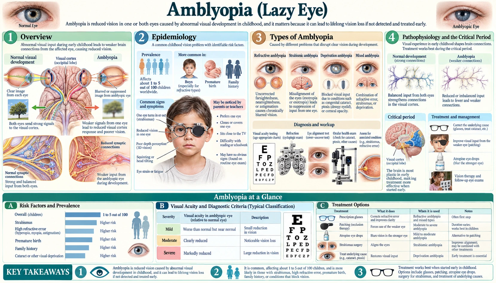
Table of Contents
- Overview
- Epidemiology
- Types of Amblyopia
- Pathophysiology and the Critical Period
- Clinical Presentation and Diagnosis
- Treatment: Occlusion Therapy (Patching)
- Treatment: Atropine Penalization
- Treatment: Dichoptic Training and Digital Approaches
- Amblyopia in Adults and Neuroplasticity
- Prognosis and Long-Term Outcomes
- Key Research Papers
- Connections
- Featured Videos
1. Overview
Amblyopia — commonly called lazy eye — is reduced vision in one eye (and in rare cases both eyes) that is not caused by any structural disease or damage to the eye itself. The eye is anatomically normal. The problem is in the brain: during early childhood, abnormal visual experience causes the visual cortex to suppress or ignore signals from one eye, and that eye's visual pathways fail to develop normally. The result is permanently reduced acuity in that eye unless treatment intervenes during the critical window of brain development.
The colloquial name "lazy eye" is somewhat misleading. The eye is not lazy — it is sending signals perfectly well. The brain is the part that has learned, through an accident of development, to favor the other eye and tune out the weaker one. This distinction matters because treatment targets the brain-eye connection, not the eye structure itself.
Amblyopia affects roughly 2–3% of the general population and is the most common cause of preventable, permanent monocular visual impairment in children and working-age adults. Because children do not know what normal vision looks like, they rarely complain — they simply adapt. This makes routine screening critically important, since early detection and treatment during the sensitive period can restore near-normal vision, while delayed treatment yields much smaller gains.
2. Epidemiology
Amblyopia is the most common vision disorder in children, with a prevalence of approximately 2–3% worldwide. In the United States alone, that translates to roughly 3 million people affected. It is the leading cause of unilateral vision loss in adults under the age of 45, outpacing all retinal diseases, glaucoma, and trauma combined in that age group.
The condition is most often detected and treated in children under 7, when the visual cortex is most plastic and the response to treatment is fastest. However, because amblyopia is generally asymptomatic in mild-to-moderate forms, a significant proportion of cases go undiagnosed until school-age vision screening or a routine eye exam.
Key risk factors include:
- Family history of amblyopia or strabismus — having a first-degree relative with either condition roughly doubles the risk.
- Premature birth — preterm infants are at elevated risk for strabismus, refractive error, and amblyopia.
- Developmental delay or neurodevelopmental conditions — cerebral palsy, Down syndrome, and other developmental conditions are associated with higher rates of amblyopia.
- Significant refractive error — high hyperopia (farsightedness), myopia (nearsightedness), or astigmatism, especially when unequal between the two eyes, is a major driver.
One of the most important epidemiological points is that children rarely self-report vision problems. A child with amblyopia in one eye sees clearly out of the other eye and does not notice anything wrong. School vision screenings and routine pediatric eye exams are the primary detection tools. Current guidelines from the American Academy of Pediatrics and the American Academy of Ophthalmology recommend vision screening at all well-child visits starting in infancy, with formal visual acuity testing by age 3–4.
3. Types of Amblyopia
There are three main types of amblyopia, defined by what causes the abnormal visual experience during the critical period. In practice, combinations can occur, and the type guides treatment decisions.
Strabismic Amblyopia
Caused by misaligned eyes (strabismus). When one eye turns inward (esotropia), outward (exotropia), or vertically (hypertropia), the two eyes point at different things and the brain receives conflicting visual signals. To avoid double vision (diplopia), the brain suppresses the image from the deviating eye. Over time, the suppression becomes habitual and the visual cortex stops developing normal connections to that eye — producing amblyopia. Strabismic amblyopia is one of the most common types and has a characteristic pattern of poor visual acuity that does not improve with glasses alone.
Refractive Amblyopia
Caused by uncorrected refractive error. Two subtypes:
- Anisometropic amblyopia — the most common refractive type. One eye has a significantly different prescription than the other (for example, one eye is substantially more farsighted or astigmatic). The blurrier eye's image is chronically suppressed. Because the eyes look normal and move in alignment, this type is particularly easy to miss without proper testing.
- Bilateral refractive amblyopia (isoametropic) — both eyes have high but roughly equal refractive error (typically high hyperopia or bilateral astigmatism). Both eyes receive blurred images during the critical period and both can develop amblyopia. This is less common but important to recognize because both eyes need correction simultaneously.
Deprivation Amblyopia
The least common but most severe form. Caused by physical obstruction of the visual axis — anything that blocks light from reaching the retina during the critical period. The main causes are congenital cataracts, ptosis (severe drooping of the eyelid covering the pupil), and corneal opacity or haziness. Because complete or near-complete visual deprivation produces the most profound amblyopia, this type requires urgent treatment — surgical correction of the underlying problem plus aggressive amblyopia therapy started as early as possible, even in the first weeks or months of life. Delay of even a few months can result in severe, difficult-to-treat amblyopia.
4. Pathophysiology and the Critical Period
Understanding why early detection and treatment of amblyopia matter requires understanding how the visual cortex develops. This is one of the most important areas of neuroscience of the twentieth century, and the work was recognized with the Nobel Prize.
The critical period. The visual cortex is not fully wired at birth — it refines its connections based on visual experience during a sensitive developmental window called the critical period. In humans, the critical period extends from birth to approximately age 7–10, with the greatest sensitivity in the first 2–3 years of life. During this window, the cortex is highly plastic: neurons in the primary visual cortex (V1) actively compete for cortical space and strengthen connections with whichever eye provides the clearest, most consistent, and most patterned input.
When one eye sends a clear signal and the other sends a blurred, suppressed, or absent signal — due to strabismus, anisometropia, or deprivation — the cortical neurons that would normally process the weaker eye's signals are "taken over" by neurons responding to the stronger eye. The columns of V1 neurons devoted to the weaker eye physically shrink. This is not a failure of the eye — it is the brain adapting to the input it receives. The result is permanently reduced acuity because the cortical processing apparatus for the amblyopic eye is underdeveloped.
The Hubel and Wiesel experiments. The foundational science was established by David Hubel and Torsten Wiesel, who won the Nobel Prize in Physiology or Medicine in 1981. Their experiments with kittens — suturing one eye shut during development — produced dramatic, permanent loss of cortical responsiveness to the closed eye, with corresponding shifts in cortical column organization visible under the microscope. These experiments directly established the critical period concept and the concept of cortical ocular dominance plasticity. The human clinical implications followed directly.
Binocular rivalry and suppression. In normal binocular vision, the brain combines the slightly different images from each eye into a single three-dimensional percept. In strabismus or significant anisometropia, the brain cannot fuse the two mismatched images. Instead of combining them, it alternates between suppressing one and then the other — binocular rivalry — but in amblyopia this rivalry resolves in favor of the dominant eye chronically. The amblyopic eye is persistently suppressed, not just occasionally. This chronic suppression, not just blurry optics, is at the heart of why amblyopia is so hard to treat after the critical period: even if vision is optically corrected with glasses, the cortical suppression pattern is already laid down.
The crowding phenomenon. A distinctive feature of amblyopia — particularly strabismic amblyopia — is the crowding effect: isolated letters or optotypes are read better than the same letters presented in a line or surrounded by other letters. This reflects abnormal spatial interactions in the amblyopic visual cortex and explains why standard Snellen charts (with full rows of letters) often reveal worse acuity than single-optotype tests. For this reason, amblyopia testing uses crowded acuity measures.
5. Clinical Presentation and Diagnosis
Most children with amblyopia have no obvious complaints. Unlike strabismus, which is visible to parents as a turned eye, mild anisometropic or deprivation amblyopia is invisible to the casual observer — the child simply prefers one eye and compensates effortlessly. Detection depends on proactive screening.
Signs to look for include:
- Reduced visual acuity in one eye that does not fully correct to normal with spectacle lenses
- Squinting, head tilting, or closing one eye — particularly in bright light or when trying to focus
- Apparent preference for one eye (child objects strongly to having one eye covered but not the other)
- Poor depth perception (stereopsis) — difficulty catching a ball, misjudging distances
- A visible eye turn (if strabismic amblyopia is present)
- An eyelid droop covering the pupil (ptosis) or a white pupil reflex (leukocoria) suggesting cataract
Formal diagnosis requires:
- Visual acuity testing with age-appropriate methods: picture charts (Allen, Lea symbols) or HOTV matching for pre-readers; Snellen or ETDRS for older children. Testing uses crowded optotypes (surrounded by flanking letters or contour bars), not isolated letters. Amblyopia is defined as best-corrected visual acuity of 20/30 or worse in the amblyopic eye, with a two-line or greater interocular difference, due to known abnormal visual experience.
- Cycloplegic refraction — the most important single test. Dilating drops (cyclopentolate or atropine) temporarily paralyze accommodation, allowing accurate measurement of the full refractive error. Without cycloplegia, young children can compensate for hyperopia with their strong accommodation and the true prescription will be underestimated. Cycloplegic refraction is a standard step before amblyopia is diagnosed or treated.
- Cover test and cover-uncover test — to detect strabismus, both manifest (tropia) and latent (phoria).
- Stereoacuity testing — the Randot, Titmus, or Lang stereotest measures depth perception; reduced stereo acuity is a sensitive marker of binocular dysfunction.
- Red reflex and anterior segment examination — to rule out media opacity (cataract, corneal scar) as a cause of deprivation amblyopia.
A period of optical correction first. Before starting active amblyopia treatment (patching or atropine), current guidelines recommend a trial of spectacle correction alone for a minimum of several weeks, particularly in refractive amblyopia. Many children improve substantially on glasses alone as the brain begins receiving a clearer signal — this is called "optical treatment" or "refractive adaptation." The residual amblyopia after optical correction is what patching or atropine then targets.
6. Treatment: Occlusion Therapy (Patching)
Patching the stronger (fellow) eye has been the cornerstone of amblyopia treatment for over a century. Covering the good eye forces the brain to use and strengthen the amblyopic eye's visual pathways. It is simple, inexpensive, and — when followed — highly effective.
What the evidence says about dosing. For most of the twentieth century, clinicians prescribed full-time (all waking hours) patching, assuming more was better. The Pediatric Eye Disease Investigator Group (PEDIG) — a large network of academic pediatric ophthalmology centers funded by the U.S. National Institutes of Health's National Eye Institute — changed this with landmark randomized controlled trials:
- For moderate amblyopia (best-corrected visual acuity 20/40–20/80), 2 hours per day of patching produced the same visual improvement as 6 hours per day (PEDIG, Arch Ophthalmol 2003: 189 children under 7; the weak eye improved by an average of 2.40 lines in both groups after 4 months). Full-time patching offered no additional benefit over part-time patching for moderate cases.
- For severe amblyopia (acuity 20/100 to 20/400), 6 hours per day was as effective as full-time (all waking hours) patching (PEDIG, Holmes et al., Ophthalmology 2003: 175 children under 7; the weak eye improved by 4.8 lines with 6 hours and 4.7 lines with full-time patching after 4 months). Even in the most severe cases, covering the good eye around the clock added nothing over 6 hours.
These findings were important not just scientifically but practically: children who need to patch for only 2 hours per day are much more likely to comply, and compliance is the single biggest predictor of outcome.
Compliance challenges. Children frequently resist patching — they are depriving themselves of their best eye, often during school hours or activities. Strategies that help include:
- Decorative, child-friendly patch designs (pirates, cartoon characters) that make the patch appealing
- Scheduling patch time during engaging activities that make the amblyopic eye work (reading, drawing, watching videos, puzzles) rather than sitting in dim light — the PEDIG dosing trials paired patching with at least 1 hour a day of near visual activities
- Adhesive cloth patches that are harder to remove than spectacle-mounted occluders
- Parental involvement, sticker reward charts, and consistent encouragement
Treatment duration and follow-up. Amblyopia treatment typically spans many months to years. Progress is monitored every 6–12 weeks. Treatment continues until vision stabilizes — either reaching normal acuity or plateauing without further improvement. After stopping, recurrence ("regression") occurs in up to 25% of children, requiring a maintenance phase or retreatment.
7. Treatment: Atropine Penalization
Atropine penalization is the main alternative to patching. Instead of covering the good eye, atropine eye drops (1% solution) are instilled into the good eye, dilating the pupil and temporarily paralyzing accommodation (the ability to focus up close). This blurs the near vision in the good eye without affecting distance, forcing the child to use the amblyopic eye for close-up tasks like reading and drawing — activities that are most valuable for cortical stimulation.
Evidence base. PEDIG's landmark atropine versus patching trial (Pediatric Eye Disease Investigator Group, Arch Ophthalmol 2002) enrolled 419 children under 7 with moderate amblyopia (20/40 to 20/100). After 6 months, atropine was about as effective as patching for improving visual acuity: the weak eye improved by 3.16 lines with patching and 2.84 lines with atropine. Patching worked faster at first, but by 6 months the authors called the difference "small and clinically inconsequential". Both treatments were well tolerated, and parents rated atropine slightly more acceptable on a questionnaire. The PEDIG 2-year follow-up (Repka et al., Arch Ophthalmol 2005) found the weak eye 3.7 lines better than at the start with patching and 3.6 lines with atropine, though on average it was still about 2 lines worse than the good eye.
Weekend atropine. A subsequent PEDIG study showed that atropine given only on weekends (Saturday and Sunday) produced comparable improvement to daily atropine for moderate amblyopia in children aged 3–7, at least in the short term. Weekend dosing further eases the burden on families.
Advantages of atropine over patching:
- No patch to apply, maintain, or resist — the drops are usually given at home in the morning, and the child has no physical reminder of treatment during the day
- Better social acceptance — no visible patch, which can be a target for teasing in school-age children
- Easier to verify compliance — parents can confirm drops were given
Disadvantages:
- Light sensitivity (photophobia) from persistent pupil dilation
- Blurred near vision in the treated (good) eye — can briefly slow reading in the fellow eye
- Systemic atropine effects are rare with eye drops at standard doses but can include flushing, rapid heart rate, and behavioral changes, which families are told about when the drops are prescribed
- Less effective for severe amblyopia (worse than 20/200), where patching is generally preferred
Atropine is considered particularly useful when patching compliance is poor, in younger children who strongly resist patches, and in families where the social stigma of a patch is a barrier to treatment.
8. Treatment: Dichoptic Training and Digital Approaches
A newer frontier in amblyopia treatment moves beyond simply depriving the good eye to actively training both eyes to work together. This approach is based on the understanding that the core deficit in amblyopia — particularly strabismic amblyopia — is not just reduced monocular acuity but abnormal binocular processing and chronic suppression of the amblyopic eye.
What dichoptic means. Dichoptic training presents different images to each eye simultaneously — typically using a VR headset, anaglyph glasses (red-blue), or specially filtered goggles. By delivering high-contrast information to the amblyopic eye and low-contrast (or degraded) information to the fellow eye, the training forces the visual system to combine both eyes' inputs rather than suppressing one. Critically, the brain has to stop suppressing the amblyopic eye to "complete the picture" (for example, the game character seen only by the amblyopic eye and the background seen only by the fellow eye must be integrated to play).
The binocular approach — research history. Robert Hess and colleagues at McGill University (Montreal Neurological Institute) pioneered this binocular treatment framework beginning around 2010. Their early studies in adults with amblyopia showed that dichoptic video-game play reduced suppression and improved stereopsis and sometimes acuity, challenging the then-prevailing view that adult amblyopia was fixed. This line of work spawned a generation of app-based and VR-based amblyopia treatments.
The PEDIG binocular iPad trial. As these approaches moved into children, PEDIG ran a large, rigorous randomized controlled trial comparing a dichoptic game on a tablet computer against patching in 385 children aged 5 to under 13 with amblyopia (Holmes et al., JAMA Ophthalmol 2016). The sobering result: at 16 weeks, the binocular game (1 hour a day) produced no greater improvement in visual acuity than 2 hours per day of patching — the weak eye improved 1.05 lines with the game and 1.35 lines with patching, a difference of 0.31 lines favoring patching, and the game did not pass the trial's test for being no worse than patching. Only 22.2% of children with game records did more than 75% of the prescribed game time. The study was widely cited as a setback for the binocular hypothesis — at least for that particular implementation.
Where the field stands now. The binocular approach has not been abandoned. Researchers argue that the contrast ratios in the early tablet game were not optimized, and newer dichoptic approaches using stronger interocular contrast differences and better game engagement show more promising preliminary results. Several commercial products have received U.S. Food and Drug Administration (FDA) authorization or breakthrough device designation. The appeal is undeniable: children play a video game or watch a movie rather than wearing a patch. Patching and atropine remain the standard of care, but two newer randomized trials have changed the picture for digital dichoptic treatment:
- A dichoptic video headset (2022). In a randomized trial of 105 children aged 4 to 7, children who watched dichoptic video content on a headset at home for 1 hour a day, 6 days a week, while wearing glasses full-time, improved by 1.8 lines in the weak eye at 12 weeks, compared with 0.8 lines in children wearing glasses alone. The trial was stopped early for success at its planned interim analysis, and no serious side effects were reported (Xiao et al., Ophthalmology 2022).
- An eye-tracking dichoptic system (2025). In a masked randomized trial of 149 children aged 4 to under 9 with anisometropic, small-angle strabismic or mixed amblyopia, an eye-tracking dichoptic home treatment used 90 minutes a day, 5 days a week for 16 weeks was non-inferior to (no worse than) 2 hours a day of patching, and in the per-protocol analysis it did slightly better. Median adherence was higher with the dichoptic treatment (94.0%) than with patching (83.9%) (Wygnanski-Jaffe et al., Am J Ophthalmol 2025).
Both trials were run by trial groups named after the products being tested, and the 2022 trial's authors wrote that future studies should compare the treatment with other methods and test it in more groups of patients.
Practical note for families. Several prescription digital therapy products are available (including a dichoptic video headset and an eye-tracking dichoptic system) that have regulatory authorization in the United States and may be covered by some insurance plans. These are typically prescribed and monitored by a pediatric ophthalmologist and used in combination with, not as a replacement for, spectacle correction and sometimes patching.
9. Amblyopia in Adults and Neuroplasticity
For most of the twentieth century, amblyopia was considered strictly a childhood condition — once the critical period closed (around age 7–10), the cortical circuits were thought to be fixed and treatment in adults was believed futile. That consensus has been substantially revised by research over the past two decades.
Emerging evidence for adult plasticity. Multiple lines of research now demonstrate that the adult visual cortex retains more plasticity than previously believed:
- Perceptual learning studies — intensive practice on visual discrimination tasks (orientation, spatial frequency, Vernier acuity) can improve visual performance in the amblyopic eye of adults. Dennis Levi and Uri Polat's work (Invest Ophthalmol Vis Sci 1997 and subsequent) established that adult amblyopes can improve on specific perceptual tasks with training, though generalization to real-world acuity has been inconsistent.
- Patching in adults — small controlled and uncontrolled studies show that even adults with longstanding amblyopia can gain one to two lines of visual acuity with intensive patching, though gains are smaller and more variable than in children.
- Noninvasive brain stimulation — transcranial direct current stimulation (tDCS) and transcranial magnetic stimulation (TMS) applied to the visual cortex have shown modest enhancement of visual plasticity in adults with amblyopia in early-phase trials. The field is still experimental.
- Animal models reopening the critical period — in rodent models, the critical period can be pharmacologically reopened (by manipulating GABAergic inhibition, brain-derived neurotrophic factor (BDNF) signaling, or the extracellular matrix) allowing recovery from amblyopia in adult animals. While not yet translatable to clinical practice, these findings establish that the critical period closure is not absolute or irreversible in principle.
Current clinical consensus. Some improvement in amblyopic vision is possible in adults — particularly for anisometropic amblyopia, which tends to be less deeply entrenched than strabismic amblyopia with dense suppression. However, the degree and reliability of improvement in adults are substantially less than in children treated during the critical period. Treatment of adult amblyopia is not part of the standard childhood protocols and is not universally offered. Ongoing clinical trials (including PEDIG studies extending into older ages) are defining the limits and practical protocols for adult treatment.
What this means for adults. For adults whose amblyopia was never treated, or was treated incompletely in childhood, the research above suggests that full acuity is unlikely to return, but some gain is possible, and improvements in binocular function and stereopsis can be meaningful for daily life. The evidence does not show that improvement in adults is impossible.
10. Prognosis and Long-Term Outcomes
The prognosis for amblyopia treated promptly during the critical period is genuinely excellent. The younger the child at treatment start, the faster and fuller the response — but treatment remains effective throughout childhood and into the preteen years, with somewhat slower gains.
Factors associated with better outcomes:
- Earlier treatment age — treatment started before age 3–4 yields the fastest cortical reorganization, though meaningful improvement occurs with treatment started through age 7–8 and beyond
- Less severe baseline amblyopia — mild-to-moderate amblyopia (20/30–20/80) recovers more fully than severe amblyopia (20/200 or worse at baseline)
- Anisometropic type — generally responds better to spectacle correction and patching than strabismic amblyopia with dense suppression
- Treatment compliance — by far the largest modifiable predictor; consistent patching or atropine use is the dominant driver of outcome
- Appropriate optical correction — ensuring the amblyopic eye has the best possible optical image with accurate glasses
Recurrence. Regression of gained acuity after stopping treatment is common, occurring in approximately 25% of children, particularly in younger children and those with strabismus. In a PEDIG observational study of 145 successfully treated children, recurrence occurred in 24% within the first year off treatment. Among children treated with 6 to 8 hours of daily patching, recurrence was more common when patching was stopped abruptly (42%) than when it was first reduced to 2 hours a day (14%). This was an observational finding, and the authors wrote that a randomized trial of weaning versus no weaning was needed to confirm it.
Long-term vision. Adults who were treated for amblyopia as children and maintained good acuity through adolescence generally retain that acuity for life. The treated amblyopic eye is typically the "weaker" eye but remains functional and useful. The most significant long-term concern is the risk to the fellow (good) eye: someone with amblyopia has reduced visual reserve — if the fellow eye is lost to injury or disease, the person is left depending on the amblyopic eye. This is why protective eyewear for the fellow eye is often discussed, and why amblyopia is treated even when it might seem "not worth the trouble" in a child with one good eye.
Deprivation amblyopia prognosis. The most severe and least reversible outcomes occur in untreated or late-treated congenital cataracts and ptosis. Dense deprivation amblyopia that is not treated within the first few months of life can result in permanently poor vision even with late treatment. This is why congenital cataracts are treated as surgical emergencies.
Key Research Papers
The following studies and resources represent the foundational and landmark evidence base for amblyopia. Real PubMed citations are linked directly where PMIDs are established; PubMed topic searches are provided where specific citation details require live verification.
- Hubel DH, Wiesel TN — Nobel Prize foundational work establishing the critical period and ocular dominance columns. — Search PubMed
- Repka MX, Beck RW, Holmes JM, et al. (PEDIG) — A randomized trial of patching regimens for treatment of moderate amblyopia in children. Arch Ophthalmol. 2003;121(5):603-611. — PubMed PMID: 12742836
- Holmes JM, Kraker RT, Beck RW, et al. (PEDIG) — A randomized trial of prescribed patching regimens for treatment of severe amblyopia in children. Ophthalmology. 2003;110(11):2075-2087. — PubMed PMID: 14597512
- Repka MX, Wallace DK, Beck RW, et al. (PEDIG) — Two-year follow-up of a 6-month randomized trial of atropine vs patching for treatment of moderate amblyopia in children. Arch Ophthalmol. 2005;123(2):149-157. — PubMed PMID: 15710809
- Pediatric Eye Disease Investigator Group — A randomized trial of atropine vs. patching for treatment of moderate amblyopia in children. Arch Ophthalmol. 2002;120(3):268-278. — PubMed PMID: 11879129
- Holmes JM, Manh VM, Lazar EL, et al. (PEDIG) — Effect of a binocular iPad game vs part-time patching in children aged 5 to 12 years with amblyopia: a randomized clinical trial. JAMA Ophthalmol. 2016;134(12):1391-1400. — PubMed PMID: 27812703
- Hess RF, Mansouri B, Thompson B — dichoptic training and binocular amblyopia treatment: Dichoptic training for amblyopia — PubMed topic search
- Levi DM, Polat U, Hu YS — Improvement in Vernier acuity in adults with amblyopia. Practice makes better. Invest Ophthalmol Vis Sci. 1997;38(8):1493-1510. — PubMed PMID: 9224277
- Critical period and visual cortex plasticity — review literature: Amblyopia: critical period, visual cortex, and plasticity — PubMed topic search. — Search PubMed
- Anisometropic amblyopia patching (PEDIG): PEDIG anisometropic amblyopia treatment randomized trials — PubMed topic search. — Search PubMed
- Prevalence and population screening for amblyopia: Amblyopia prevalence and screening — PubMed topic search
- Transcranial stimulation and adult amblyopia neuroplasticity: tDCS/TMS and adult amblyopia plasticity — PubMed topic search. — Search PubMed
- Holmes JM, Beck RW, Kraker RT, et al. (PEDIG) — Risk of amblyopia recurrence after cessation of treatment. J AAPOS. 2004;8(5):420-428. — PubMed PMID: 15492733
- Xiao S, Angjeli E, Wu HC, Gaier ED, Gomez S, Travers DA, Binenbaum G, Langer R, Hunter DG, Repka MX — Randomized controlled trial of a dichoptic digital therapeutic for amblyopia. Ophthalmology. 2022;129(1):77-85. — PubMed PMID: 34534556
- Wygnanski-Jaffe T, Kushner BJ, Moshkovitz A, Belkin M, Yehezkel O — High-adherence dichoptic treatment versus patching in anisometropic and small angle strabismus amblyopia: a randomized controlled trial. Am J Ophthalmol. 2025;269:293-302. — PubMed PMID: 39179129
Connections
- Ophthalmology
- Strabismus — misaligned eyes are the most common cause of strabismic amblyopia; correcting the strabismus is often the first step in treatment.
- Cataracts — congenital cataracts cause deprivation amblyopia if not surgically corrected urgently in the first weeks of life; even brief visual deprivation during the critical period can cause permanent vision loss.
- Glaucoma — optic nerve disease that can co-occur with amblyopia; elevated intraocular pressure must be managed carefully in amblyopic eyes.
- Macular Degeneration — another cause of central vision loss; important to distinguish from amblyopia, which affects the cortex rather than the retina.
- Dry Eye Disease — a common condition managed by ophthalmologists; patients with amblyopia may also develop dry eye, particularly if one eye has been surgically treated.
- Keratoconus — corneal thinning and distortion causing progressive visual blur; like amblyopia, it presents with reduced acuity not fully correctable with standard glasses.
- Retinitis Pigmentosa — progressive retinal degeneration affecting rod then cone function; must be distinguished from amblyopia as a cause of reduced vision, particularly in children.
- All Conditions — browse the full list of health topics.