Endometrial Cancer
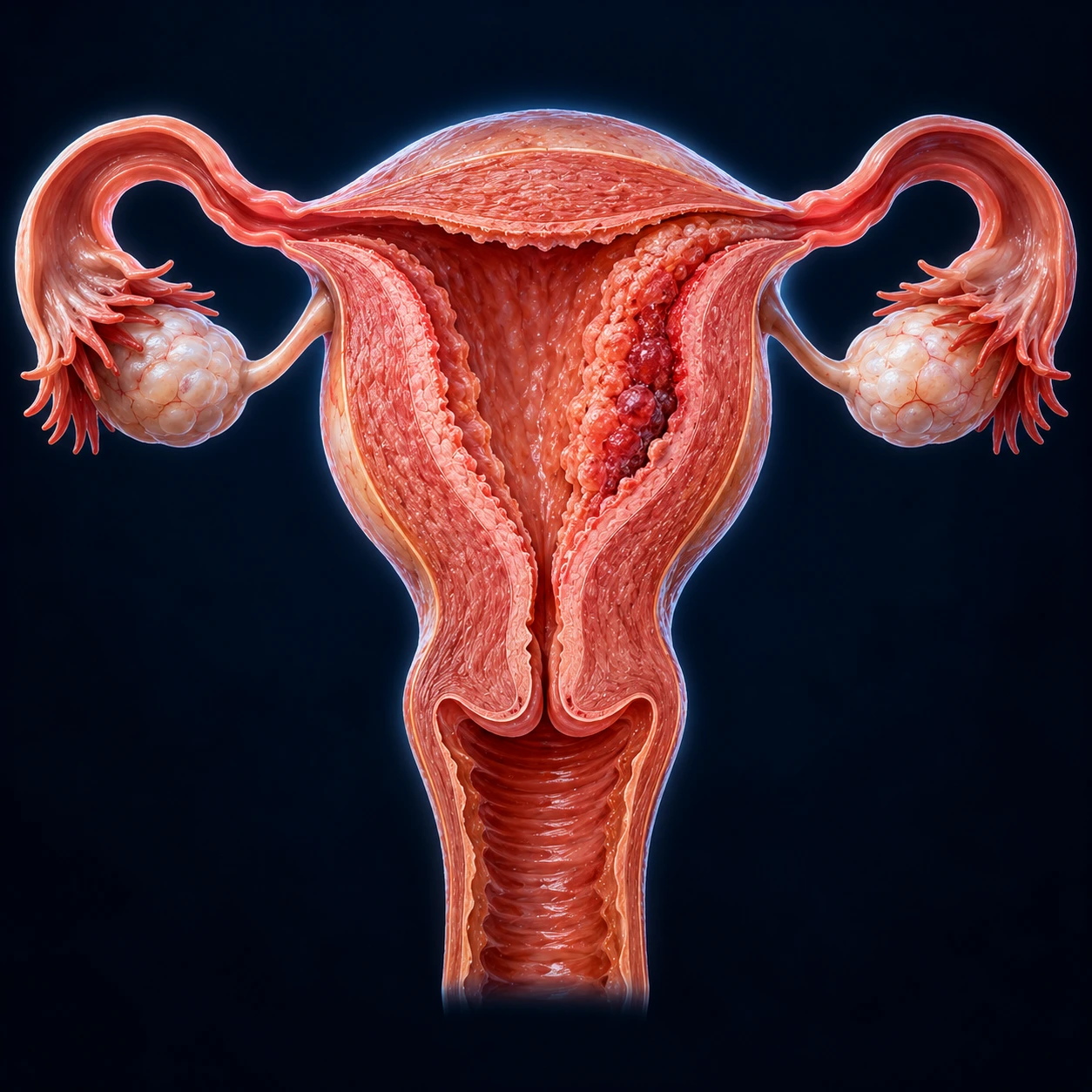
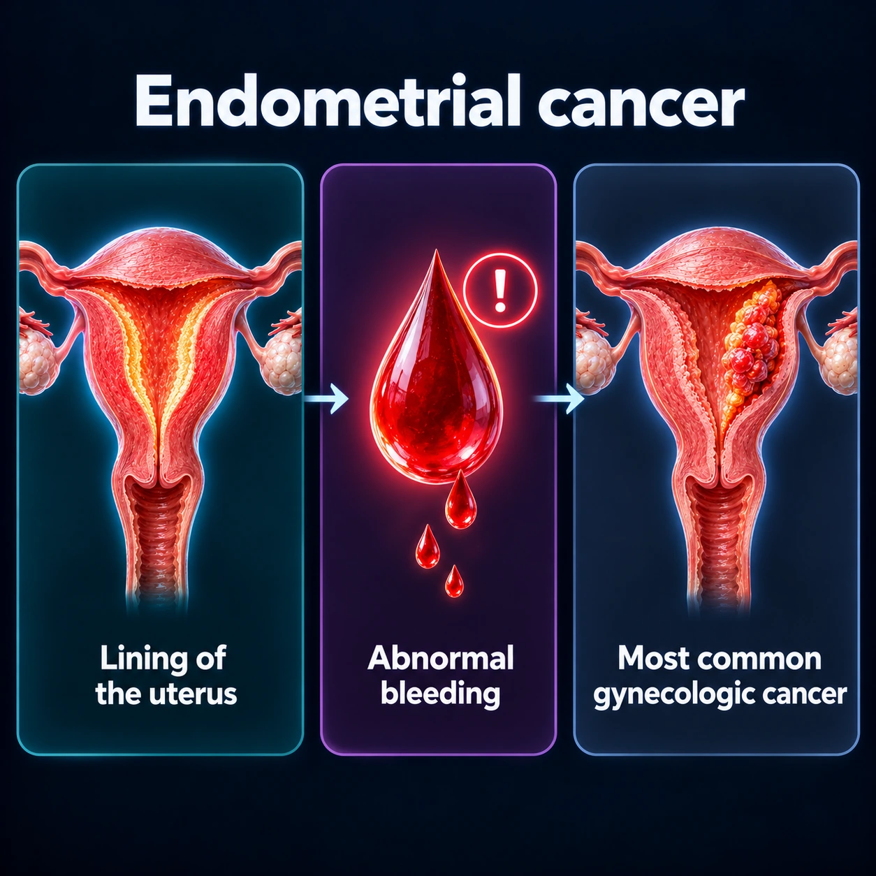
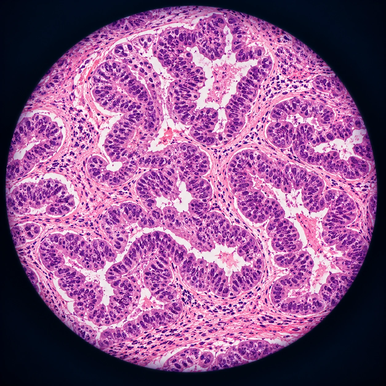
Table of Contents
1. Overview
Endometrial cancer is cancer of the endometrium — the lining of the uterus (womb) that thickens each month and sheds as a menstrual period. It is the most common gynecologic cancer in the United States, with roughly 67,000 new cases and about 13,000 deaths each year. People often call it “uterine cancer,” and the two terms are used almost interchangeably, because the great majority of cancers that start in the body of the uterus begin in the endometrium.
There is good news and bad news in the same sentence. The good news: about 9 in 10 women with endometrial cancer notice abnormal vaginal bleeding early, which is exactly why most cases are caught at an early, highly curable stage. The bad news: endometrial cancer is one of the few cancers in the U.S. whose incidence AND death rate are both rising, driven largely by the obesity epidemic and an aging population. So the disease is becoming more common even as we get better at treating it.
The single most important idea on this page is a hormone story. Most endometrial cancer is driven by estrogen that is not balanced by progesterone — what doctors call unopposed estrogen. Understanding that one mechanism explains almost every risk factor, every prevention strategy, and the logic behind the most common treatment. We will explain it in plain language below.
2. Epidemiology
Endometrial cancer is the fourth most common cancer in women in the United States and the most common cancer of the female reproductive tract — more common than ovarian and cervical cancer combined. A woman’s lifetime risk is roughly 1 in 36 (about 3%). Most cases are diagnosed after menopause, with a median age around 60–63 years, although a meaningful minority of cases occur in younger women, particularly those with obesity or polycystic ovary syndrome.
What makes endometrial cancer unusual among major cancers is its trajectory. While many cancers (lung, colorectal, stomach) are becoming less deadly over time, endometrial cancer incidence in the U.S. has been rising by roughly 1–2% per year, and — unlike most cancers — mortality is rising too. Two forces drive this: the obesity epidemic (more women carry the metabolic risk factor), and a relative increase in aggressive, non–estrogen-driven subtypes that are harder to treat.
There is a stark and honest racial disparity that deserves to be stated plainly. Black women in the U.S. are diagnosed at similar or slightly lower rates than White women but die from endometrial cancer at roughly twice the rate. The reasons are layered: Black women are more likely to develop the aggressive serous and other high-grade (“non-endometrioid”) subtypes, are more often diagnosed at a later stage, and face unequal access to timely biopsy, specialist surgery, and clinical trials. This gap is one of the widest racial survival gaps in all of oncology, and closing it is an active priority in research and policy.
3. Pathophysiology
To understand endometrial cancer, picture the endometrium as a lawn that responds to two hormones. Estrogen is the fertilizer — it tells the lining to grow and thicken. Progesterone is the gardener — after ovulation it matures the lining, stops the growth, and prepares it either to support a pregnancy or to shed cleanly as a period. In a normal monthly cycle, estrogen builds the lining in the first half and progesterone reins it in during the second half.
Now imagine fertilizer with no gardener. When estrogen acts on the endometrium month after month without progesterone to balance it, the lining keeps proliferating. Over years, that relentless growth can progress through a recognized sequence: normal endometrium → endometrial hyperplasia (overgrowth) → hyperplasia with atypia (overgrowth with abnormal-looking cells) → endometrial cancer. The crucial point: hyperplasia with atypia is a true precancer. Left untreated, a large fraction progresses to or already harbors cancer, which is why it is treated aggressively (see Treatment).
Where does the unopposed estrogen come from in a postmenopausal woman who no longer ovulates? Often from her own body fat. Fat tissue (adipose tissue) is hormonally active. It contains the enzyme aromatase, which converts androgens (made by the adrenal glands and ovaries) into estrogen. More fat means more aromatase, more estrogen, and — because there is no ovulation and therefore no progesterone after menopause — that estrogen is entirely unopposed. This single biochemical fact links obesity directly to endometrial cancer.
Pathologists historically divided endometrial cancer into two clinical types, a framework introduced by Bokhman in 1983 that remains a useful first cut:
- Type 1 (about 80% of cases): the estrogen-driven, “endometrioid” type. Tends to be lower grade, caught earlier, and has a good prognosis. This is the type tied to obesity, polycystic ovary syndrome (PCOS), and the hormone story above.
- Type 2 (about 20%): not driven by estrogen. Includes serous carcinoma and clear cell carcinoma, which are higher grade, more likely to have spread at diagnosis, and carry a worse prognosis. Serous tumors frequently carry mutations in the p53 tumor-suppressor gene. These tumors can arise even in thin women without classic risk factors.
This two-type model is honest but simplified. Modern molecular profiling (see Diagnosis and Recent Research) has refined it considerably.
4. Etiology and Risk Factors
Almost every well-established risk factor traces back to the same root: more lifetime exposure to estrogen unopposed by progesterone. Hold that lens up and the list below stops looking random.
- Obesity — the strongest modifiable risk factor. Each increase in body-mass index meaningfully raises risk because fat tissue manufactures estrogen via aromatase. Risk can be several-fold higher in women with severe obesity.
- Polycystic ovary syndrome (PCOS) and chronic anovulation — when a woman does not ovulate regularly, she does not make the monthly surge of progesterone, so her endometrium sees estrogen without its counterbalance for years. This is why younger women with polycystic ovary syndrome, especially with infrequent or absent periods, carry elevated risk.
- Early menarche and late menopause — more menstrual years means more cumulative estrogen exposure.
- Never having been pregnant (nulliparity) — pregnancy is a prolonged progesterone-rich state that gives the endometrium a protective “rest” from unopposed estrogen.
- Estrogen-only hormone replacement therapy (HRT) in a woman who still has her uterus — a textbook example of unopposed estrogen. (Adding a progestin removes this excess risk; see Prevention.)
- Tamoxifen — this breast-cancer drug blocks estrogen in breast tissue but acts like estrogen on the uterine lining, modestly raising endometrial cancer risk. The benefit for breast cancer usually outweighs this, and abnormal bleeding on tamoxifen is a recognized warning sign that doctors evaluate.
- Type 2 diabetes and insulin resistance — high insulin and related growth factors promote endometrial cell growth and are tightly intertwined with obesity.
- Older age — most cases occur after menopause.
- Lynch syndrome — the most important inherited cause (see below).
Lynch syndrome deserves its own paragraph. It is an inherited condition caused by a fault in one of the DNA mismatch-repair (MMR) genes (MLH1, MSH2, MSH6, PMS2) and accounts for roughly 3% of all endometrial cancers — and a higher share among women diagnosed young. The risk depends on the gene: in a 2011 French study, the risk of endometrial cancer by age 70 was about 54% with an MLH1 fault, 21% with MSH2 and 16% with MSH6, while the risk by age 40 did not exceed 2%; and for many of these women, endometrial cancer is the first cancer they develop — a “sentinel” cancer that can lead to testing of the whole family. Lynch syndrome also dramatically raises the risk of colorectal cancer and others, so identifying it protects not just the patient but her parents, siblings, and children. Because of this, most pathology labs now perform universal MMR/microsatellite-instability testing on every endometrial tumor, and abnormal results trigger referral for genetic counseling and testing. Women diagnosed before age 50, or with a strong family history of colon or uterine cancer, are especially likely to be offered testing.
5. Clinical Presentation
The key fact on this page: any bleeding after menopause is treated as abnormal until proven otherwise. Even a single episode of spotting, even a few drops, even “just once”, is evaluated by doctors. About 90% of women with endometrial cancer have abnormal bleeding as their first and often only symptom, which is precisely why this cancer is so often caught early and cured.
The reassurance: the large majority of postmenopausal bleeding turns out to be benign — thinning of the lining (atrophy), polyps, or hormonal causes. Only a minority is cancer. But there is no way to tell the difference without an evaluation, and waiting only lets a treatable cancer grow. Because postmenopausal bleeding is the first sign in about 9 in 10 women with this cancer, clinics evaluate it as urgent, and prompt evaluation is how most of these cancers are found while still curable.
In premenopausal women, the warning signs are different and easier to overlook because some irregular bleeding is normal. Patterns that warrant evaluation include: bleeding between periods, unusually heavy or prolonged periods, and a marked change in a long-standing pattern — especially in a woman with obesity, polycystic ovary syndrome, infrequent periods, or a family history of Lynch-related cancers. A young woman with polycystic ovary syndrome who goes many months without a period is exactly the person whose endometrium may be quietly overgrowing, and persistent abnormal bleeding in that setting is a recognized warning sign, not “just hormones.”
Other, later symptoms can include watery or blood-tinged vaginal discharge, pelvic pain or pressure, and pain with intercourse — but these usually appear after bleeding has already started.
6. Diagnosis
The work-up for suspected endometrial cancer is relatively quick and mostly done in the office.
- Transvaginal ultrasound is often the first test. It measures the endometrial stripe — the thickness of the lining. In a postmenopausal woman with bleeding, a thin stripe (commonly a threshold around 4–5 mm or less) makes cancer very unlikely, while a thickened lining prompts a biopsy. (This threshold does not apply to premenopausal women, whose lining normally varies through the cycle.)
- Endometrial biopsy is the key test and the one that gives the diagnosis. A thin flexible tube is passed through the cervix to sample the lining — no anesthesia, done in a few minutes in the office. An honest heads-up: it is crampy. Many women find the brief cramp intense, similar to a strong menstrual cramp. Many women take ibuprofen beforehand to ease the cramp, and the discomfort passes quickly. The biopsy gives the diagnosis in a few uncomfortable minutes.
- Hysteroscopy with dilation and curettage (D&C) is used when the office biopsy is inconclusive, technically impossible, or when bleeding continues despite a “normal” biopsy. A thin camera (hysteroscope) lets the surgeon look directly inside the uterus and take targeted samples, usually under sedation or anesthesia.
Once cancer is confirmed, the pathologist determines the type and grade, and the tumor is tested with molecular and immunohistochemical markers. Two are routine now: MMR/microsatellite-instability testing (to flag Lynch syndrome and to predict response to immunotherapy) and p53 staining (to flag aggressive serous-like tumors). Imaging such as magnetic resonance imaging (MRI) or computed tomography (CT) may be added to assess spread, but endometrial cancer is ultimately surgically staged — the true extent is confirmed at the time of the operation.
7. Treatment
Surgery is the cornerstone. The standard operation is a hysterectomy (removal of the uterus and cervix) plus bilateral salpingo-oophorectomy (removal of both fallopian tubes and ovaries, abbreviated BSO), usually with sentinel lymph node mapping to check whether cancer has reached the lymph nodes.
Importantly — and unlike cervical cancer — minimally invasive surgery (laparoscopic or robotic) is fully appropriate for endometrial cancer. The landmark Gynecologic Oncology Group (GOG) LAP2 trial found that laparoscopic staging had fewer moderate-to-severe complications after surgery than open surgery (14% vs 21%) and far fewer hospital stays longer than 2 days (52% vs 94%). Its later report on cancer outcomes found a 3-year recurrence rate of 11.4% with laparoscopy and 10.2% with open surgery — just short of the trial’s formal test of non-inferiority — and nearly identical 5-year survival (89.8% in both groups). The FIRES trial validated sentinel node mapping: among 36 women whose lymph nodes held cancer and who had at least one sentinel node mapped, the sentinel nodes found the cancer in 35 (sensitivity 97.2%, negative predictive value 99.6%), and at least one sentinel node was mapped in 86% of women. Sentinel mapping spares many women a full lymph-node dissection and its risk of leg swelling (lymphedema). So most women today can have a smaller, gentler operation.
After surgery, treatment is tailored to risk:
- Radiation. For early-stage tumors with intermediate-to-high-risk features, vaginal brachytherapy (internal radiation to the top of the vagina) reduces local recurrence with fewer side effects than whole-pelvis radiation. The PORTEC-2 and GOG-249 trials helped define who benefits, showing brachytherapy is enough for many women who once received broader pelvic radiation. In PORTEC-2 (427 women with high-intermediate-risk disease), the 5-year rate of cancer returning in the vagina was 1.8% with vaginal brachytherapy and 1.6% with whole-pelvis radiation, while mild-to-moderate bowel side effects at the end of radiation were 12.6% versus 53.8%. The earlier PORTEC-1 trial had found that pelvic radiation after surgery lowered pelvic and vaginal recurrence (4% vs 14% at 5 years) without improving 5-year survival (81% vs 85%).
- Chemotherapy. For higher-risk or advanced disease, carboplatin plus paclitaxel is the standard backbone. The PORTEC-3 trial (660 women with high-risk disease) tested adding chemotherapy to radiation. Its 2018 report found better 5-year failure-free survival (75.5% vs 68.6%) but no significant gain in overall survival (81.8% vs 76.7%, p = 0.11); the longer 2019 analysis found improved 5-year overall survival (81.4% vs 76.1%, hazard ratio 0.70, p = 0.034), and its authors pointed especially to women with stage III or serous cancers.
- Immunotherapy. This is the most exciting recent advance, particularly for tumors that are mismatch-repair deficient (dMMR / MSI-high). The RUBY trial showed that adding the immunotherapy drug dostarlimab to chemotherapy dramatically improved outcomes in advanced disease, with the largest benefit in dMMR tumors. Its 2024 overall-survival analysis (494 patients) found that adding dostarlimab lowered the risk of death by 31% in all patients (hazard ratio 0.69) and by 68% in mismatch-repair-deficient tumors (hazard ratio 0.32), with a smaller trend in mismatch-repair-proficient tumors (hazard ratio 0.79). The NRG-GY018 trial showed a similar, striking benefit for pembrolizumab plus chemotherapy. For previously treated advanced cancers, pembrolizumab plus lenvatinib (the KEYNOTE-775 regimen) is an option regardless of MMR status. This is why the molecular testing described above directly changes treatment.
Fertility-sparing treatment is possible for a carefully selected group of young women who want to preserve the chance of pregnancy. The criteria are strict: a low-grade (grade 1) endometrioid cancer confined to the lining, with no evidence of deeper invasion or spread on imaging, and a willingness to be monitored closely. Instead of hysterectomy, these women are treated with high-dose progestin therapy — oral progestins and/or a levonorgestrel intrauterine device (IUD) — with repeat biopsies every few months. In pooled data, roughly three-quarters of well-selected women achieve a complete response, but relapse is common (a substantial fraction, often around a third, recur), so this is a temporary, closely watched strategy — not a cure — and hysterectomy is generally recommended once childbearing is complete.
Finally, the precancer matters here too. Endometrial hyperplasia with atypia is most definitively treated by hysterectomy, but in women who want to preserve fertility or who cannot have surgery, progestin therapy — particularly the levonorgestrel IUD — can reverse it in a large share of cases and is a well-established alternative.
8. Complications
Complications come from both the disease and its treatment.
- From the cancer: if not caught early, spread to the cervix, ovaries, lymph nodes, abdominal lining, lungs, or other organs (see Metastatic Cancers). Anemia from chronic bleeding is common at presentation.
- From surgery: the usual surgical risks (bleeding, infection, blood clots), plus immediate surgical menopause in premenopausal women whose ovaries are removed — hot flashes, bone-density loss, and vaginal dryness that may need management.
- Lymphedema: swelling of the legs after lymph-node removal. Sentinel-node mapping was specifically adopted to reduce this.
- From radiation: vaginal narrowing or dryness, bladder and bowel irritation; pelvic radiation carries more of these than vaginal brachytherapy.
- From chemotherapy and immunotherapy: the usual chemo effects (low blood counts, neuropathy, hair loss); immunotherapy can cause immune-related inflammation of the thyroid, gut, skin, or other organs, which is usually manageable but needs monitoring.
- Emotional and sexual impact: loss of fertility, changes in sexual function, and the psychological weight of a cancer diagnosis are real and deserve support — not afterthoughts.
9. Prognosis
Overall, endometrial cancer has a favorable prognosis compared with most cancers, precisely because early bleeding leads to early diagnosis. The honest numbers:
- Overall 5-year survival is roughly 81%.
- When caught while still confined to the uterus (localized), 5-year survival is about 95%. This is the majority of cases.
- Survival drops with spread — to roughly the high-60s percent for regional (lymph-node) involvement and the teens to ~20% for distant metastatic disease.
Prognosis depends heavily on type, grade, and molecular profile, not just stage. Serous and clear cell carcinomas and p53-abnormal (copy-number-high) tumors do worse stage-for-stage, while tumors with POLE mutations do remarkably well (see below). And the survival statistics are not experienced equally: the racial disparity described earlier means averages can hide meaningfully worse outcomes for Black women, a gap driven by tumor biology and access rather than by any difference in the women themselves.
10. Prevention
Several of the strongest risk factors are modifiable, and the protective strategies follow directly from the hormone story.
- Weight management is the single most powerful lever. Because body fat manufactures the estrogen that drives most endometrial cancer, achieving and maintaining a healthy weight lowers risk more than any other lifestyle change. Physical activity helps independently as well. This is the strongest evidence-based prevention message on this page.
- Combined oral contraceptives (“the pill”) are strongly protective. They supply progestin, which protects the endometrium. The protection is large and durable: long-term use is associated with roughly a 30–50% reduction in endometrial cancer risk, and — this is the genuinely remarkable part — the protection persists for decades after stopping the pill. This is an honest benefit worth weighing alongside the pill’s other risks and benefits with a clinician.
- The levonorgestrel IUD is also protective, because it delivers progestin directly to the uterine lining — useful for women who cannot take the pill, and the same device used to treat precancer.
- Protecting the endometrium in polycystic ovary syndrome. In women with polycystic ovary syndrome who do not have regular periods, the endometrium can be protected — for example with cyclic or continuous progestin or a levonorgestrel IUD — so the lining is not exposed to years of unopposed estrogen.
- Progestin paired with estrogen in hormone replacement therapy for women who still have a uterus. Estrogen-only hormone replacement therapy is used after hysterectomy, but in a woman with a uterus, the estrogen is balanced by a progestin, which removes the excess endometrial cancer risk of estrogen alone.
- Family history and Lynch testing. Women with Lynch syndrome can be offered enhanced surveillance and, when childbearing is complete, risk-reducing hysterectomy — a powerful prevention option for the highest-risk group.
11. Recent Research and Advances
The biggest shift in how doctors think about endometrial cancer came from molecular classification. In 2013, The Cancer Genome Atlas (TCGA) project showed that endometrial cancer is not two types but four distinct molecular groups, each with a different prognosis. A practical, clinic-friendly version of this system (often called ProMisE, the Proactive Molecular Risk Classifier for Endometrial Cancer) now sorts tumors into:
- POLE-ultramutated — tumors with a mutation in the POLE gene that gives them an enormous number of mutations. Counterintuitively, these have an excellent prognosis, so well that researchers are studying whether some of these patients can safely skip extra treatment after surgery.
- Mismatch-repair deficient (MMRd / MSI-high) — intermediate prognosis, and crucially the group most likely to respond to immunotherapy. This overlaps with Lynch syndrome.
- Copy-number-high (p53-abnormal) — includes most serous cancers; the worst-prognosis group, the one most likely to need chemotherapy and the most active research target.
- Copy-number-low (“no specific molecular profile”) — the large middle group with intermediate outcomes.
Why does this matter to a patient? Because two tumors that look identical under the microscope can behave completely differently depending on their molecular group — and treatment is increasingly chosen on that basis. The same testing that identifies the immunotherapy-responsive MMRd group also flags Lynch syndrome. The arrival of immunotherapy (dostarlimab in RUBY, pembrolizumab in NRG-GY018, pembrolizumab plus lenvatinib in KEYNOTE-775) represents the first major survival improvement in advanced endometrial cancer in decades, and the benefit is greatest in exactly the molecular group the new testing identifies. Research now focuses on de-escalating treatment for the favorable groups (sparing women unnecessary radiation and chemotherapy) while intensifying it for the aggressive p53-abnormal group.
In 2023 the International Federation of Gynecology and Obstetrics (FIGO) rewrote its staging system for endometrial cancer — the first update since 2009 — adding the tumor’s cell type and, when known, its molecular group to the stage itself. Molecular testing (POLE-mutated, mismatch-repair deficient, no specific molecular profile, p53-abnormal) is encouraged for all endometrial cancers, and in stage I and II disease a p53-abnormal result moves the cancer up a stage while a POLE mutation moves it down.
Key Research Papers
Historical Background
Endometrial cancer played a pivotal role in the history of women’s health and pharmacovigilance. In the 1970s, an epidemic of endometrial cancer followed the popular use of estrogen-only replacement therapy in menopausal women. In 1975, two landmark papers in the New England Journal of Medicine — by Smith and colleagues and by Ziel and Finkle — established that exogenous (estrogen-only) hormone therapy sharply increased endometrial cancer risk. The response — adding progestins to estrogen therapy for women with a uterus — remains standard practice and is one of medicine’s clearest examples of identifying and reversing a drug-caused cancer. In 1983, Bokhman proposed the influential type I / type II framework distinguishing estrogen-driven from non–estrogen-driven tumors. Three decades later, in 2013, The Cancer Genome Atlas reclassified the disease into four molecular subgroups, ushering in today’s era of biology-guided treatment.
- Ziel HK, Finkle WD. Increased Risk of Endometrial Carcinoma among Users of Conjugated Estrogens. New England Journal of Medicine. 1975;293(23):1167-1170. — doi:10.1056/NEJM197512042932303
- Smith DC, Prentice R, Thompson DJ, Herrmann WL. Association of Exogenous Estrogen and Endometrial Carcinoma. New England Journal of Medicine. 1975;293(23):1164-1167. — doi:10.1056/NEJM197512042932302
- Bokhman JV. Two pathogenetic types of endometrial carcinoma. Gynecologic Oncology. 1983;15(1):10-17. — doi:10.1016/0090-8258(83)90111-7
- The Cancer Genome Atlas Research Network. Integrated genomic characterization of endometrial carcinoma. Nature. 2013;497(7447):67-73. — doi:10.1038/nature12113
- Talhouk A, McConechy MK, Leung S, et al. A clinically applicable molecular-based classification for endometrial cancers. British Journal of Cancer. 2015;113(2):299-310. — doi:10.1038/bjc.2015.190
- Crosbie EJ, Kitson SJ, McAlpine JN, et al. Endometrial cancer. The Lancet. 2022;399(10333):1412-1428. — doi:10.1016/S0140-6736(22)00323-3
- Lu KH, Broaddus RR. Endometrial Cancer. New England Journal of Medicine. 2020;383(21):2053-2064. — doi:10.1056/NEJMra1514010
- Clarke MA, Devesa SS, Harvey SV, Wentzensen N. Hysterectomy-Corrected Uterine Corpus Cancer Incidence Trends and Differences in Relative Survival Reveal Racial Disparities and Rising Rates of Nonendometrioid Cancers. Journal of Clinical Oncology. 2019;37(22):1895-1908. — doi:10.1200/JCO.19.00151
- Renehan AG, Tyson M, Egger M, Heller RF, Zwahlen M. Body-mass index and incidence of cancer: a systematic review and meta-analysis of prospective observational studies. The Lancet. 2008;371(9612):569-578. — doi:10.1016/S0140-6736(08)60269-X
- Collaborative Group on Epidemiological Studies on Endometrial Cancer. Endometrial cancer and oral contraceptives: an individual participant meta-analysis of 27 276 women with endometrial cancer from 36 epidemiological studies. The Lancet Oncology. 2015;16(9):1061-1070. — doi:10.1016/S1470-2045(15)00212-0
- Bonadona V, Bonaiti B, Olschwang S, et al. Cancer Risks Associated With Germline Mutations in MLH1, MSH2, and MSH6 Genes in Lynch Syndrome. JAMA. 2011;305(22):2304-2310. — doi:10.1001/jama.2011.743
- Creutzberg CL, van Putten WL, Koper PC, et al. Surgery and postoperative radiotherapy versus surgery alone for patients with stage-1 endometrial carcinoma: multicentre randomised trial. PORTEC Study Group. Post Operative Radiation Therapy in Endometrial Carcinoma. The Lancet. 2000;355(9213):1404-1411. — doi:10.1016/s0140-6736(00)02139-5
- Nout RA, Smit VT, Putter H, et al. Vaginal brachytherapy versus pelvic external beam radiotherapy for patients with endometrial cancer of high-intermediate risk (PORTEC-2): an open-label, non-inferiority, randomised trial. The Lancet. 2010;375(9717):816-823. — doi:10.1016/S0140-6736(09)62163-2
- Walker JL, Piedmonte MR, Spirtos NM, et al. Laparoscopy Compared With Laparotomy for Comprehensive Surgical Staging of Uterine Cancer: Gynecologic Oncology Group Study LAP2. Journal of Clinical Oncology. 2009;27(32):5331-5336. — doi:10.1200/JCO.2009.22.3248
- Walker JL, Piedmonte MR, Spirtos NM, et al. Recurrence and survival after random assignment to laparoscopy versus laparotomy for comprehensive surgical staging of uterine cancer: Gynecologic Oncology Group LAP2 Study. Journal of Clinical Oncology. 2012;30(7):695-700. — doi:10.1200/JCO.2011.38.8645
- Rossi EC, Kowalski LD, Scalici J, et al. A comparison of sentinel lymph node biopsy to lymphadenectomy for endometrial cancer staging (FIRES trial): a multicentre, prospective, cohort study. The Lancet Oncology. 2017;18(3):384-392. — doi:10.1016/S1470-2045(17)30068-2
- de Boer SM, Powell ME, Mileshkin L, et al. Adjuvant chemoradiotherapy versus radiotherapy alone for women with high-risk endometrial cancer (PORTEC-3): final results of an international, open-label, multicentre, randomised, phase 3 trial. The Lancet Oncology. 2018;19(3):295-309. — doi:10.1016/S1470-2045(18)30079-2
- de Boer SM, Powell ME, Mileshkin L, et al. Adjuvant chemoradiotherapy versus radiotherapy alone in women with high-risk endometrial cancer (PORTEC-3): patterns of recurrence and post-hoc survival analysis of a randomised phase 3 trial. The Lancet Oncology. 2019;20(9):1273-1285. — doi:10.1016/S1470-2045(19)30395-X
- Mirza MR, Chase DM, Slomovitz BM, et al. Dostarlimab for Primary Advanced or Recurrent Endometrial Cancer. New England Journal of Medicine. 2023;388(23):2145-2158. — doi:10.1056/NEJMoa2216334
- Powell MA, Bjørge L, Willmott L, et al. Overall survival in patients with endometrial cancer treated with dostarlimab plus carboplatin-paclitaxel in the randomized ENGOT-EN6/GOG-3031/RUBY trial. Annals of Oncology. 2024;35(8):728-738. — doi:10.1016/j.annonc.2024.05.546
- Eskander RN, Sill MW, Beffa L, et al. Pembrolizumab plus Chemotherapy in Advanced Endometrial Cancer. New England Journal of Medicine. 2023;388(23):2159-2170. — doi:10.1056/NEJMoa2302312
- Makker V, Colombo N, Casado Herráez A, et al. Lenvatinib plus Pembrolizumab for Advanced Endometrial Cancer. New England Journal of Medicine. 2022;386(5):437-448. — doi:10.1056/NEJMoa2108330
- Berek JS, Matias-Guiu X, Creutzberg C, et al. FIGO staging of endometrial cancer: 2023. Journal of Gynecologic Oncology. 2023;34(5):e85. — doi:10.3802/jgo.2023.34.e85
PubMed Topic Searches
Endometrial cancer is a fast-moving research field, with active work on molecular classification, immunotherapy, fertility-sparing care, and the racial survival gap. The PubMed searches below open current peer-reviewed literature in a new tab so you can explore the evidence yourself.
- PubMed: Endometrial cancer treatment
- PubMed: Endometrial cancer molecular classification
- PubMed: Endometrial cancer immunotherapy (dostarlimab)
- PubMed: Endometrial cancer and obesity
- PubMed: Endometrial cancer and Lynch syndrome
- PubMed: Endometrial hyperplasia progestin treatment
- PubMed: Endometrial cancer fertility-sparing therapy
- PubMed: Endometrial cancer sentinel lymph node
- PubMed: Endometrial cancer racial disparities
- PubMed: Endometrial cancer postmenopausal bleeding
- PubMed: Endometrial cancer, oral contraceptives, prevention
- PubMed: Serous endometrial carcinoma and p53