Pompe Disease
- Overview and Classification
- Pathophysiology and GAA Deficiency
- Classic Infantile-Onset Pompe Disease (IOPD)
- Late-Onset Pompe Disease (LOPD)
- Cardiac Manifestations
- Respiratory Failure: The Defining Feature
- Diagnosis and Newborn Screening
- Treatment: Enzyme Replacement Therapy
- Treatment Outcomes and Monitoring
- Key Research Papers
- Connections
Overview and Classification
Pompe disease — also known as glycogen storage disease type II (GSD II) or acid maltase deficiency — is a rare, autosomal recessive metabolic disorder caused by mutations in the GAA gene located on chromosome 17q25.3. This gene encodes the lysosomal enzyme acid alpha-glucosidase (acid maltase), which is responsible for breaking down glycogen within lysosomes. When GAA activity is absent or severely reduced, glycogen accumulates progressively inside lysosomes throughout the body, with the most devastating consequences occurring in striated muscle — both cardiac and skeletal.
The disease was first described in 1932 by Dutch pathologist J.C. Pompe, who documented a seven-month-old infant with massive cardiomegaly and profound generalized weakness — a clinical picture now recognized as classic infantile-onset Pompe disease. A 1999 Dutch genetic screen of newborns predicted a frequency of about 1 in 40,000 births for all forms together — about 1 in 138,000 for the infantile form and 1 in 57,000 for the adult form — two to four times higher than earlier estimates, though population-based newborn screening data suggest the overall prevalence may be higher.
Pompe disease is categorized into two broad clinical forms based on age of onset and residual enzyme activity: infantile-onset Pompe disease (IOPD) and late-onset Pompe disease (LOPD). These are not truly distinct subtypes but rather represent a continuum of severity — from the severe classic infantile form presenting at birth with near-zero enzyme activity, to mild adult-onset disease with years of preserved function. Pompe disease holds a unique position among the glycogen storage diseases as the only one with primary lysosomal involvement. It was also the first GSD to receive an approved enzyme replacement therapy: alglucosidase alfa received U.S. Food and Drug Administration (FDA) approval in 2006. Today, Pompe disease is included on the US Recommended Uniform Screening Panel (RUSP), with newborn screening active in more than 40 states.
Pathophysiology and GAA Deficiency
The fundamental defect in Pompe disease is mutation of the GAA gene, which encodes lysosomal acid alpha-glucosidase — the enzyme responsible for hydrolyzing glycogen within the lysosomal compartment. More than 600 distinct GAA mutations have been identified, ranging from null (nonsense, frameshift, large deletion) mutations producing no functional enzyme to missense mutations yielding residual enzyme activity. This allelic heterogeneity explains the wide clinical spectrum of the disease.
In the absence of adequate GAA activity, glycogen cannot be degraded within lysosomes and accumulates steadily in the lysosomes of virtually all tissues. The burden is highest in striated muscle — cardiac and skeletal — as well as smooth muscle. Lysosomes become progressively engorged with glycogen, leading to organelle enlargement, membrane rupture, and spillage of glycogen into the cytoplasm. Cellular dysfunction follows from both mechanical disruption and a secondary buildup of autophagic vacuoles that impairs the normal protein quality-control machinery of the cell.
The residual GAA enzyme activity strongly predicts clinical phenotype. Classic IOPD typically involves near-complete loss of function (less than 1% residual activity), while LOPD patients generally retain 1–20% of normal enzyme activity. Even a small amount of residual activity appears sufficient to prevent cardiac glycogen accumulation, which is why cardiomyopathy is absent in LOPD.
An important immunological concept in Pompe disease is "cross-reactive immunologic material" (CRIM) status. CRIM-positive patients make some endogenous GAA protein (even if non-functional) and therefore do not mount an immune response against infused recombinant enzyme. CRIM-negative patients produce no GAA protein at all and, when exposed to enzyme replacement therapy (ERT), develop high-titer antibodies against the infused enzyme — dramatically reducing its efficacy and potentially causing life-threatening infusion reactions. Prophylactic immune tolerance induction (ITI) using rituximab, methotrexate, and IVIG is now standard practice for CRIM-negative IOPD patients before initiating ERT.
Classic Infantile-Onset Pompe Disease (IOPD)
Classic infantile-onset Pompe disease (IOPD) is the most severe form, presenting within the first weeks to months of life — typically between one and six months of age. GAA enzyme activity is near zero (less than 1% of normal). Without treatment, IOPD is fatal in nearly all infants. In a natural-history study of 168 infants, symptoms began at a median age of 2 months, breathing support was first needed at a median 5.9 months, and death came at a median 8.7 months (range 0.3 to 73.4 months); only 25.7% were alive at 12 months and 12.3% at 18 months.
The hallmark of classic IOPD is the combination of profound generalized hypotonia and massive hypertrophic cardiomegaly. Affected infants present as "floppy babies" — severely hypotonic, with generalized muscle weakness, poor head control, absent deep tendon reflexes, and marked feeding difficulties from weak sucking and swallowing. The degree of weakness is often described as the most severe encountered in any metabolic myopathy.
The cardiomegaly of IOPD is a direct consequence of glycogen accumulation in cardiomyocytes, producing a hypertrophic cardiomyopathy that enlarges both ventricles. Chest radiograph reveals a massively enlarged cardiac silhouette — the "boot-shaped" or "egg-on-a-side" configuration — with a cardiothoracic ratio exceeding 0.5. The electrocardiogram shows two pathognomonic findings: a markedly shortened PR interval (caused by glycogen infiltration of the atrioventricular conduction system, which accelerates conduction) and extremely high-voltage QRS complexes (from massive ventricular hypertrophy). This pattern superficially resembles Wolff-Parkinson-White syndrome but lacks the delta wave and pre-excitation pattern of true WPW.
Additional features include hepatomegaly from hepatic glycogen storage, macroglossia from glycogen accumulation in the tongue, and rapidly progressive respiratory failure requiring mechanical ventilatory support. Early initiation of enzyme replacement therapy (ideally within the first weeks of life through newborn screening) produces dramatic regression of cardiomyopathy and substantially prolonged survival, though motor and respiratory outcomes vary depending on how quickly treatment begins and the patient's CRIM status.
Late-Onset Pompe Disease (LOPD)
Late-onset Pompe disease encompasses a broad spectrum of patients with symptom onset from childhood through late adulthood — including individuals first diagnosed after age 60. The unifying feature is residual GAA enzyme activity (typically 1–20% of normal), which is sufficient to prevent cardiac glycogen accumulation but insufficient to protect skeletal and respiratory muscles from progressive glycogen buildup over decades.
The cardinal manifestation of LOPD is proximal limb-girdle weakness, particularly of the hip flexors, hip extensors, and thigh muscles, followed by shoulder girdle involvement. Patients report difficulty rising from low chairs, climbing stairs, and lifting arms overhead. On examination, many demonstrate a Gowers maneuver (using arms to "walk up" the legs to rise from the floor), scapular winging, and absent or diminished deep tendon reflexes in the legs. A subset develops rigid spine syndrome.
Crucially, respiratory failure in LOPD often precedes or overshadows the limb weakness in clinical presentation. Diaphragm involvement is disproportionately severe relative to limb muscles in Pompe disease. Patients may present to pulmonologists with unexplained restrictive respiratory failure, to sleep specialists with obstructive or central sleep apnea and nocturnal hypoventilation, or to neurologists with a limb-girdle muscular dystrophy phenotype. The characteristic history includes orthopnea — difficulty breathing when lying flat — due to the inability of the weakened diaphragm to lift abdominal contents in the supine position. A drop in forced vital capacity (FVC) of more than 10% from upright to supine is highly suggestive of diaphragm weakness.
One of the most frustrating aspects of LOPD is the long diagnostic delay — averaging five to ten years or more from symptom onset to correct diagnosis. Patients are commonly misdiagnosed with limb-girdle muscular dystrophy, polymyositis, or idiopathic respiratory failure. Awareness of LOPD among pulmonologists, neurologists, and sleep medicine specialists is critical for timely diagnosis and treatment initiation.
Cardiac Manifestations
Cardiac involvement in Pompe disease differs sharply between the two clinical forms and has important diagnostic and therapeutic implications. In classic IOPD, hypertrophic cardiomyopathy is a defining and life-threatening feature. Glycogen-laden cardiomyocytes throughout both ventricles produce massive concentric hypertrophy that can cause left ventricular outflow tract obstruction, right ventricular outflow tract obstruction, and severe diastolic dysfunction. Echocardiography in untreated IOPD reveals dramatically increased left ventricular mass index — often more than ten times the upper limit of normal — and systolic dysfunction in advanced cases.
The electrocardiographic signature of IOPD cardiomyopathy is both pathognomonic and diagnostically useful. The shortened PR interval (below 0.10–0.12 seconds) results from glycogen infiltration of the atrioventricular node, which accelerates atrioventricular conduction. The massively elevated QRS voltages reflect the degree of ventricular hypertrophy. Together these findings — short PR interval plus high QRS voltage — in an infant with hypotonia and cardiomegaly are the classic signal for GAA enzyme activity testing. Ventricular arrhythmias, including ventricular tachycardia, may occur in severely affected infants. This pattern is distinguished from true Wolff-Parkinson-White syndrome: WPW shows a delta wave and a slurred QRS upstroke representing true pre-excitation, which is absent in IOPD.
Enzyme replacement therapy produces dramatic and relatively rapid reversal of IOPD cardiomyopathy. Left ventricular mass normalizes within six to twelve months of ERT initiation in most treated infants, and systolic function recovers. This cardiac response is one of the most striking benefits of ERT in IOPD and translates directly to reduced early mortality.
In LOPD, cardiomyopathy is absent — this is a key distinguishing feature from IOPD. Late-onset patients may develop minor valvular thickening or subtle conduction abnormalities, but clinically significant cardiac disease does not occur. Serial echocardiography is a cornerstone of IOPD monitoring but is not routinely required for cardiac surveillance in LOPD.
Respiratory Failure: The Defining Feature
Respiratory failure is the defining and ultimately lethal complication of Pompe disease across both clinical forms. In untreated IOPD, progressive respiratory muscle weakness combined with pulmonary compression from the massively enlarged heart leads to respiratory failure requiring mechanical ventilation, typically within the first year of life. Even with ERT, respiratory function in IOPD is one of the outcomes that responds less completely than cardiac function, and many treated patients require eventual ventilatory support.
In LOPD, diaphragm weakness is disproportionately and characteristically severe relative to the degree of limb weakness. The diaphragm is rich in slow-twitch type I muscle fibers, which are preferentially affected by lysosomal glycogen accumulation in Pompe disease. The physiological consequence of diaphragm weakness is greatest in the supine position: lying flat requires the diaphragm to both contract downward and resist the upward pressure of abdominal viscera, a task that an already-weakened diaphragm cannot sustain. This produces the characteristic symptom of orthopnea — breathlessness when supine — and leads to nocturnal hypoventilation beginning in REM sleep (when accessory respiratory muscles are most inhibited) and later progressing to NREM hypoventilation and daytime hypercapnia.
The trajectory of respiratory decline in LOPD typically follows a predictable path: nocturnal hypoxemia and hypercapnia → morning headaches, unrefreshing sleep, and daytime somnolence → daytime hypercapnia → acute-on-chronic hypercapnic respiratory failure. Key pulmonary function measurements include forced vital capacity (FVC) — both upright and supine — and sniff nasal inspiratory pressure (Sniff NIP), which is a sensitive and practical bedside measure of diaphragm strength. A supine FVC more than 10% lower than the upright FVC is considered highly suggestive of Pompe disease in the appropriate clinical context. Non-invasive ventilation (BiPAP) is indicated when nocturnal hypoventilation is documented or FVC falls below 50% of predicted, and it substantially improves quality of life and survival.
Diaphragm ultrasound — measuring diaphragm thickness and excursion — is increasingly used to detect and monitor diaphragmatic atrophy in Pompe disease. Many LOPD patients in the pre-ERT era reached total ventilator dependence as their terminal event, underscoring the importance of early diagnosis and respiratory monitoring.
Diagnosis and Newborn Screening
The diagnostic approach to Pompe disease has been transformed over the past two decades by the introduction of dried blood spot (DBS) enzyme activity assays and the expansion of newborn screening programs. The first-line test in any patient suspected of Pompe disease — infant or adult — is measurement of acid alpha-glucosidase (GAA) enzyme activity in a dried blood spot. This assay is simple, inexpensive, and highly sensitive; low GAA activity on DBS prompts confirmatory testing.
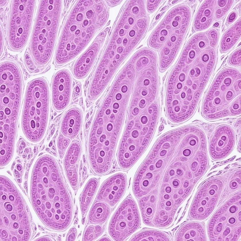
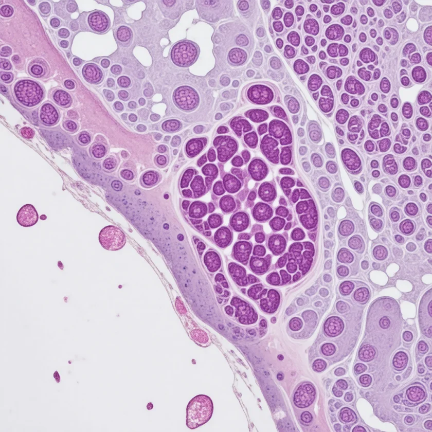
Confirmatory testing includes GAA enzyme activity measured in leukocytes or skin fibroblasts (the traditional gold-standard assay), and GAA gene sequencing to identify specific mutations and predict CRIM status. Muscle biopsy, once the cornerstone of diagnosis, is now rarely required when enzyme and genetic testing are available. When performed, it demonstrates a vacuolar myopathy with PAS-positive (periodic acid-Schiff-positive) glycogen deposits in enlarged lysosomes; electron microscopy confirms the lysosomal origin of the stored glycogen, distinguishing Pompe disease from other glycogenoses with cytoplasmic glycogen excess.
Newborn screening for Pompe disease, now active in more than 40 US states and several other countries, identifies affected infants before symptom onset. Presymptomatic ERT initiation in screen-detected IOPD has dramatically improved motor and respiratory outcomes compared with post-symptomatic diagnosis. The newborn screen measures GAA enzyme activity in the same DBS card used for other metabolic screens, using fluorometric or tandem mass spectrometry-based assays.
Useful biomarkers beyond enzyme activity include serum creatine kinase (CK) — elevated in most patients, sometimes markedly in IOPD and moderately in LOPD — and urinary glucose tetrasaccharide (Glc4), a glycogen degradation product that is elevated in Pompe disease and serves as a useful biomarker for disease burden and treatment monitoring. Liver transaminases (aspartate aminotransferase (AST), alanine aminotransferase (ALT)) are often elevated, reflecting muscle disease rather than hepatic pathology. Transient asymptomatic hyperCKemia in an adult, unexplained respiratory failure, or a limb-girdle weakness pattern are all recognised reasons for GAA enzyme activity testing.
Treatment: Enzyme Replacement Therapy
The treatment of Pompe disease has been revolutionized since 2006 by the development and successive improvement of enzyme replacement therapy (ERT), which delivers recombinant human acid alpha-glucosidase directly into the bloodstream for uptake into lysosomes via the mannose-6-phosphate receptor pathway.
Alglucosidase alfa — U.S. Food and Drug Administration approved 2006: The first approved treatment for Pompe disease; a recombinant human GAA produced in Chinese hamster ovary (CHO) cells. Administered as an intravenous infusion every two weeks at 20 mg/kg. In IOPD, alglucosidase alfa produces dramatic regression of cardiomyopathy and substantially prolonged survival. In LOPD, the landmark 2010 randomized trial (van der Ploeg et al., NEJM; 90 people aged 8 or older who could walk and were not on invasive ventilation) found that after 78 weeks the treated group walked 28.1 m farther on the 6-minute walk test and had a forced vital capacity 3.4 percentage points better than the placebo group — improved walking and stable lung function over 18 months. That trial did not measure time to ventilator dependence or loss of walking. The main limitation of first-generation ERT is suboptimal mannose-6-phosphate (M6P) content on the glycan chains, resulting in relatively poor skeletal muscle uptake.
Avalglucosidase alfa — U.S. Food and Drug Administration approved 2021: A next-generation ERT engineered with approximately 15-fold higher content of bis-mannose-6-phosphate glycans compared with alglucosidase alfa. This enhanced M6P content produces dramatically greater receptor-mediated uptake into skeletal muscle lysosomes. In the COMET trial, 100 people with late-onset Pompe disease who had never been treated were randomly given avalglucosidase alfa or alglucosidase alfa. After 49 weeks, breathing capacity (upright forced vital capacity, percent of predicted) improved by 2.89 points with the newer enzyme versus 0.46 with the older one. This showed the newer enzyme was at least as good (non-inferior); being better (superiority) was narrowly missed (p=0.063). The newer-enzyme group walked 30 m farther on the 6-minute walk test, and serious side effects occurred in 16% versus 25%. The authors described the results as a clinically meaningful improvement and named the aim of making it the new standard treatment for late-onset disease.
Cipaglucosidase alfa + miglustat — U.S. Food and Drug Administration approved 2023: The first combination ERT plus pharmacological chaperone regimen. Miglustat is an iminosugar pharmacological chaperone that binds to and stabilizes the cipaglucosidase alfa enzyme during transit to lysosomes, improving its thermal stability and extending the window for receptor binding and lysosomal targeting. In the PROPEL trial (123 adults with late-onset Pompe disease, some new to treatment and some on alglucosidase alfa for at least 2 years), the 6-minute walk improved by 20.8 m with cipaglucosidase alfa plus miglustat versus 7.2 m with alglucosidase alfa plus a dummy pill; the 13.6 m difference did not reach statistical superiority. The authors pointed to possible benefit for breathing and for people already on enzyme therapy for more than 2 years, from secondary and subgroup analyses. The regimen pairs the infusion with an oral pill.
CRIM-negative IOPD — immune tolerance induction (ITI): CRIM-negative patients who receive ERT without prior ITI develop high-titer immunoglobulin G (IgG) antibodies that neutralize the infused enzyme, severely limiting efficacy. The current standard is prophylactic ITI using rituximab (anti-CD20), methotrexate, and intravenous immunoglobulin (IVIG), initiated before the first ERT infusion. This approach has substantially improved outcomes in CRIM-negative IOPD.
Gene therapy: Multiple clinical trials are ongoing evaluating adeno-associated virus (AAV)-mediated delivery of functional GAA sequences to muscle and liver. Preliminary results are encouraging, and gene therapy may offer a one-time durable treatment option in the future.
Treatment Outcomes and Monitoring
The prognosis of Pompe disease has been transformed by enzyme replacement therapy, particularly for IOPD. In the pre-ERT era, classic IOPD was fatal in nearly all infants, most within the first two years of life. With ERT, survival beyond ten years has been documented in many patients; cardiomyopathy resolves rapidly and dramatically in the majority of treated IOPD infants. Motor outcomes are more variable — some children treated early achieve independent walking, while others remain wheelchair-dependent or ventilator-dependent despite treatment, particularly those treated after symptom establishment or those who are CRIM-negative without adequate ITI.
Newborn screening has had a profound impact on IOPD outcomes: presymptomatic diagnosis and ERT initiation within the first weeks of life — before irreversible muscle damage occurs — produces substantially better motor and respiratory outcomes than treatment begun after symptomatic presentation. This makes Pompe disease one of the most compelling examples of the benefit of universal newborn screening for treatable inherited disorders.
For LOPD, ERT slows but does not halt disease progression. Patients on ERT maintain ambulation and ventilator independence longer than untreated historical controls, but gradual functional decline continues over years. In the COMET trial the newer enzyme avalglucosidase alfa proved at least as good as alglucosidase alfa, with gains in breathing and walking that narrowly missed statistical superiority; in PROPEL, the possible benefit of cipaglucosidase alfa plus miglustat was seen mainly in people who had already been on enzyme therapy for more than 2 years.
Monitoring of Pompe disease patients on ERT involves a multidisciplinary approach. In IOPD: serial echocardiography to track cardiomyopathy regression; pulmonary function testing (FVC, maximum inspiratory pressure); motor function assessments; and GAA antibody titers in CRIM-negative patients. In LOPD: pulmonary function tests including supine FVC and Sniff NIP at least every six months; six-minute walk test; motor function scales (Pompe-specific scales such as the Rotterdam Pompe Functional Scale and the Quick Motor Function Test); and urinary Glc4 as a disease burden biomarker.
Diet and rehabilitation are important adjuncts to ERT. A high-protein diet has been studied as a strategy to reduce muscle catabolism and may offer modest benefit. Physical therapy — including resistance exercise training — is safe and beneficial in LOPD; contrary to concerns raised early in the Pompe disease field, exercise does not accelerate lysosomal glycogen accumulation or worsen muscle damage. Respiratory therapy, including airway clearance techniques and training in the use of non-invasive ventilation, is essential for maintaining respiratory health across both disease forms.
Key Research Papers
- van den Hout JM, et al. Long-term intravenous treatment of Pompe disease with recombinant human alpha-glucosidase from milk. Pediatrics. 2004;113(5):e448-57. — PubMed PMID: 15121988
- Kishnani PS, et al. A retrospective, multinational, multicenter study on the natural history of infantile-onset Pompe disease. J Pediatr. 2006;148(5):671-676. — PubMed PMID: 16737883
- Kishnani PS, et al. Recombinant human acid alpha-glucosidase: major clinical benefits in infantile-onset Pompe disease. Neurology. 2007;68(2):99-109. — PubMed PMID: 17151339
- van der Ploeg AT, et al. A randomized study of alglucosidase alfa in late-onset Pompe's disease. N Engl J Med. 2010;362(15):1396-1406. — PubMed PMID: 20393176
- Schoser B, et al. Survival and long-term outcomes in late-onset Pompe disease following alglucosidase alfa treatment: a systematic review and meta-analysis. J Neurol. 2017;264(4):621-630. — PubMed PMID: 27372449
- Chien YH, et al. Early detection of Pompe disease by newborn screening is feasible: results from the Taiwan screening program. Pediatrics. 2008;122(1):e39-45. — PubMed PMID: 18519449
- Berrier KL, et al. CRIM-negative infantile Pompe disease: characterization of immune responses in patients treated with ERT monotherapy. Genet Med. 2015;17(11):912-918. — PubMed PMID: 25741864
- Koeberl DD, et al. Enhanced efficacy of enzyme replacement therapy in Pompe disease through mannose-6-phosphate receptor expression in skeletal muscle. Mol Genet Metab. 2011;103(2):107-112. (Mouse study.) — PubMed PMID: 21397538
- Diaz-Manera J, et al. Safety and efficacy of avalglucosidase alfa versus alglucosidase alfa in patients with late-onset Pompe disease (COMET): a phase 3, randomised, multicentre trial. Lancet Neurol. 2021;20(12):1012-1026. — PubMed PMID: 34800399
- Schoser B, et al. Safety and efficacy of cipaglucosidase alfa plus miglustat versus alglucosidase alfa plus placebo in late-onset Pompe disease (PROPEL): an international, randomised, double-blind, parallel-group, phase 3 trial. Lancet Neurol. 2021;20(12):1027-1037. — PubMed PMID: 34800400
- Ausems MG, et al. Frequency of glycogen storage disease type II in The Netherlands: implications for diagnosis and genetic counselling. Eur J Hum Genet. 1999;7(6):713-716. — PubMed PMID: 10482961