Gaucher Disease
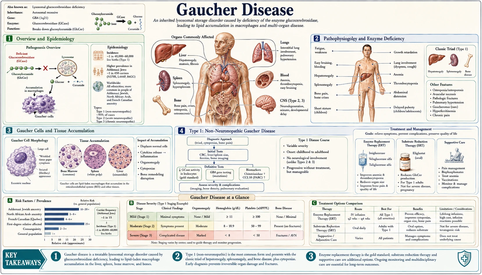
Overview and Epidemiology
Gaucher disease is the most common lysosomal storage disorder in the world, arising from autosomal recessive mutations in the GBA1 gene (written GBA in older papers) located on chromosome 1q21. This gene encodes the lysosomal enzyme glucocerebrosidase (acid beta-glucosidase), which is responsible for breaking down the sphingolipid glucocerebroside (glucosylceramide). When this enzyme is deficient or absent, glucocerebroside accumulates within the lysosomes of macrophages throughout the reticuloendothelial system, leading to progressive organ damage.
In the general population, Gaucher disease affects approximately 1 in 40,000 individuals, but prevalence is dramatically higher in the Ashkenazi Jewish community — reaching about 1 in 800 births (a 2017 review; higher figures, up to 1 in 400, are also quoted), with a carrier frequency of approximately 1 in 15. This makes it one of the most prevalent genetic diseases in that population and a strong candidate for carrier screening programs. The disease was first described in 1882 by French physician Philippe Charles Ernest Gaucher, who initially believed the lipid-laden cells he observed in a patient's spleen represented a primary splenic neoplasm; the metabolic basis was not elucidated until the mid-twentieth century.
Three main clinical types are recognized based on the presence and severity of neurological involvement. Type 1 (non-neuronopathic) accounts for most cases, about 90–95% in Europe and North America and fewer in other regions, and is characterized by the complete absence of primary neurological features. Type 2 (acute neuronopathic) is the most severe and lethal form, presenting in infancy with rapidly progressive brainstem disease. Type 3 (subacute or chronic neuronopathic) follows a slower course with variable neurological involvement. Gaucher disease holds a landmark position in medicine as one of the first inherited metabolic diseases to be successfully treated with enzyme replacement therapy, transforming the prognosis for Type 1 patients from progressive disability to near-normal life expectancy.
Pathophysiology and Enzyme Deficiency
The molecular basis of Gaucher disease lies in loss-of-function mutations in the GBA1 gene, which encodes lysosomal acid beta-glucosidase (glucocerebrosidase). This enzyme normally cleaves the glucose moiety from glucocerebroside (glucosylceramide), a sphingolipid that is a normal intermediate in the turnover of complex glycosphingolipids derived from cell membrane recycling — particularly from senescent red blood cells and white blood cells phagocytosed by macrophages. When glucocerebrosidase activity falls below a critical threshold, glucocerebroside accumulates progressively within lysosomes, predominantly in macrophages of the reticuloendothelial system: the spleen, liver (Kupffer cells), bone marrow, and lungs.
More than 400 distinct pathogenic GBA1 mutations have been identified, including missense, nonsense, frameshift, and splice-site variants, as well as recombinant alleles arising from gene conversion with the highly homologous pseudogene GBAP1 located 16 kilobases downstream. In the Ashkenazi Jewish population, a handful of mutations account for the vast majority of disease alleles. The N370S substitution is the most prevalent pathogenic allele worldwide in this population and is uniquely associated with Type 1 disease — individuals homozygous for N370S or compound heterozygous with another mild allele never develop neuronopathic involvement. The L444P mutation, by contrast, is strongly associated with neuronopathic disease: homozygous L444P is the classic genotype for Type 3, while compound heterozygosity with other severe alleles can produce Type 2 or Type 3 phenotypes.
The genotype-phenotype correlation in Gaucher disease is imperfect and sometimes frustratingly unpredictable, particularly within Type 1. Even individuals sharing identical genotypes — including siblings — can exhibit widely divergent disease severity, ranging from completely asymptomatic individuals discovered incidentally to patients with massive splenomegaly, severe bone disease, and debilitating cytopenias. This phenotypic variability implies important contributions from genetic modifier loci, epigenetic factors, and environmental influences that remain incompletely understood. The secondary lipid glucosylsphingosine (lyso-Gb1, the deacylated form) also accumulates and is increasingly recognized as a bioactive mediator that contributes to inflammation, neuronal toxicity in neuronopathic forms, and the GBA1-Parkinson's disease connection.
Gaucher Cells and Tissue Accumulation
The cellular hallmark of Gaucher disease is the Gaucher cell — a lipid-engorged macrophage with a distinctive and highly recognizable morphological appearance on bone marrow biopsy or tissue sections. Gaucher cells are large (20–100 micrometers), with an eccentric nucleus and abundant pale cytoplasm that displays a characteristic "crumpled tissue paper" or "wrinkled crepe paper" fibrillar pattern on light microscopy. This appearance results from the massive accumulation of glucocerebroside within lysosomes, which pack the cytoplasm with elongated, tubular inclusions. The cells stain strongly with periodic acid-Schiff (PAS) reagent due to their glycolipid content.
While highly characteristic, Gaucher cells are not strictly pathognomonic — morphologically similar "pseudo-Gaucher cells" can appear in conditions with high rates of leukocyte turnover and membrane glycolipid generation, including chronic myelogenous leukemia (CML), thalassemia, multiple myeloma, and other hematological malignancies. Confirmation with enzyme activity assay or genetic testing is therefore always required for a definitive diagnosis. Gaucher cells accumulate preferentially in organs with high macrophage content: the spleen (causing massive splenomegaly), liver (Kupffer cell replacement causing hepatomegaly), bone marrow (causing cytopenias and bone disease), and less commonly the lungs and lymph nodes.
Gaucher cells cause organ dysfunction through two principal mechanisms. First, they physically displace functional tissue — in the spleen, normal lymphoid architecture and red pulp function are replaced by sheets of storage cells; in bone marrow, normal hematopoietic precursors and trabecular bone are crowded out, leading to cytopenias and skeletal complications. Second, activated Gaucher cells produce a range of pro-inflammatory mediators including interleukin-1 (IL-1), interleukin-6 (IL-6), tumor necrosis factor-alpha (TNF-α), and chemokines that drive systemic inflammation, activate complement, and recruit additional macrophages — creating a self-amplifying inflammatory cycle. This inflammatory milieu contributes significantly to constitutional symptoms (fatigue, weight loss), the elevated acute-phase reactants seen in patients, and may drive the increased risk of hematological malignancies — particularly multiple myeloma, with a relative risk of at least 5.9 (95% CI 2.8–10.8) — observed in Gaucher disease patients. A 2017 review says the risk of other cancers is still under discussion.
Type 1: Non-Neuronopathic Gaucher Disease
Type 1 Gaucher disease accounts for about 90–95% of diagnosed cases in Europe and North America, fewer in other regions, and virtually all cases in the Ashkenazi Jewish population. The defining feature is the absence of primary neurological involvement — its substrate, glucocerebroside (glucosylceramide), does not accumulate in neurons in Type 1, and patients do not develop the progressive central nervous system (CNS) disease that characterizes Types 2 and 3. Instead, clinical manifestations arise entirely from visceral and skeletal involvement, with a severity spectrum that ranges from completely asymptomatic individuals discovered incidentally during family screening to patients with severely debilitating multi-system disease.
The cardinal clinical features of Type 1 include splenomegaly, hepatomegaly, cytopenias, and bone disease. Splenomegaly is typically the most dramatic finding and is often the presenting complaint — the spleen may grow to 1,500–3,000 grams (normal approximately 150g) or even larger, extending well below the iliac crest and occupying much of the abdominal cavity. Massive splenomegaly causes abdominal discomfort, early satiety, and mechanical complications. Hypersplenism from the enlarged spleen, combined with direct bone marrow infiltration by Gaucher cells, produces cytopenias — thrombocytopenia is the most common (predisposing to bleeding), followed by anemia (causing fatigue and exertional limitation), with leukopenia less prominent.
Hepatomegaly occurs in most symptomatic patients, usually to a lesser degree than splenomegaly. Liver function tests are often mildly elevated; frank cirrhosis is uncommon but occurs in severe disease. Pulmonary involvement is rare but can manifest as hepatopulmonary syndrome or, most seriously, pulmonary hypertension — a complication associated with significant morbidity and mortality. Constitutional symptoms including fatigue, cachexia, and growth retardation in children are frequent in undertreated disease. Bone disease (discussed separately) is often the most disabling long-term complication. The wide phenotypic variability within Type 1 — even among patients with identical genotypes — underscores the need for individualized monitoring and treatment planning rather than genotype-driven protocols alone.
Types 2 and 3: Neuronopathic Forms
Type 2 (Acute Neuronopathic Gaucher Disease) is the rarest and most devastating form of the disease, representing a pediatric neurological emergency with no effective treatment for its neurological component. Onset occurs in early infancy, typically between 3 and 6 months of age. Visceral involvement (splenomegaly, hepatomegaly) is present, but neurological deterioration rapidly dominates the clinical picture. The neurological syndrome is characterized by progressive brainstem dysfunction: bulbar palsy manifesting as difficulty swallowing and feeding (leading to aspiration and failure to thrive), trismus (jaw stiffness), and retroflexion of the neck (opisthotonus-like posturing). Oculomotor abnormalities including strabismus, abnormal saccades, and failure of fixation are early signs. Pyramidal signs, generalized hypotonia evolving to spasticity, and refractory seizures develop. Death from respiratory failure or aspiration pneumonia typically occurs before 2 years of age. Enzyme replacement therapy corrects visceral disease but does not cross the blood-brain barrier, leaving the neurological component untreatable with currently approved therapies.
Type 3 (Subacute or Chronic Neuronopathic Gaucher Disease) presents a more heterogeneous picture with neurological involvement that is slower in onset and more variable in severity than Type 2. Onset may occur in childhood or adolescence, and survival into adulthood is achievable with supportive care and treatment of visceral disease. The pathognomonic neurological finding in Type 3 is horizontal supranuclear gaze palsy — the inability to voluntarily move the eyes horizontally, with preservation of reflex (doll's eye) horizontal movements, caused by damage to horizontal gaze centers in the brainstem. This finding distinguishes Type 3 from Type 1 and, when present, is diagnostic of neuronopathic Gaucher disease. Additional neurological features include cerebellar ataxia, progressive myoclonic epilepsy (stimulus-sensitive myoclonus with epilepsy that can be highly refractory to treatment), pyramidal signs, and variable cognitive decline.
Type 3 Gaucher disease has a particular geographic distribution, with a historically high prevalence in the Norrbottnian region of northern Sweden attributable to a founder effect: patients from Norrbotten carry a single exon 10 leucine-to-proline mutation (the NciI or L444P mutation), and a 1990 study found further patients homozygous for it. High prevalence is also reported in East Asian populations. The L444P homozygous genotype is classically associated with Type 3, though the correlation is imperfect. Type 3 is sometimes further subdivided into Type 3a (predominantly neurological with mild visceral disease), Type 3b (predominantly visceral with limited neurological involvement, often horizontal gaze palsy only), and Type 3c (a cardiovascular variant with aortic and mitral valve calcification, oculomotor palsy, and corneal opacities associated with D409H homozygosity). Visceral disease in Type 3 responds well to enzyme replacement therapy, though neurological progression continues regardless of treatment.
No therapy is approved for the neurological disease. In a small 2023 open-label phase 2 trial (LEAP) of 11 adults with Type 3, the brain-reaching experimental drug venglustat was added to imiglucerase for a year. In spinal fluid, glucosylceramide fell a median 81% and glucosylsphingosine 70%, and ataxia improved slightly in nine patients, yet thinking and daily-function scores still worsened over time, and the authors call the signals preliminary.
Bone Disease and Skeletal Complications
Bone disease is frequently the most disabling aspect of Gaucher disease and, historically, was one of the most refractory to treatment — even after enzyme replacement therapy became available, skeletal improvement lags years behind improvement in visceral disease. The skeleton is affected through progressive infiltration of bone marrow by Gaucher cells, which disrupts normal hematopoiesis, impairs bone remodeling, causes ischemia of bone tissue, and creates a pro-inflammatory milieu that favors bone resorption over formation.
The Erlenmeyer flask deformity is the classic radiological sign of Gaucher bone disease, best seen at the distal femur. Normally, the distal femur tapers inward from the metaphysis toward the diaphysis (normal tubulation); Gaucher cell infiltration prevents this normal cortical modeling, leaving the bone with a widened, flask- or funnel-shaped metaphysis resembling an Erlenmeyer flask in a chemistry laboratory. While visually striking, this deformity itself is not necessarily painful, but it reflects diffuse marrow infiltration and predicts risk of other complications. Avascular necrosis (osteonecrosis) is one of the most painful and disabling complications, caused by ischemia of subchondral bone when Gaucher cell packing raises intramedullary pressure and compromises the vascular supply. The femoral head is most commonly affected, followed by the humeral head; collapse of necrotic bone leads to severe joint destruction requiring total joint replacement.
Bone crises — also called "pseudo-osteomyelitis" episodes — are acute, extremely painful events characterized by severe localized bone pain, warmth, swelling, fever, and elevated inflammatory markers, indistinguishable clinically from acute osteomyelitis. They reflect acute infarction of bone and surrounding periosteum and can last days to weeks before resolving. Differentiation from true osteomyelitis requires magnetic resonance imaging (MRI) and bone scan, and cultures are obtained when infection cannot be excluded. Bone crises can be precipitated by physical stress, infection, or occur without identifiable trigger. Chronic complications also include diffuse osteoporosis and osteopenia (with low bone mineral density monitored by dual-energy X-ray absorptiometry predisposing to vertebral compression fractures and fragility fractures throughout the skeleton) and direct cortical destruction leading to pathological fractures. Comprehensive skeletal monitoring — including magnetic resonance imaging of the femora (the most sensitive imaging modality for marrow infiltration and early avascular necrosis) and serial dual-energy X-ray absorptiometry — is the usual practice described for symptomatic patients.
Diagnosis and Biomarkers
The diagnostic gold standard for Gaucher disease is measurement of leukocyte glucocerebrosidase (acid beta-glucosidase) enzyme activity in peripheral blood leukocytes. Activity below 15% of mean normal confirms the diagnosis with high specificity. This test is done before any bone marrow biopsy (which is no longer required for diagnosis), as the enzyme assay is simpler, less invasive, and more definitive. A dried blood spot (DBS) version of the enzyme assay is available for newborn screening programs and facilitates diagnosis in resource-limited settings, though leukocyte-based assays are more reliable for borderline results.
GBA1 gene sequencing usually accompanies or follows the enzyme assay. Identifying the specific pathogenic mutations serves multiple critical functions: confirming the diagnosis with molecular precision, predicting the likely clinical type (particularly distinguishing N370S-associated Type 1 from L444P-associated neuronopathic disease), guiding prognosis and counseling regarding the risk of neurological involvement, and enabling accurate carrier testing for family members. Given the complexity of GBA1/GBAP1 pseudogene recombinants, sequencing is described as most reliable in a laboratory with specific expertise in this locus, using methods that distinguish the functional gene from the pseudogene (e.g., long-range polymerase chain reaction (PCR) or gene-specific amplification prior to sequencing).
Disease activity biomarkers are essential for monitoring treatment response and disease progression. Chitotriosidase (a chitinase secreted by activated macrophages, markedly elevated in Gaucher cells) was the first validated disease biomarker and remains widely used — levels may be elevated 1,000-fold above normal in severe disease and fall dramatically with effective treatment, making it an excellent monitor of macrophage burden. However, a 24-base pair duplication in the chitotriosidase gene (CHIT1) leaves about 6% of the general population, those with two copies of it, with total deficiency of the enzyme, which makes the test uninformative, and activity is low and difficult to interpret in a third of patients with one copy (a 2017 review; figures up to 35% in some populations are also quoted). Alternative biomarkers are used in these individuals. CCL18/PARC (a CC-chemokine also secreted by Gaucher cells) serves as the standard alternative. Plasma glucosylsphingosine (lyso-Gb1) is a newer biomarker whose sensitivity and specificity are reported to be superior to those of chitotriosidase and CCL18 (the 2017 review adds that it had yet to be assessed on a larger scale). It is described as sensitive across all Gaucher disease types, including neuronopathic forms, and is now recommended as a primary biomarker in several international guidelines. Angiotensin-converting enzyme (ACE) and ferritin are less specific and not routinely used.
Treatment: Enzyme Replacement and Substrate Reduction Therapy
Enzyme replacement therapy is the standard of care for symptomatic Type 1 Gaucher disease and for the visceral manifestations of Type 3. All available enzyme products are delivered by intravenous infusion every two weeks and work by supplying exogenous recombinant glucocerebrosidase targeted (via mannose receptor-mediated uptake) to macrophage lysosomes. Enzyme replacement is highly effective: over 1–2 years of treatment, spleen and liver volumes decrease substantially (often 30–50%), cytopenias resolve in most patients with improvement in platelet counts and hemoglobin, bone pain and bone crises diminish, and bone mineral density improves — though skeletal recovery is slower and less complete than visceral improvement. A critical limitation is that the infused enzyme does not cross the blood-brain barrier; it has no effect on the neurological manifestations of Types 2 and 3.
Three enzyme replacement products are approved for Gaucher disease. Imiglucerase (approved 1994) was the first recombinant enzyme product, produced in Chinese hamster ovary (CHO) cells with mannose residue exposure by enzymatic modification; it replaced the earlier alglucerase, which was extracted from human placental tissue. Velaglucerase alfa (approved 2010) is produced in a human fibroblast cell line (HT-1080 cells), yielding naturally high mannose content without post-processing modification. Taliglucerase alfa (approved 2012) is uniquely produced in genetically modified carrot plant cells — the first plant-derived recombinant protein therapeutic approved by the U.S. Food and Drug Administration (FDA). All three are considered clinically equivalent in efficacy and safety.
Substrate reduction therapy is an oral alternative strategy: rather than supplying the missing enzyme, it inhibits the upstream enzyme (glucosylceramide synthase) responsible for synthesizing glucocerebroside, thereby reducing substrate accumulation to a level that the residual deficient glucocerebrosidase can handle. Eliglustat (approved 2014) is the preferred oral first-line agent for eligible adult Type 1 patients — it is a highly specific, potent inhibitor of glucosylceramide synthase. Eliglustat is metabolized by cytochrome P450 2D6 (CYP2D6), and the U.S. Food and Drug Administration label selects patients with an FDA-cleared test of that gene. It is indicated for extensive, intermediate and poor metabolizers. The label says ultra-rapid metabolizers may not achieve adequate drug levels and that no dose can be recommended when the type cannot be determined. In the placebo-controlled ENGAGE trial of 40 previously untreated adults, spleen volume fell 27.77% on eliglustat and rose 2.26% on placebo at 9 months. In the ENCORE trial of adults stable on infusions for 3 years or more, 85% of those switched to eliglustat stayed stable against 94% on imiglucerase at 12 months, within the trial’s non-inferiority margin. Miglustat, an older, less selective substrate reduction drug, is associated with more gastrointestinal side effects and modest efficacy; it is used when enzyme replacement and eliglustat are unsuitable, and notably has an indication for neurological progression in Niemann-Pick disease Type C. Emerging therapies include pharmacological chaperones (small molecules that stabilize misfolded mutant enzyme in the endoplasmic reticulum) and gene therapy approaches currently in clinical trials.
GBA1 Mutations and Parkinson's Disease Risk
One of the most significant discoveries in Parkinson's disease genetics over the past two decades is the recognition that heterozygous GBA1 mutations — the carrier state for Gaucher disease — represent the single most common genetic risk factor for Parkinson's disease identified to date. A 2009 multicenter study of 5,691 patients and 4,898 controls gave an odds ratio of 5.43 for any GBA1 mutation in patients against controls, which is a comparison of patients with controls and not a lifetime risk; a 3- to 5-fold increased lifetime risk for carriers is often quoted. In homozygous individuals (those with actual Gaucher disease), the risk is even higher. Testing only the two common mutations, the same study found them in 15% of Ashkenazi Jewish patients against 3% of controls, and in 3% of other patients against less than 1% of controls; full sequencing in other patients found mutations in 7%. A 2026 trial protocol puts the share at 10–15% of patients with Parkinson’s disease, and figures of 5–15% in general populations and 15–20% in the Ashkenazi Jewish population are also quoted.
The mechanistic link between GBA1 mutations and Parkinson's disease centers on lysosomal dysfunction and impaired alpha-synuclein clearance. Glucocerebrosidase plays a critical role in the normal lysosomal autophagy pathway responsible for degrading alpha-synuclein, the key protein whose aggregation in Lewy bodies defines Parkinson's disease pathology. Even a single pathogenic GBA1 allele (heterozygous carrier state) reduces glucocerebrosidase activity enough to impair lysosomal function, slow alpha-synuclein turnover, and promote its progressive aggregation. This creates a bidirectional vicious cycle: alpha-synuclein oligomers in turn further inhibit glucocerebrosidase activity and lysosomal trafficking, accelerating the process. Accumulation of lyso-Gb1 may further amplify neuronal alpha-synuclein pathology through direct lipid-protein interactions.
Clinically, GBA1-associated Parkinson's disease has a characteristic profile distinct from idiopathic Parkinson's disease: earlier age of onset (typically 5–10 years younger than idiopathic Parkinson's disease), faster motor progression, higher burden of non-motor symptoms — particularly cognitive impairment and dementia (with Parkinson's disease dementia or Lewy body dementia occurring more frequently and earlier), and worse overall prognosis. The severity correlates partially with the specific GBA1 variant: mutations associated with severe Gaucher disease (L444P, 84GG, an extra base at position 84 of the gene) confer higher Parkinson's disease risk and more rapid progression than milder alleles (N370S). This connection has major therapeutic implications: small molecule pharmacological chaperones designed to increase residual glucocerebrosidase activity (such as ambroxol, currently in clinical trials) represent a mechanistically targeted Parkinson's disease treatment strategy. A phase 3 trial, ASPro-Parkinson's disease, plans to enrol 330 people with Parkinson’s disease and known GBA1 status to test whether high-dose ambroxol slows progression; the 2026 paper is its protocol and reports no results. Specialist practice commonly includes telling people with Gaucher disease and confirmed mutation carriers about this risk, asking about early motor or cognitive symptoms, and offering neurological follow-up.
Key Research Papers
- Stirnemann J, et al. A Review of Gaucher Disease Pathophysiology, Clinical Presentation and Treatments. Int J Mol Sci. 2017;18(2):441. — PubMed PMID: 28218669
- Mistry PK, et al. Gaucher disease: progress and ongoing challenges. Mol Genet Metab. 2017;120(1-2):8-21. — PubMed PMID: 27916601
- Dvir H, et al. X-ray structure of human acid-beta-glucosidase, the defective enzyme in Gaucher disease. EMBO Rep. 2003;4(7):704-709. — Search PubMed
- Weinreb NJ, et al. Long-term clinical outcomes in type 1 Gaucher disease following 10 years of imiglucerase treatment. J Inherit Metab Dis. 2013;36(3):543-553. — PubMed PMID: 22976765
- Pastores GM, et al. Therapeutic goals in the treatment of Gaucher disease. Semin Hematol. 2004;41(4 Suppl 5):4-14. — Search PubMed
- Cox TM, et al. Eliglustat compared with imiglucerase in patients with Gaucher's disease type 1 stabilised on enzyme replacement therapy. Lancet. 2015;385(9985):2355-2362. — PubMed PMID: 25819691
- Sidransky E, et al. Multicenter analysis of glucocerebrosidase mutations in Parkinson's disease. N Engl J Med. 2009;361(17):1651-1661. — PubMed PMID: 19846850
- Nalls MA, et al. Large-scale meta-analysis of genome-wide association data identifies six new risk loci for Parkinson's disease. Nat Genet. 2014;46(9):989-993. — PubMed PMID: 25064009
- Reczek D, et al. LIMP-2 is a receptor for lysosomal mannose-6-phosphate-independent targeting of beta-glucocerebrosidase. Cell. 2007;131(4):770-783. — PubMed PMID: 18022370
- Mistry PK, Lukina E, Ben Turkia H, et al. Effect of oral eliglustat on splenomegaly in patients with Gaucher disease type 1: the ENGAGE randomized clinical trial. JAMA. 2015;313(7):695-706. — PubMed PMID: 25688781
- Dahl N, Lagerström M, Erikson A, Pettersson U. Gaucher disease type III (Norrbottnian type) is caused by a single mutation in exon 10 of the glucocerebrosidase gene. Am J Hum Genet. 1990;47(2):275-278. — PubMed PMID: 2378352
- Schiffmann R, Cox TM, Dedieu JF, et al. Venglustat combined with imiglucerase for neurological disease in adults with Gaucher disease type 3: the LEAP trial. Brain. 2023;146(2):461-474. — PubMed PMID: 36256599
- Toffoli M, Menozzi E, Cullen M, et al. Protocol of ASPro-PD: a phase 3 trial of ambroxol to slow progression in genetically stratified Parkinson's disease. J Neurol. 2026;273(2):151. — PubMed PMID: 41708985