Hemoglobin A1c, Fructosamine, and Glycation Markers
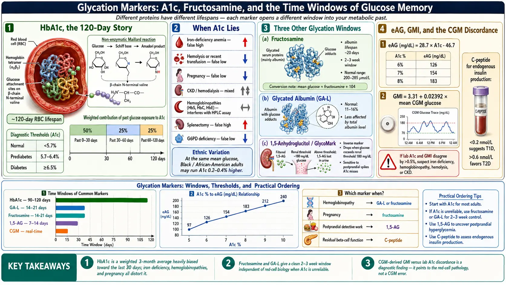
Table of Contents
- Why We Measure Glycation at All
- Hemoglobin A1c — The 3-Month Average
- Diagnostic Thresholds and Treatment Targets
- Hemoglobin A1c to Estimated Average Glucose (eAG) — Translating Percent to mg/dL
- NGSP and IFCC — Why Your Hemoglobin A1c Is Comparable
- When Hemoglobin A1c Lies
- Ethnic and Genetic Variation in Hemoglobin A1c
- Fructosamine — The 2- to 3-Week Window
- Glycated Albumin (GA)
- 1,5-Anhydroglucitol (1,5-AG)
- GMI — The CGM-Derived Hemoglobin A1c Estimate
- C-Peptide — Measuring Your Own Insulin
- Autoantibodies — What They Show
- AGEs, RAGE, and Skin Autofluorescence
- Which Test Fits Which Situation
- Key Research Papers
- Connections
- Featured Videos
Why We Measure Glycation at All
Sugar sticks to proteins. Not metaphorically — literally. When glucose circulates in the blood at elevated levels, it slowly and irreversibly attaches itself to the amino groups of proteins in a non-enzymatic reaction called glycation. The higher your average glucose, the more of your proteins carry these glucose tags. Glycation markers are simply measurements of how much sugar has stuck to specific proteins over specific time windows. Different proteins have different lifespans, so different markers open different windows into your metabolic past.
A fingerstick or fasting glucose value is a snapshot — a single frame. Glycation markers are time exposures. Each one averages your blood sugar over the life of the protein it sticks to: hemoglobin for three months, albumin for three weeks, and so on. Understanding which marker answers which question is one of the most useful skills a person with diabetes can develop.
Hemoglobin A1c — The 3-Month Average
Hemoglobin A1c (HbA1c) is glycated hemoglobin. Hemoglobin lives inside red blood cells (RBCs), which circulate for approximately 120 days before the spleen removes them. While they circulate, the hemoglobin inside them picks up glucose molecules in proportion to ambient blood sugar. Hemoglobin A1c is the fraction of hemoglobin carrying a glucose tag on the beta chain's N-terminal valine, reported as a percentage of total hemoglobin.
A 120-day average sounds uniform, but it isn't. Recent glucose matters more than old glucose because newer RBCs make up a larger share of the currently circulating pool and because glycation accumulates over time. In practice, roughly 50% of your hemoglobin A1c value reflects the last 30 days, about 25% reflects days 30–60, and the remaining 25% reflects days 60–120. That weighting is why a hemoglobin A1c drawn eight weeks after a serious treatment change already captures most of the benefit — you don't have to wait the full three months.
Diagnostic Thresholds and Treatment Targets
The American Diabetes Association (ADA) and the International Expert Committee (2009 report) share one diagnostic hemoglobin A1c cut-off: 6.5% or higher means diabetes. They draw the lower, "at-risk" band differently. The ADA's Standards of Care in Diabetes—2026 list:
- <5.7% — normal.
- 5.7–6.4% — prediabetes (increased risk).
- ≥6.5% — diabetes (confirmed on a repeat test unless unambiguous hyperglycemia is present).
The 5.7–6.4% prediabetes band is the ADA's own. The International Expert Committee did not use it: its report described people with hemoglobin A1c from 6.0% to just under 6.5% as the group at highest risk of going on to diabetes, called for proven prevention programmes for people at 6.0% or above, and noted that people below that range may still be at risk.
Diagnosis is one question. Treatment targets are another, and they are individualized:
- <7.0% — the general adult target for most people with diabetes.
- <6.5% — reasonable for younger adults, those newly diagnosed, those with long life expectancy, and people planning pregnancy.
- <8.0% — acceptable for older adults, those with hypoglycemia unawareness, limited life expectancy, advanced complications, or when tighter control would be dangerous.
- Pregnancy — <6.0–6.5% if achievable without hypoglycemia. Pregnancy also shortens RBC turnover, which complicates interpretation (see below).
A target is a negotiation between the benefit of tighter control (fewer microvascular complications) and the cost of pushing too hard (hypoglycemia, weight gain, polypharmacy). The ADA's 2026 Standards tie lower goals (such as <6.5%) to good health and low treatment risk, and less strict goals (up to 8%) to frailty, serious other illness, limited life expectancy, or a greater risk of harm from treatment.
Hemoglobin A1c to Estimated Average Glucose (eAG) — Translating Percent to mg/dL
Hemoglobin A1c is a percentage; fingerstick meters and continuous glucose monitors (CGMs) report milligrams per deciliter. The estimated Average Glucose (eAG) equation, derived by Nathan and colleagues in 2008, converts between them:
eAG (mg/dL) = 28.7 × hemoglobin A1c − 46.7
A few reference points worth memorizing:
- Hemoglobin A1c 5.0% → eAG ~97 mg/dL
- Hemoglobin A1c 6.0% → eAG ~126 mg/dL
- Hemoglobin A1c 6.5% → eAG ~140 mg/dL
- Hemoglobin A1c 7.0% → eAG ~154 mg/dL
- Hemoglobin A1c 8.0% → eAG ~183 mg/dL
- Hemoglobin A1c 9.0% → eAG ~212 mg/dL
- Hemoglobin A1c 10.0% → eAG ~240 mg/dL
The eAG is handy for translating lab reports into the units your meter speaks. It is not the same as your CGM's mean glucose — see the GMI section below for why those two numbers can disagree.
NGSP and IFCC — Why Your Hemoglobin A1c Is Comparable
Before the early 2000s, hemoglobin A1c values from different labs were not directly comparable. Two global standardization programs fixed that:
- NGSP (National Glycohemoglobin Standardization Program) — reports hemoglobin A1c as a percentage (e.g., 7.0%) and anchors values to the reference method of the Diabetes Control and Complications Trial (DCCT). This is the number you see on U.S. lab reports.
- IFCC (International Federation of Clinical Chemistry) — reports hemoglobin A1c in mmol/mol (e.g., 53 mmol/mol = 7.0% NGSP). Used in much of Europe and on some dual-unit reports.
Any modern hemoglobin A1c assay in the U.S., U.K., or EU is certified to one or both of these standards. A point-of-care hemoglobin A1c from a non-certified device may not be traceable to the NGSP standard, so its result is not directly comparable with a certified lab value.
When Hemoglobin A1c Lies
Hemoglobin A1c assumes normal red blood cells living a normal 120-day life. When that assumption breaks, the number breaks with it. These are the conditions that falsely raise or lower hemoglobin A1c:
- Iron-deficiency anemia. Old RBCs stay in circulation longer than 120 days because the bone marrow cannot replace them fast enough. Hemoglobin has more time to accumulate glucose tags, so hemoglobin A1c reads falsely high. Treating the iron deficiency can drop hemoglobin A1c by 0.5–1.0% without any change in actual glucose.
- Recent blood loss, hemolysis, or hemolytic anemia. Young RBCs dominate, average hemoglobin age is lower, and hemoglobin A1c reads falsely low.
- Recent transfusion. You are now carrying someone else's glycated (or not-glycated) hemoglobin. Unreliable for at least 3–4 months.
- Hemoglobinopathies. HbS (sickle trait or disease), HbC, HbE, and beta-thalassemia with persistent fetal hemoglobin (HbF) can interfere with certain hemoglobin A1c assay chemistries (especially some high-performance liquid chromatography (HPLC) methods). The usual workarounds are an NGSP-certified HbF-tolerant method, or fructosamine or glycated albumin instead.
- Pregnancy. Accelerated RBC turnover in the second and third trimesters lowers hemoglobin A1c by roughly 0.3–0.5%. Fructosamine or glycated albumin is preferred when fine-tuning glycemia near delivery.
- Chronic kidney disease and hemodialysis. Multiple competing effects: reduced erythropoiesis lengthens RBC life (raises hemoglobin A1c), but uremia, carbamylation of hemoglobin, and erythropoietin therapy shorten it (lower hemoglobin A1c). Net effect is unpredictable, which is why glycated albumin is often used alongside it.
- Splenectomy. Without the spleen culling old RBCs, average RBC age rises and hemoglobin A1c reads falsely high.
- High altitude and chronic hypoxia. Higher hemoglobin turnover patterns and erythropoietin drive produce small upward shifts.
- Chronic liver disease and cirrhosis. Hypersplenism shortens RBC survival; hemoglobin A1c reads falsely low.
- Glucose-6-phosphate dehydrogenase (G6PD) deficiency. Oxidative hemolysis episodes shorten RBC life; hemoglobin A1c reads low during and after flares.
- Severe iron deficiency without anemia. Even before hemoglobin drops, iron deficiency appears to shift glycation kinetics upward.
- Erythropoietin or iron therapy. Young RBC cohorts flood in; hemoglobin A1c temporarily reads low.
If your self-monitored glucose and your hemoglobin A1c tell different stories, the first question to ask is not "which one is lying" but "which one is right for me." See the anemia page and chronic kidney disease for the conditions that most commonly distort hemoglobin A1c in practice.
Ethnic and Genetic Variation in Hemoglobin A1c
Even in healthy people with identical mean glucose, hemoglobin A1c differs slightly by ancestry. A U.S. Centers for Disease Control and Prevention (CDC) analysis and subsequent multi-ethnic studies found that Black and African-American adults run hemoglobin A1c values approximately 0.2–0.4% higher than White adults at the same measured mean glucose. Smaller upward shifts have been reported in Hispanic and Asian populations. The mechanism probably involves differences in RBC lifespan, intracellular glucose transport, and glycation kinetics — not differences in diabetes control.
A 2017 U.S. study at 10 diabetes centres gives a measured figure. It followed 104 Black and 104 White people aged 8 or older with type 1 diabetes who wore continuous glucose monitors for 12 weeks. At the same measured average glucose, hemoglobin A1c ran 0.4 percentage points higher in Black participants (95% confidence interval 0.2 to 0.6). Glycated albumin and fructosamine showed no racial difference in their link to average glucose. The authors note that race explained only part of the A1c gap between the groups, and that too few participants had A1c below 6.5% to apply the result to people in that range.
The practical implication is modest but real: a Black patient with a hemoglobin A1c of 6.6% may have the same average glucose as a White patient with a hemoglobin A1c of 6.3%. This does not change diagnostic thresholds — American Diabetes Association guidelines use one hemoglobin A1c cutoff for everyone — but it is a reason to pair hemoglobin A1c with direct glucose measurements (fingersticks, CGM, fructosamine) whenever a diagnosis or target feels borderline.
Fructosamine — The 2- to 3-Week Window
Fructosamine measures all glycated serum proteins, which are mostly albumin with smaller contributions from globulins and lipoproteins. Albumin has a half-life of about 20 days, so fructosamine reflects your average glucose over roughly the past 2–3 weeks. It is cheap (often under $20 as a cash lab), widely available, and completely independent of hemoglobin — which is exactly what you want when hemoglobin A1c is unreliable.
Typical reference values (exact cutoffs vary by lab):
- 200–285 µmol/L — normal range in adults with normal albumin.
- >285 µmol/L — consistent with diabetes.
- A rough conversion: mean glucose (mg/dL) ≈ fructosamine (µmol/L) + 104. Use it cautiously — the relationship is less tight than hemoglobin A1c-to-eAG.
Because fructosamine scales with total serum protein, it needs an albumin correction when albumin is abnormal. A low-albumin state (nephrotic syndrome, cirrhosis, severe malnutrition) depresses fructosamine artificially, and a high-albumin state (dehydration) inflates it. Also affected:
- Thyroid disease. Hyperthyroidism speeds albumin turnover and lowers fructosamine; hypothyroidism does the opposite.
- Hyperbilirubinemia. Interferes with some colorimetric assays.
- High-dose vitamin C. Can reduce the nitroblue tetrazolium assay chemistry.
Fructosamine shines in three situations: pregnancy (when hemoglobin A1c is misleading and rapid adjustment matters), hemoglobinopathies (when hemoglobin A1c chemistry is compromised), and rapid treatment-change assessment (when you need to know in three weeks, not three months, whether a new insulin regimen is working).
Glycated Albumin (GA)
Glycated albumin (GA) is a refinement of the fructosamine idea. Instead of measuring all glycated serum proteins lumped together, the glycated albumin test measures only the glycated fraction of albumin and reports it as a percentage of total albumin (normal is roughly 11–16%). Because it is expressed as a ratio, it is less sensitive to the total albumin level and provides a more standardized window into the past 2–3 weeks.
Glycated albumin is especially useful in hemodialysis patients (where hemoglobin A1c is notoriously unreliable and fructosamine is confounded by protein losses) and in pregnancy. It is more common in Japanese and Korean clinical practice than in the U.S., but U.S. reference labs now offer it. If your hemoglobin A1c is distrusted for any of the reasons in the When hemoglobin A1c Lies section, glycated albumin is one of the alternatives.
1,5-Anhydroglucitol (1,5-AG)
1,5-anhydroglucitol (1,5-AG) is the odd marker out. It does not measure glycated anything. It measures a dietary sugar-alcohol normally present in blood at stable levels. When blood glucose exceeds the renal threshold of about 180 mg/dL, 1,5-AG competes with glucose for reabsorption in the kidney and is spilled into the urine. Serum 1,5-AG therefore drops when glucose spikes above 180. It is an inverse marker.
The window is short — roughly the past 1–2 weeks — and 1,5-AG is particularly sensitive to postprandial spikes that hemoglobin A1c smooths away. A patient whose hemoglobin A1c sits at 6.8% but whose CGM shows daily post-meal spikes to 240 mg/dL will often have a low 1,5-AG, flagging glycemic variability that the hemoglobin A1c is hiding.
1,5-AG is the answer to "my hemoglobin A1c looks fine but I feel terrible after meals." It is also useful for assessing whether a new prandial insulin strategy is flattening postprandial excursions. Reference ranges vary by kit; levels above roughly 10 µg/mL are typical in non-diabetic adults, and values that drop over time suggest worsening postprandial control.
GMI — The CGM-Derived Hemoglobin A1c Estimate
The Glucose Management Indicator (GMI) is a CGM-derived estimate of what your hemoglobin A1c should be, given the mean glucose your sensor has measured over the reporting period. It replaced an earlier, less rigorous "estimated hemoglobin A1c" concept. The Beck 2018 formula is:
GMI (%) = 3.31 + 0.02392 × mean glucose (mg/dL)
Your CGM report shows GMI automatically. Here is where it gets useful: lab hemoglobin A1c and GMI frequently disagree by 0.3–0.5%, and sometimes by more than a full point. That gap is a diagnostic finding, not a flaw.
- Lab hemoglobin A1c higher than GMI by 0.5%+ — possible causes include iron deficiency, splenectomy, slower RBC turnover, or ethnic variation pushing hemoglobin A1c up.
- Lab hemoglobin A1c lower than GMI by 0.5%+ — possible causes include hemolysis, recent blood loss, hemoglobinopathy, pregnancy, liver disease, or erythropoietin therapy.
- Persistent mismatch with no obvious cause — a hemoglobin electrophoresis test can settle it. A silent HbS, HbC, or HbE trait is a common and often-missed explanation.
See the CGM and Time in Range article for how to use GMI alongside Time-in-Range and coefficient of variation to describe your glycemia in three dimensions instead of one.
C-Peptide — Measuring Your Own Insulin
Insulin is stored inside beta cells as proinsulin, a single chain that is cleaved into two fragments at the moment of release: insulin (the active hormone) and C-peptide (the connecting peptide). The two are secreted in equal molar amounts, but C-peptide has a much longer half-life and is not extracted by the liver, so it gives a more stable readout of endogenous insulin production. Crucially, C-peptide is not present in injected insulin — so it measures only what your own pancreas is still making, even in patients already on insulin therapy.
Common ways to order it:
- Random (non-fasting) C-peptide with a simultaneous glucose — the quickest screen. Interpret in the context of the glucose: a low C-peptide at glucose 50 mg/dL means little; a low C-peptide at glucose 250 mg/dL is meaningful.
- Fasting C-peptide + fasting glucose — standard pairing for evaluating residual beta-cell function.
- Stimulated C-peptide — measured 6 minutes after intravenous (IV) glucagon or 90 minutes after a mixed-meal tolerance test. The most sensitive way to detect preserved endogenous insulin.
Broad interpretation (units vary; nmol/L shown here, multiply by 3.0 for ng/mL):
- <0.2 nmol/L — consistent with type 1 diabetes; little to no endogenous insulin production.
- 0.2–0.6 nmol/L — indeterminate; possibilities include latent autoimmune diabetes in adults (LADA), long-standing type 2 diabetes with partial beta-cell failure, or early type 1 diabetes.
- >0.6 nmol/L — meaningful endogenous production; favors type 2 diabetes, maturity-onset diabetes of the young (MODY), or other non-autoimmune forms.
C-peptide is one of the single most clinically useful tests in diabetes and one of the most under-ordered. In an adult labeled "type 2" who is progressing rapidly, losing weight, or failing oral therapy, a C-peptide and an autoantibody panel are the tests that tell these forms apart. See Types: T1, T2, LADA, MODY for the full differential.
Autoantibodies — What They Show
Five antibodies define autoimmune (type 1 and LADA) diabetes:
- GAD65 — glutamic acid decarboxylase; the most common positive in adult-onset autoimmune diabetes (LADA).
- IA-2 (ICA512) — islet antigen 2; common in pediatric-onset type 1 diabetes.
- ZnT8 — zinc transporter 8; adds sensitivity when the others are negative.
- Insulin autoantibody (IAA) — useful mainly before insulin injections begin; once a person injects insulin, the body can make antibodies to the injected insulin, so a positive result can no longer be read as a sign of autoimmunity.
- ICA — islet cell antibodies by indirect immunofluorescence; an older broad-spectrum test.
The sibling article on diabetes types covers these antibodies in depth, including the thresholds, ordering strategy, and genetic testing that distinguishes MODY from LADA.
AGEs, RAGE, and Skin Autofluorescence
Advanced glycation end products (AGEs) are what glycated proteins and lipids eventually become if they are not cleared — stable, cross-linked molecules that accumulate in long-lived tissues like skin, cartilage, lens of the eye, and arterial wall. They signal through the receptor for AGEs (RAGE), which drives oxidative stress and inflammation, and they are mechanistically implicated in diabetic retinopathy, nephropathy, neuropathy, and cardiovascular disease.
In clinical research, AGEs are estimated non-invasively with a skin autofluorescence reader that shines ultraviolet (UV) light on the forearm and measures tissue fluorescence proportional to skin AGE content. The technique is painless, takes under a minute, and correlates with long-term cumulative glycemic burden better than a single hemoglobin A1c. It is not yet standard clinical care in the U.S. — think of it as a biomarker of decades of glycemic exposure, potentially useful for cardiovascular risk stratification. Dietary AGEs (from high-heat cooking of meats and fats) contribute modestly; endogenous production from hyperglycemia contributes more.
Which Test Fits Which Situation
Glycation markers are not a one-size-fits-all panel. This is how the tests are commonly matched to the question:
- New diagnosis of diabetes in any adult. A common workup includes hemoglobin A1c + C-peptide + GAD65 + IA-2 + ZnT8. The added marginal cost is small; the diagnostic clarity is enormous. Missing LADA in a "type 2" patient delays insulin by years and harms beta cells.
- Pregnancy. Fructosamine or glycated albumin is often used as the main tracking marker, alongside frequent CGM/fingerstick data. Hemoglobin A1c is typically drawn once at confirmation and at the start of each trimester for context, but it is not used to adjust treatment near delivery.
- Hemoglobin A1c disagrees with CGM/GMI by ≥0.5%. Fructosamine serves as an independent check. If fructosamine agrees with the CGM, A1c is the outlier, and the usual search for the cause is a complete blood count (CBC) + iron studies + reticulocyte count + hemoglobin electrophoresis.
- Good hemoglobin A1c, bad symptoms. Fatigue after meals, weight regain, or neuropathy progressing despite "fine" labs. 1,5-AG can capture hidden postprandial spikes, and fructosamine can corroborate.
- Hemodialysis or advanced chronic kidney disease (CKD). Hemoglobin A1c carries less weight here. Glycated albumin plus CGM is the usual primary monitoring pair. See chronic kidney disease.
- Known hemoglobinopathy (HbS/C/E trait, thalassemia). An accurate result needs an NGSP-certified HbF-tolerant method, and fructosamine or glycated albumin is often preferred for routine monitoring.
- Rapid therapy change (new insulin regimen, glucagon-like peptide-1 (GLP-1) start, bariatric surgery). Fructosamine at 3 weeks gives an answer sooner than waiting 3 months for hemoglobin A1c.
- Unexplained low hemoglobin A1c with symptoms of hyperglycemia. Possible causes are hemolysis, hypersplenism, liver disease, or recent blood loss. A complete blood count with reticulocytes is the usual first step. See anemia.
- Screening insulin resistance in a non-diabetic. Hemoglobin A1c alone is insensitive. Pairing it with fasting insulin and homeostatic model assessment of insulin resistance (HOMA-IR) gives a metabolic picture years before hemoglobin A1c rises.
No single marker fits every case. Each answers a different question, and knowing which one fits matters most when the default test stops telling the truth.
Key Research Papers
The following peer-reviewed papers anchor the science of hemoglobin A1c, fructosamine, glycated albumin, the Glucose Management Indicator (GMI), and advanced glycation end products discussed throughout this article.
- The Diabetes Control and Complications Trial Research Group. The Effect of Intensive Treatment of Diabetes on the Development and Progression of Long-Term Complications in Insulin-Dependent Diabetes Mellitus (DCCT). New England Journal of Medicine. 1993;329(14):977-986. — doi:10.1056/NEJM199309303291401
- UK Prospective Diabetes Study (UKPDS) Group. Intensive Blood-Glucose Control with Sulphonylureas or Insulin Compared with Conventional Treatment and Risk of Complications in Patients with Type 2 Diabetes (UKPDS 33). Lancet. 1998;352(9131):837-853. — doi:10.1016/S0140-6736(98)07019-6
- American Diabetes Association Professional Practice Committee for Diabetes. 2. Diagnosis and Classification of Diabetes: Standards of Care in Diabetes—2026. Diabetes Care. 2026;49(Suppl 1):S27-S49. — doi:10.2337/dc26-S002 · PubMed PMID: 41358893
- American Diabetes Association Professional Practice Committee for Diabetes. 6. Glycemic Goals, Hypoglycemia, and Hyperglycemic Crises: Standards of Care in Diabetes—2026. Diabetes Care. 2026;49(Suppl 1):S132-S149. — doi:10.2337/dc26-S006 · PubMed PMID: 41358894
- Nathan DM, Kuenen J, Borg R, Zheng H, Schoenfeld D, Heine RJ. Translating the A1C Assay into Estimated Average Glucose Values (ADAG Study). Diabetes Care. 2008;31(8):1473-1478. — doi:10.2337/dc08-0545
- Bergenstal RM, Beck RW, Close KL, et al. Glucose Management Indicator (GMI): A New Term for Estimating A1C from Continuous Glucose Monitoring. Diabetes Care. 2018;41(11):2275-2280. — doi:10.2337/dc18-1581
- Selvin E, Steffes MW, Zhu H, et al. Glycated Hemoglobin, Diabetes, and Cardiovascular Risk in Nondiabetic Adults. New England Journal of Medicine. 2010;362(9):800-811. — doi:10.1056/NEJMoa0908359
- The International Expert Committee. International Expert Committee Report on the Role of the A1C Assay in the Diagnosis of Diabetes. Diabetes Care. 2009;32(7):1327-1334. — doi:10.2337/dc09-9033
- Goldstein DE, Little RR, Lorenz RA, et al. Tests of Glycemia in Diabetes. Diabetes Care. 2004;27(7):1761-1773. — doi:10.2337/diacare.27.7.1761
- Armbruster DA. Fructosamine: Structure, Analysis, and Clinical Usefulness. Clinical Chemistry. 1987;33(12):2153-2163. — doi:10.1093/clinchem/33.12.2153
- Singh R, Barden A, Mori T, Beilin L. Advanced Glycation End-Products: A Review. Diabetologia. 2001;44(2):129-146. — doi:10.1007/s001250051591
- Brownlee M. Biochemistry and Molecular Cell Biology of Diabetic Complications. Nature. 2001;414(6865):813-820. — doi:10.1038/414813a
- Vlassara H, Uribarri J. Advanced Glycation End Products (AGE) and Diabetes: Cause, Effect, or Both? Current Diabetes Reports. 2014;14(1):453. — doi:10.1007/s11892-013-0453-1
- Cohen RM, Franco RS, Khera PK, et al. Red Cell Life Span Heterogeneity in Hematologically Normal People Is Sufficient to Alter HbA1c. Blood. 2008;112(10):4284-4291. — doi:10.1182/blood-2008-04-154112
- Battelino T, Danne T, Bergenstal RM, et al. Clinical Targets for Continuous Glucose Monitoring Data Interpretation: Recommendations from the International Consensus on Time in Range. Diabetes Care. 2019;42(8):1593-1603. — doi:10.2337/dci19-0028
- Bergenstal RM, Gal RL, Connor CG, Gubitosi-Klug R, Kruger D, Olson BA, et al. Racial Differences in the Relationship of Glucose Concentrations and Hemoglobin A1c Levels. Annals of Internal Medicine. 2017;167(2):95-102. — doi:10.7326/M16-2596 · PubMed PMID: 28605777
PubMed Topic Searches
Live PubMed queries that update as new papers are indexed.
- PubMed: HbA1c standardization, NGSP, and IFCC
- PubMed: HbA1c interference by hemoglobinopathies
- PubMed: HbA1c and iron deficiency anemia
- PubMed: HbA1c ethnic variation and African-American cohorts
- PubMed: Fructosamine for diabetes monitoring
- PubMed: Glycated albumin in hemodialysis
- PubMed: 1,5-Anhydroglucitol and postprandial glucose excursions
- PubMed: Glucose Management Indicator (GMI) from CGM
- PubMed: C-peptide in LADA, type 1, and type 2
- PubMed: GAD65, IA-2, and ZnT8 autoantibodies
- PubMed: Advanced glycation end products and skin autofluorescence
- PubMed: HbA1c in pregnancy and red cell turnover
Connections
- Endocrinology
- Glycation & Hemoglobin A1c: What That Number Actually Measures — interactive animation
- Blood Sugar & Insulin — interactive animation
- Diabetes Overview
- Type 1 vs Type 2 vs LADA and MODY
- CGM and Time-in-Range
- Complications: Retinopathy, Nephropathy, Neuropathy
- Hypoglycemia Awareness and Prevention
- Insulin Types and Dosing Strategies
- Remission and Reversal Strategies
- Cardio-Renal Protection: Sodium-Glucose Cotransporter-2 and Glucagon-Like Peptide-1
- Diabetic Foot Care and Ulcer Prevention
- Insulin Resistance
- Hemoglobin A1C Test
- Fasting Insulin and Homeostatic Model Assessment of Insulin Resistance
- Kidney Disease
- Anemia
- Iron Deficiency Anemia
- Metabolic Syndrome
- Lab Tests