Wolff-Parkinson-White Syndrome
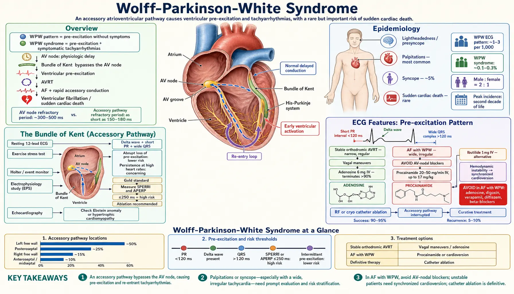
Table of Contents
- Overview
- Epidemiology
- The Bundle of Kent (Accessory Pathway)
- Electrocardiogram Features: Pre-excitation Pattern
- Arrhythmias in WPW
- Sudden Cardiac Death Risk
- Clinical Presentation
- Diagnosis and Risk Stratification
- Acute Management
- Catheter Ablation — Curative Treatment
- Asymptomatic WPW Management
- Key Research Papers
- Connections
- Featured Videos
1. Overview
Wolff-Parkinson-White (WPW) syndrome is defined as the combination of ventricular pre-excitation on the resting electrocardiogram (ECG) (delta wave, short PR interval, wide QRS) with symptomatic tachyarrhythmias. It is caused by an abnormal accessory atrioventricular pathway — the Bundle of Kent — that bypasses the AV node's physiologic conduction delay and directly connects the atrial myocardium to ventricular myocardium across the AV groove.
The distinction between WPW pattern (electrocardiogram findings without symptoms) and WPW syndrome (electrocardiogram pattern + documented tachyarrhythmias) is clinically important: asymptomatic individuals with a WPW pattern require risk stratification but not necessarily immediate treatment.
WPW syndrome is the most common cause of atrioventricular reentrant tachycardia (AVRT) and carries a unique risk: when atrial fibrillation occurs in the setting of a rapidly conducting accessory pathway (refractory period <250 ms), extremely rapid ventricular rates can develop, potentially degenerating into ventricular fibrillation and sudden cardiac death. Catheter ablation of the accessory pathway is curative in the vast majority of cases.
2. Epidemiology
The WPW ECG pattern is present in approximately 1–3 per 1,000 individuals in the general population. WPW syndrome (symptomatic pre-excitation with tachyarrhythmias) has a prevalence of approximately 0.1–0.3%. It is more common in men (male-to-female ratio approximately 2:1) and in young adults, with a peak incidence in the second decade of life.
The annual risk of sudden cardiac death (SCD) in WPW syndrome is approximately 0.1–0.6% (higher in symptomatic patients, lower in those with intermittent pre-excitation). In a community-based Olmsted County cohort, sudden cardiac death occurred at 0.15% per patient-year (2 deaths in 1,338 patient-years), and in none of the patients who were asymptomatic at diagnosis (Munger 1993); a meta-analysis of asymptomatic pre-excitation estimated 1.25 per 1,000 person-years (Obeyesekere 2012), making SCD risk stratification a key aspect of management.
Familial WPW may be associated with PRKAG2 mutations (glycogen storage cardiomyopathy) or other congenital heart disease (Ebstein anomaly, whose accessory pathways are typically right-sided, and hypertrophic cardiomyopathy).
3. The Bundle of Kent (Accessory Pathway)
The accessory pathway (AP) is a strand of embryologically retained myocardial tissue crossing the normally insulating AV fibrous ring. These pathways can be located anywhere around the AV groove:
- Left free wall (most common, ~50%): left lateral accessory pathway
- Posteroseptal (~25%): near coronary sinus ostium, ablation challenging due to proximity to coronary arteries and AV node
- Right free wall (~15%)
- Anteroseptal/midseptal (~10%): parahisian location, highest AV block risk with ablation
The AP conducts bidirectionally in most patients: antegrade (atrium → ventricle, producing delta wave) and retrograde (ventricle → atrium, enabling AVRT). Some APs conduct only retrograde ("concealed bypass tract" — no delta wave, only capable of supporting orthodromic AVRT).
AV nodal physiology vs. accessory pathway: The AV node slows conduction (physiologic delay) and has a refractory period of ~300–500 ms. Accessory pathways bypass this delay and may have refractory periods as short as 150–180 ms — enabling rapid conduction rates during atrial fibrillation (AF) that would otherwise be blocked by the AV node.
4. Electrocardiogram Features: Pre-excitation Pattern
The resting 12-lead electrocardiogram in WPW shows three characteristic features:
- Short PR interval (<120 ms): The AV node's conduction delay is bypassed, so atrial impulses activate the ventricle earlier.
- Delta wave: A slurred, initial upstroke of the QRS complex, representing early ventricular activation via the accessory pathway (before the main wavefront arrives through the His-Purkinje system).
- Wide QRS complex (>120 ms): The combined result of delta wave + normal His-Purkinje activation ("fusion complex" — part accessory pathway, part normal conduction).
Secondary ST-T wave changes: Abnormal repolarization in pre-excited zones produces ST depression and T-wave inversions in leads where the delta wave is positive, and ST elevation where the delta wave is negative.
Localization of accessory pathway using delta wave polarity:
- Negative delta wave in lead I, positive in V1 → left free wall pathway
- Negative delta wave in aVF → posteroseptal pathway
- Positive delta in I, negative in V1 → right free wall pathway
Intermittent pre-excitation (delta wave intermittently present or disappearing with exercise/increased heart rate) suggests a pathway with a relatively long antegrade refractory period and lower SCD risk.
5. Arrhythmias in WPW
Orthodromic AVRT (most common, ~95% of AVRT in WPW):
Antegrade conduction proceeds normally through the AV node → His-Purkinje → ventricles (narrow QRS). Retrograde conduction returns via the accessory pathway to the atria. Electrocardiogram: narrow complex tachycardia, retrograde P wave visible in ST segment (RP interval ~100–160 ms), HR 150–250 bpm. Regular, abrupt onset/offset.
Antidromic AVRT (~5% of AVRT in WPW):
Antegrade conduction down the accessory pathway → ventricles (wide, fully pre-excited QRS). Retrograde conduction up the AV node or another accessory pathway. Electrocardiogram: wide complex regular tachycardia, maximally pre-excited QRS. Can resemble ventricular tachycardia. Requires differentiation — patients with antidromic AVRT are at particularly high risk for SCD.
Atrial Fibrillation with WPW (life-threatening emergency):
When atrial fibrillation occurs in a WPW patient, atrial impulses can conduct rapidly over the accessory pathway (bypassing AV nodal rate limitation). If the accessory pathway has a short antegrade refractory period (<250 ms), ventricular rates can exceed 300 bpm → ventricular fibrillation → sudden cardiac death.
Electrocardiogram signature of atrial fibrillation with WPW: wide complex, irregular tachycardia with varying QRS morphologies (fusion between pathway-conducted and normally conducted beats). Ventricular rate >200 bpm with irregular RR intervals is a medical emergency.
6. Sudden Cardiac Death Risk
SCD in WPW results from atrial fibrillation conducting rapidly over the accessory pathway, triggering ventricular fibrillation. Risk factors for SCD in WPW:
- Shortest pre-excited RR interval (SPERRI) ≤250 ms during atrial fibrillation: highest-risk criterion; indicates pathway capable of extremely rapid conduction
- Multiple accessory pathways: increased arrhythmia burden
- Prior cardiac arrest or syncope: highest risk — absolute indication for ablation
- Symptomatic SVT (AVRT episodes): indicates pathway capable of sustaining reentry
- Ebstein anomaly or hypertrophic cardiomyopathy: structural substrates with higher arrhythmia burden
Low-risk features: Intermittent pre-excitation (spontaneous loss of delta wave during sinus rhythm or with exercise), inducible PR normalization with procainamide or adenosine, SPERRI >250 ms during induced atrial fibrillation.
7. Clinical Presentation
WPW syndrome presents primarily through its tachyarrhythmias:
- Palpitations: Most common; regular (AVRT) or irregular (atrial fibrillation with pre-excitation) rapid heartbeat, abrupt onset/offset
- Lightheadedness and presyncope: From rapid rates reducing diastolic filling
- Syncope: True syncope (~5% of WPW patients); when syncope is the first symptom, it may indicate rapid atrial fibrillation with pre-excitation and requires urgent evaluation
- Sudden cardiac death: Rare but devastating first presentation; estimates range from about 0.1% a year in asymptomatic pre-excitation (Obeyesekere 2012) to 0.15% a year in a community cohort (Munger 1993), and are higher in high-risk patients
Asymptomatic WPW (no palpitations, no documented tachyarrhythmia) may be discovered incidentally on electrocardiogram obtained for sports/military screening, insurance, or unrelated cardiac evaluation.
8. Diagnosis and Risk Stratification
Resting 12-lead electrocardiogram: Confirms pre-excitation (delta wave, short PR, wide QRS).
Exercise stress test: Abrupt loss of pre-excitation during exercise (at a specific heart rate threshold) indicates the accessory pathway's antegrade refractory period exceeds the sinus cycle length at that rate — suggests relatively benign pathway. Persistence of pre-excitation at high heart rates (pathologic finding) suggests short refractory period.
Holter/event monitor: Documents clinical SVT or atrial fibrillation episodes.
Electrophysiology study (EPS): Gold standard for risk stratification:
- Induces atrial fibrillation and measures SPERRI (shortest pre-excited RR interval during atrial fibrillation)
- Measures accessory pathway antegrade effective refractory period (APERP)
- SPERRI ≤250 ms or APERP ≤250 ms = high risk → ablation recommended
- EPS also localizes pathway for ablation in the same session
Echocardiography: Excludes associated structural disease (Ebstein anomaly, hypertrophic cardiomyopathy).
9. Acute Management
Orthodromic AVRT (narrow complex, stable):
- Vagal maneuvers (Valsalva, carotid massage) — first line
- Adenosine 6 mg intravenous (IV) rapid push — terminates AVRT in >90%
- If adenosine fails: intravenous verapamil or diltiazem (only if no pre-excitation on baseline electrocardiogram)
- Electrical cardioversion for hemodynamic instability
Atrial fibrillation with WPW (wide complex irregular tachycardia, potential emergency):
- AVOID intravenous adenosine, digoxin, verapamil, diltiazem, and beta-blockers — these block the AV node and may paradoxically accelerate conduction over the accessory pathway, precipitating VF
- Procainamide 20–50 mg/min intravenous (up to 17 mg/kg): drug of choice — slows conduction in both the AV node and accessory pathway
- Electrical cardioversion (synchronized DC shock) for hemodynamic instability or pre-syncope/syncope: immediate and definitive
- Ibutilide 1 mg intravenous (alternative to procainamide if unavailable)
10. Catheter Ablation — Curative Treatment
Catheter ablation of the accessory pathway is the definitive, curative treatment for WPW syndrome:
- Success rate: 90–95% across all pathway locations; left free wall pathways have highest success rates (~95%); parahisian pathways have lower rates (~80–90%) due to AV block risk
- Procedure: Radiofrequency (RF) energy or cryoablation applied at the atrial or ventricular insertion point of the accessory pathway, guided by 3D electroanatomical mapping
- Cryoablation advantage for parahisian pathways: Reversible cryo-lesions allow testing before permanent ablation, reducing AV block risk
- Recurrence rate: 5–10%; successful re-ablation in most cases
- Complications: AV block (<1% for non-septal pathways, 1–5% for parahisian), cardiac tamponade (<1%), stroke (<0.5%)
- Indications: All symptomatic WPW syndrome (Class I); asymptomatic WPW with SPERRI ≤250 ms or high-risk occupation (competitive athlete, pilot, professional driver) (Class IIa)
11. Asymptomatic WPW Management
Management of incidentally found WPW pattern without symptoms is debated:
- ACC/AHA guidelines: EPS reasonable for risk stratification in asymptomatic patients >5 years old; ablation reasonable if EPS demonstrates high-risk features
- High-risk asymptomatic patients (young age, competitive athlete, high-risk occupation, family history of SCD with WPW, SPERRI ≤250 ms on EPS): ablation strongly favored
- Low-risk asymptomatic patients (older age, incidental finding, intermittent pre-excitation): observation acceptable; annual reassessment
- Pediatric patients (<10 years): Conservative management (most pathways have longer refractory periods in infants/young children); EPS deferred until school age unless symptomatic
Key Research Papers
- Klein GJ, Bashore TM, Sellers TD, et al. (1979). Ventricular fibrillation in the Wolff-Parkinson-White syndrome. The New England Journal of Medicine — PubMed PMID: 492252
- Munger TM, Packer DL, Hammill SC, et al. (1993). A population study of the natural history of Wolff-Parkinson-White syndrome in Olmsted County, Minnesota, 1953-1989. Circulation — PubMed PMID: 8443907
- Obeyesekere MN, Leong-Sit P, Massel D, et al. (2012). Risk of arrhythmia and sudden death in patients with asymptomatic preexcitation: a meta-analysis. Circulation — PubMed PMID: 22532593
- Pappone C, Santinelli V, Manguso F, et al. (2003). A randomized study of prophylactic catheter ablation in asymptomatic patients with the Wolff-Parkinson-White syndrome. The New England Journal of Medicine — PubMed PMID: 14602878
- Jackman WM, Wang XZ, Friday KJ, et al. (1991). Catheter ablation of accessory atrioventricular pathways (Wolff-Parkinson-White syndrome) by radiofrequency current. The New England Journal of Medicine — PubMed PMID: 2030716
- Gollob MH, Green MS, Tang AS, et al. (2001). Identification of a gene responsible for familial Wolff-Parkinson-White syndrome. The New England Journal of Medicine — PubMed PMID: 11407343
- Cohen MI, Triedman JK, Cannon BC, et al. (2012). PACES/HRS expert consensus statement on the management of the asymptomatic young patient with a Wolff-Parkinson-White (WPW, ventricular preexcitation) electrocardiographic pattern: developed in partnership between the Pediatric and Congenital Electrophysiology Society (PACES) and the Heart Rhythm Society (HRS). Endorsed by the governing bodies of PACES, HRS, the American College of Cardiology Foundation (ACCF), the American Heart Association (AHA), the American Academy of Pediatrics (AAP), and the Canadian Heart Rhythm Society (CHRS). Heart Rhythm — PubMed PMID: 22579340
- Page RL, Joglar JA, Caldwell MA, et al. (2016). 2015 ACC/AHA/HRS Guideline for the Management of Adult Patients With Supraventricular Tachycardia: A Report of the American College of Cardiology/American Heart Association Task Force on Clinical Practice Guidelines and the Heart Rhythm Society. Journal of the American College of Cardiology — PubMed PMID: 26409259
PubMed Topic Searches
The following PubMed topic searches return current peer-reviewed literature relevant to this condition. Each link opens a live PubMed query.
- PubMed: Wolff-Parkinson-White syndrome epidemiology
- PubMed: WPW accessory pathway electrophysiology
- PubMed: WPW sudden cardiac death risk
- PubMed: WPW catheter ablation outcomes
- PubMed: Atrial fibrillation WPW ventricular fibrillation
- PubMed: Orthodromic AVRT Wolff-Parkinson-White
- PubMed: Delta wave pre-excitation ECG
- PubMed: Asymptomatic WPW risk stratification
- PubMed: Procainamide WPW atrial fibrillation
- PubMed: WPW cryoablation parahisian pathway
- PubMed: PRKAG2 familial WPW cardiomyopathy
- PubMed: WPW guidelines ACC AHA 2019