Copper for Neurological Health
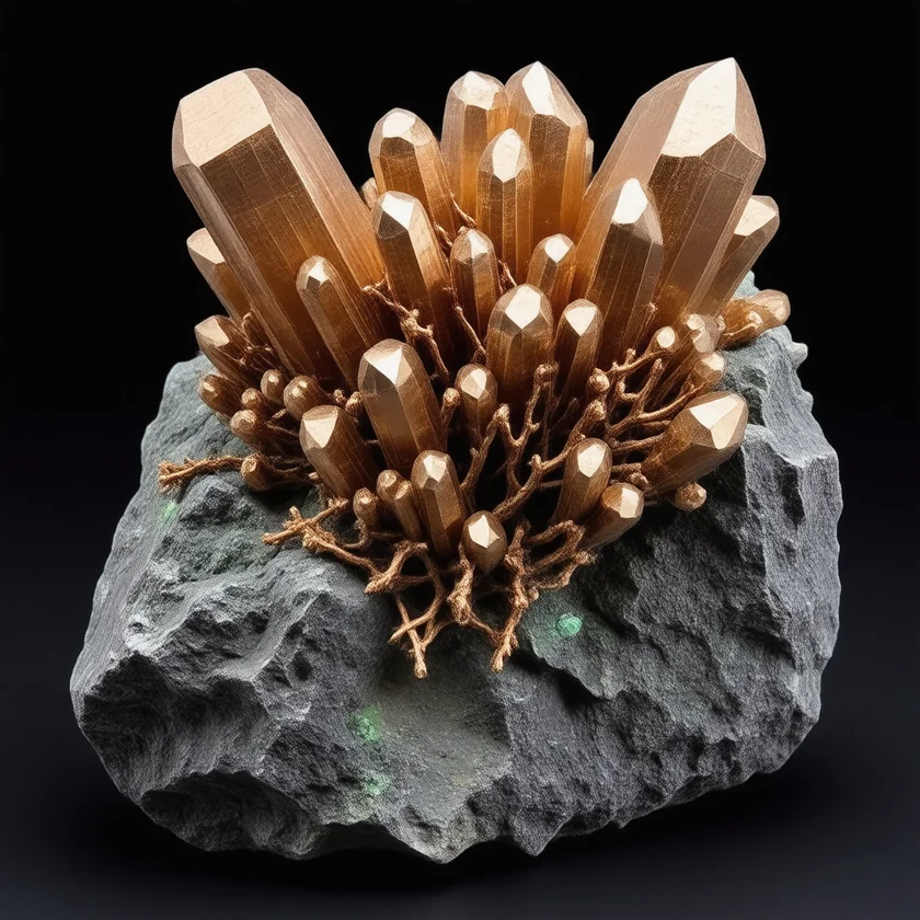
Copper is the catalytic atom in dopamine beta-hydroxylase (DBH), the enzyme inside synaptic vesicles of noradrenergic neurons and adrenal chromaffin cells that converts dopamine to norepinephrine. Copper deficiency literally blocks norepinephrine synthesis. Copper is also the catalytic atom in tyrosinase, the rate-limiting enzyme of melanin synthesis (the pigment of the substantia nigra, the eye, and the skin). And copper is required for myelin formation through several less-direct mechanisms, including cytochrome c oxidase (oligodendrocytes have very high mitochondrial demand), peptidylglycine alpha-amidating monooxygenase (PAM, which amidates neuropeptide hormones), and ceruloplasmin-mediated iron handling in oligodendrocytes. The clinical syndromes that result from copper deficiency in the nervous system are dramatic and consequential: copper-deficiency myelopathy (an acquired adult syndrome from zinc overdose, denture cream, or bariatric surgery that almost perfectly mimics Vitamin B12 subacute combined degeneration), Menkes disease (the X-linked infantile encephalopathy from ATP7A loss-of-function), aceruloplasminemia (brain iron overload from inadequate ferroxidase activity), and the proposed contribution of copper dyshomeostasis to Alzheimer disease and Parkinson disease. This page walks through the mechanisms, the syndromes, the diagnostic workup, and the treatment of copper's role in neurological health.
Table of Contents
- Dopamine Beta-Hydroxylase — Copper and Norepinephrine Synthesis
- Tyrosinase — Melanin Synthesis and Neuromelanin
- Myelin, Cytochrome c Oxidase, and Oligodendrocyte Energy
- Peptidylglycine Alpha-Amidating Monooxygenase (PAM)
- Copper-Deficiency Myelopathy — the Acquired Adult Syndrome
- Zinc-Induced Copper Deficiency (Supplement, Denture Cream)
- Post-Bariatric Copper Deficiency Myelopathy
- Mimicking B12 Subacute Combined Degeneration
- Menkes Disease Neurology
- Copper, Alzheimer Disease, and Parkinson Disease
- Diagnosis and Treatment
- Key Research Papers
- Connections
- Featured Videos
Dopamine Beta-Hydroxylase — Copper and Norepinephrine Synthesis
Dopamine beta-hydroxylase (DBH, EC 1.14.17.1) is a copper-dependent monooxygenase that catalyzes the hydroxylation of dopamine at the beta carbon to produce norepinephrine. The reaction is:
dopamine + ascorbate + O&sub2; → norepinephrine + semidehydroascorbate + H&sub2;O
DBH is a homotetramer of 290 kilodaltons total, expressed exclusively in the dense-core vesicles of noradrenergic and adrenergic neurons (locus coeruleus, lateral medullary tegmentum, sympathetic ganglia) and the chromaffin cells of the adrenal medulla. Each subunit contains two catalytic copper atoms (CuA and CuB) and uses ascorbate (Vitamin C) as the electron donor — one of the clearest examples in human biochemistry of a direct Vitamin-C-and-copper enzymatic partnership.
The location of DBH inside synaptic vesicles is significant. Dopamine is synthesized in the cytosol by aromatic amino acid decarboxylase (DDC, converting L-DOPA to dopamine), then packaged into vesicles by the vesicular monoamine transporter VMAT2. Inside the vesicle, the acidic pH and presence of ascorbate plus the copper-dependent DBH convert dopamine to norepinephrine before exocytotic release at the synapse. In noradrenergic neurons (and adrenal chromaffin cells producing epinephrine), this DBH step is essential — without it, the vesicles release dopamine where norepinephrine should appear.
Copper deficiency reduces DBH activity (because newly synthesized DBH cannot be loaded with copper), reducing norepinephrine production. The clinical consequences are subtle in mild deficiency but include autonomic dysfunction (orthostatic hypotension, hypothermia), cognitive slowing (locus coeruleus norepinephrine is essential for vigilance and attention), and depression-like symptoms (reduced central noradrenergic drive). In Menkes disease, the DBH failure contributes to the hypothermia, hypotonia, and feeding difficulties of the affected infant.
The very rare genetic syndrome of congenital DBH deficiency (loss-of-function mutations in the DBH gene itself) produces a clean phenotype of severe orthostatic hypotension from birth, with detectable dopamine in plasma but virtually undetectable norepinephrine. The treatment is L-DOPS (droxidopa), a synthetic precursor that bypasses the DBH step and is decarboxylated directly to norepinephrine. The genetic DBH-deficient patients prove the absolute requirement for the copper-containing enzyme.
Tyrosinase — Melanin Synthesis and Neuromelanin
Tyrosinase (EC 1.14.18.1) is a copper-dependent oxidase that catalyzes the first two steps of melanin synthesis:
- Hydroxylation of tyrosine to L-DOPA (the rate-limiting step)
- Oxidation of L-DOPA to dopaquinone
Tyrosinase contains two copper atoms (CuA and CuB) in its active site, both essential for catalysis. Loss-of-function tyrosinase mutations are the most common cause of oculocutaneous albinism type 1 (OCA1), which is characterized by absence of melanin in skin, hair, and eyes from birth. OCA1 patients have nystagmus, photophobia, severely reduced visual acuity, and white-to-pale-yellow hair with pale skin and blue or violet irises.
In Menkes disease (ATP7A loss-of-function, systemic copper deficiency), tyrosinase fails because the apoprotein cannot be loaded with copper in the trans-Golgi network. The Menkes infant has the characteristically pale skin and lustreless hair of tyrosinase deficiency, alongside the connective-tissue and neurologic failures from the other copper-dependent enzymes.
In the brain, a related pigment called neuromelanin accumulates in the dopaminergic neurons of the substantia nigra pars compacta and the noradrenergic neurons of the locus coeruleus. Neuromelanin is not synthesized by tyrosinase (it is produced by spontaneous oxidation of dopamine and norepinephrine and accumulates in autophagic vacuoles), but it does contain copper, iron, and other transition metals bound to its melanin scaffold. The loss of neuromelanin-containing neurons is the pathognomonic feature of Parkinson disease, and the iron and copper bound to neuromelanin in surviving neurons has been proposed as a contributor to the oxidative stress of the dopaminergic neurodegeneration process — see the section on Alzheimer and Parkinson disease below.
Myelin, Cytochrome c Oxidase, and Oligodendrocyte Energy
Myelin — the lipid-rich insulating sheath wrapped around axons by oligodendrocytes (in the central nervous system) and Schwann cells (in the peripheral nervous system) — depends on copper indirectly through several mechanisms. The most important is the very high mitochondrial energy demand of oligodendrocytes, which synthesize and maintain an enormous mass of myelin membrane per cell. Cytochrome c oxidase (complex IV of the mitochondrial electron transport chain) is the terminal enzyme of oxidative phosphorylation and its catalytic core contains three copper atoms — two forming the CuA site and a third at the CuB site — plus heme a and heme a3. Copper deficiency reduces COX activity, impairs adenosine triphosphate (ATP) generation in oligodendrocytes, and contributes to the demyelination of copper-deficiency myelopathy.
A second, more direct mechanism involves ceruloplasmin and brain iron handling. Iron is required for oligodendrocyte differentiation and myelin synthesis (the iron-containing enzymes of cholesterol synthesis and fatty-acid desaturation are essential for the lipid biosynthesis that builds myelin). Brain ceruloplasmin (a GPI-anchored membrane form, distinct from the soluble plasma form) is expressed by astrocytes and is required for proper iron handling in the brain. Aceruloplasminemia produces brain iron overload with neurodegeneration, while copper deficiency reduces brain ceruloplasmin activity and impairs iron mobilization to oligodendrocytes.
The third mechanism involves the longstanding observation that copper-deficient animals (sheep, cattle) develop a demyelinating syndrome called "enzootic ataxia" or "swayback" — named for the rear-limb weakness and ataxia of affected lambs. The syndrome shares pathophysiologic features with human copper-deficiency myelopathy and provided the early veterinary literature that informed the human clinical description.
Peptidylglycine Alpha-Amidating Monooxygenase (PAM)
Peptidylglycine alpha-amidating monooxygenase (PAM) is a less-famous but biologically important copper-dependent enzyme that performs the C-terminal amidation of a wide range of bioactive neuropeptides and peptide hormones. Many neuropeptides require C-terminal amidation for full biological activity, including:
- Substance P
- Calcitonin and calcitonin gene-related peptide (CGRP)
- Vasopressin and oxytocin
- Cholecystokinin (CCK)
- Neuropeptide Y
- Gastrin
- Thyrotropin-releasing hormone (TRH)
- Melanin-concentrating hormone
- Glucagon-like peptide 1
The monooxygenase part of PAM contains two copper atoms (called CuH and CuM, about 11 Ångströms apart) and uses ascorbate as the cofactor (like DBH). Severe copper deficiency reduces PAM activity, which can theoretically contribute to subtle endocrine and neuropeptide dysregulation in copper-deficient patients. The clinical signal is hard to isolate because PAM affects so many hormone systems, but it is part of the broader picture of why copper deficiency produces such polyphenotypic disease.
Copper-Deficiency Myelopathy — the Acquired Adult Syndrome
The most clinically important neurologic manifestation of copper deficiency in adults is copper-deficiency myelopathy, first reported as a single case by Schleper and Stuerenburg (2001, a 46-year-old woman), then characterized in case series by Kumar and colleagues at Mayo Clinic (2003 onward) and reviewed by Jaiser and Winston (2010, 55 published cases). In that review the syndrome most often presented in the fifth and sixth decades of life (people in their 40s and 50s) with:
- Gait ataxia — from posterior column (proprioceptive) dysfunction; patients describe difficulty walking in the dark or with eyes closed
- Sensory loss — particularly position and vibration sense in the feet, with relative preservation of pain and temperature
- Spasticity — from lateral column (corticospinal tract) dysfunction; brisk reflexes, Babinski sign, leg stiffness
- Romberg sign — positive, reflecting the proprioceptive deficit
- Variable peripheral neuropathy — large-fiber sensorimotor neuropathy with reduced ankle reflexes
- Optic neuropathy — in some cases, with reduced visual acuity and central scotomata
- Hematologic abnormalities — concomitant anemia (often microcytic) and neutropenia, with sometimes thrombocytopenia. The combination of myelopathy plus unexplained cytopenias is a major diagnostic clue
Magnetic resonance imaging (MRI) of the spinal cord often shows characteristic T2 hyperintensity in the posterior columns of the cervical and upper thoracic cord — the same imaging finding seen in Vitamin B12 subacute combined degeneration. The radiologic resemblance is the source of the most common diagnostic pitfall: assuming the patient has B12 deficiency and treating with B12, while the actual etiology is copper.
The 2010 review of 55 published cases puts numbers on this picture. Women outnumbered men 3.6 to 1. Low blood counts (cytopenias, mostly anemia) were found in 78%. Spinal MRI was abnormal in only 47%, so a normal scan does not rule the condition out; in the 2004 Mayo series of 13 patients, the spinal-cord change showed on MRI in just three. After copper treatment, blood counts returned to normal, while nerve function improved or stabilized but any recovery was partial.
Zinc-Induced Copper Deficiency (Supplement, Denture Cream)
Zinc excess and upper-gut surgery are the two best-known causes of copper-deficiency myelopathy in adults, although in many cases no cause is found: in the 2004 Mayo series of 13 patients the cause "remained unexplained in most", and in the 2010 review of 55 cases no cause was established in 20%. Zinc and copper compete for absorption through the intestinal metallothionein system — high luminal zinc induces large amounts of metallothionein in the enterocyte, and the metallothionein then preferentially binds incoming copper and prevents its export into the portal circulation. The sequestered copper is shed with the enterocyte at the end of its lifespan, producing a steady drain of body copper.
The risk threshold is roughly 50 mg/day of supplemental zinc sustained over months to years. The common settings include:
- "Immune support" zinc lozenges — chronic users taking multiple lozenges per day during cold season or year-round (each lozenge typically contains 10–25 mg zinc)
- Macular degeneration (AREDS-formula supplements) — the original AREDS formulation contained 80 mg zinc plus 2 mg copper (the copper was specifically added to prevent zinc-induced copper deficiency, and this is a model for safe high-dose zinc supplementation). Generic "eye health" formulations may include zinc without the protective copper.
- Acrodermatitis enteropathica / wound healing — legitimate high-dose zinc therapy for these conditions requires concurrent copper monitoring
- Zinc-containing denture adhesive creams — some formulations contained zinc as a binding agent. Patients applying large amounts several times per day for years accumulated systemic zinc burden sufficient to deplete copper. A 2008 study of four such patients measured about 17,000 to 34,000 micrograms of zinc per gram in the creams they used; serum zinc improved in three after they stopped using the cream.
- Taste disorder treatment — oral zinc was historically prescribed for idiopathic dysgeusia; chronic use produced cases of copper deficiency
- Occupational zinc exposure — rare, in welders and metal-foundry workers (and even rarer with modern personal protective equipment)
The case series measure serum zinc alongside copper and ceruloplasmin; in the 2004 Mayo series, zinc was high or high-normal in 7 of the 11 patients tested. Elevated zinc with low copper points strongly to the zinc-induced syndrome, although the 2010 review also found high zinc in some cases not thought to be primary zinc overload. Treatment in the reported cases combined copper with removing the risk factor, such as the zinc source.
Post-Bariatric Copper Deficiency Myelopathy
The second major cause of copper-deficiency myelopathy in adults is bariatric surgery, particularly Roux-en-Y gastric bypass and biliopancreatic diversion with duodenal switch. These procedures bypass the duodenum and proximal jejunum, where dietary copper absorption is concentrated. A 2025 meta-analysis of 43 studies found copper deficiency in 16%, 28%, 21% and 16% of patients at 1, 2, 3 and 4 years after metabolic and bariatric surgery, most often after biliopancreatic diversion, with some patients presenting decades after the original surgery. In a Mayo Clinic review of 40 patients whose copper deficiency showed up as blood-count problems (1970–2005), 10 (25%) had had bariatric surgery and another 14 (35%) other stomach or bowel surgery, most often partial removal of the stomach; in 12 no cause was found, and most also had nerve findings, usually myeloneuropathy.
The clinical picture is identical to the zinc-induced syndrome — progressive myelopathy with gait ataxia, sensory loss, spasticity, and concomitant cytopenias. The diagnostic clue is the surgical history. Bariatric programs now routinely prescribe prophylactic copper supplementation (~2 mg/day) and monitor copper status periodically, but many patients fall out of bariatric follow-up over time and present years later with established myelopathy.
Recovery after diagnosis and treatment is similar across causes: hematologic indices usually normalize within weeks to months of copper repletion, but neurologic recovery is often partial because axonal demyelination has already occurred. Because any neurological recovery in the reported cases was partial, reviewers stress early recognition, before significant neurologic deficit develops.
Mimicking B12 Subacute Combined Degeneration
Copper-deficiency myelopathy is the textbook mimic of Vitamin B12 deficiency subacute combined degeneration. The two syndromes are almost indistinguishable on clinical and radiographic grounds:
- Both produce posterior-column and lateral-column dysfunction with sensory ataxia and spasticity
- Both show T2 hyperintensity in the dorsal columns of the cervical-thoracic cord on magnetic resonance imaging
- Both have concurrent megaloblastic / macrocytic hematologic abnormalities (though copper deficiency more often microcytic with neutropenia, while B12 deficiency more often macrocytic with hypersegmented neutrophils)
- Both can be associated with optic neuropathy and peripheral neuropathy
- Both occur in adults with risk factors (pernicious anemia and gastric surgery for B12; zinc overdose and bariatric surgery for copper)
Critically, B12 supplementation in a copper-deficient patient does not improve the syndrome and may delay diagnosis. Reviews of the case literature describe measuring copper, ceruloplasmin, and zinc at the same time as B12 in suspected B12-deficiency myelopathy, and turning to copper status when B12 levels are normal or B12 repletion does not improve symptoms.
The reverse mistake is equally important: a copper-deficient patient with concomitant B12 deficiency (the two can coexist, particularly in post-bariatric patients) has both deficiencies treated in the reported cases. The hematologic improvement from B12 alone may mask the persistent copper-deficient neurologic syndrome.
Menkes Disease Neurology
Menkes disease, the X-linked recessive disorder caused by loss-of-function ATP7A mutations described on the Connective Tissue page, produces a devastating infantile encephalopathy that demonstrates the simultaneous failure of multiple copper-dependent enzymes in the developing brain:
- Intractable seizures — emerging in the first months of life, often refractory to multiple anticonvulsants. The seizure substrate involves cytochrome c oxidase failure (mitochondrial dysfunction in neurons), neurotransmitter dysregulation (DBH and PAM failures), and oxidative stress.
- Developmental regression — affected infants typically appear normal at birth and may meet early milestones, then progressively regress through the first year of life
- Hypotonia — from both central and peripheral mechanisms
- Feeding difficulties — from impaired swallowing coordination plus the systemic metabolic dysfunction
- Hypothermia — from DBH failure (sympathetic norepinephrine is essential for thermoregulation)
- Vascular tortuosity — including intracranial vessels, with cerebral hemorrhage risk
- White-matter abnormalities — from oligodendrocyte dysfunction in the energy-dependent myelination process
- Death typically by 3 years of age in untreated classical Menkes disease
The treatment is copper histidinate (also called copper histidine), injected under the skin every day, because people with Menkes disease cannot absorb copper from the gut. The injections can normalize blood copper and, depending on the specific ATP7A variant, may replete brain copper. In January 2026 the U.S. Food and Drug Administration (FDA) approved copper histidinate for the treatment of Menkes disease in children — its first approval.
Timing decides much of the outcome. In a 2008 study at the U.S. National Institutes of Health (NIH), 12 newborns who began copper injections within 22 days of birth had 92% survival at a median follow-up of 4.6 years, compared with 13% (median follow-up 1.8 years) in a historical group of 15 children diagnosed and treated late; two of the 12 developed normally, and both carried ATP7A variants that kept some gene function. The same study found that a blood test of dopamine, norepinephrine and their breakdown products — which reflects the copper enzyme DBH — identified affected newborns with high sensitivity and specificity. In the 60-child phase I/II trial of copper histidine, death by age 3 was 28.6% in children treated before symptoms began and 50% in those already older and symptomatic when treatment started, and the early-treated group showed significantly better motor, language and social development.
The intervention requires neonatal screening (currently performed in a small number of pilot programs) because the diagnostic window is so narrow — by the time the classical kinky-hair phenotype appears at 2–4 months of age, the brain copper deficit is usually too advanced to fully reverse.
Copper, Alzheimer Disease, and Parkinson Disease
The role of copper in neurodegenerative disease is an active area of research and a source of substantial controversy. Two propositions are reasonably well-established:
- Total brain copper content is altered in Alzheimer disease and Parkinson disease tissue — with some studies finding reduced copper in surviving neurons and others finding elevated copper in plaques and Lewy bodies. The data are heterogeneous.
- Elevated bioavailable (non-ceruloplasmin) copper in plasma is associated with Alzheimer disease in cross-sectional studies (Squitti et al., meta-analyses). The "non-ceruloplasmin copper" is the loosely-bound serum copper that is not on its protein chaperone; some authors interpret this as evidence of copper dyshomeostasis in Alzheimer disease.
The mechanistic proposals are several:
- Copper binds amyloid-beta peptide with high affinity, and the copper-amyloid complex is redox-active — capable of generating hydrogen peroxide and hydroxyl radicals through Fenton-type chemistry. The oxidative damage may contribute to plaque toxicity and synaptic loss in Alzheimer disease.
- Copper binds alpha-synuclein (the protein that forms Lewy bodies in Parkinson disease) and modulates its aggregation. Copper-bound alpha-synuclein is more prone to oligomer formation, with implications for how Parkinson disease develops.
- Brain ceruloplasmin and iron handling — loss of brain ceruloplasmin activity (whether from genetic aceruloplasminemia or from acquired dysfunction) produces brain iron overload with parkinsonian and other neurodegenerative phenotypes.
- Mitochondrial dysfunction — cytochrome c oxidase deficiency is documented in both Alzheimer and Parkinson disease brain tissue, and copper is required for COX activity.
The therapeutic implications are unsettled. PBT2, an 8-hydroxyquinoline compound that binds copper and zinc (a metal ionophore) and was developed to target amyloid beta, was tested in the 12-month IMAGINE study of people with prodromal or mild Alzheimer disease (40 completed the double-blind phase). PBT2 lowered the amyloid PET signal by 3% (p = 0.048), but there was no significant difference from controls at 12 months, and none in brain imaging, blood amyloid markers, cognition or daily function. Copper supplementation in Alzheimer disease has not been studied rigorously, and on current evidence the research does not show a benefit from copper supplements for cognitive aging in people whose copper status is normal.
For more on these conditions, see our pages on Alzheimer Disease and Parkinson Disease.
Diagnosis and Treatment
The tests described in the case literature for suspected copper-deficiency myelopathy:
- Serum copper — typically low (<70 mcg/dL)
- Ceruloplasmin — typically low (<15 mg/dL), with parallel reduction in ferroxidase activity
- Serum zinc — elevated in the zinc-induced cases
- Complete blood count with differential — microcytic anemia and neutropenia are characteristic; thrombocytopenia in severe cases
- Vitamin B12, methylmalonic acid, homocysteine — to rule out concomitant B12 deficiency
- Magnetic resonance imaging of the cervical and thoracic spinal cord — T2 hyperintensity in the dorsal columns
- Nerve conduction studies and electromyography (EMG) — characterize the peripheral neuropathy component
- Dietary and surgical history — zinc supplements, denture cream, bariatric surgery, malabsorption
Treatment as reported in case series and reviews (the 2010 review summarizes it as copper supplementation plus modification of any risk factors):
- Removing the cause — stopping zinc supplements or zinc-containing denture cream, correcting parenteral nutrition, treating underlying malabsorption
- Oral copper repletion — 2–8 mg elemental copper per day (the Recommended Dietary Allowance (RDA) is 0.9 mg/day; repletion doses are 2–10× Recommended Dietary Allowance for 6–12 weeks then taper to maintenance)
- Intravenous (IV) copper — reserved for severe deficiency with poor oral tolerance or absorption
- Whole-food sources — beef liver, oysters, dark chocolate, cashews, sunflower seeds; see Whole Food Copper Sources
- Cofactor nutrients — practitioners also point to Vitamin A (retinol form, for hepatic ceruloplasmin synthesis), magnesium, and Vitamin C (DBH cofactor)
- Concurrent B12 if dual deficiency — common in post-bariatric patients
- Follow-up testing — serum copper, ceruloplasmin, zinc, and complete blood count (CBC), commonly repeated at 6 weeks and 3 months. Hematologic recovery is usually rapid; neurologic recovery is variable and often incomplete
Key Research Papers
- Schleper B, Stuerenburg HJ (2001). Copper deficiency-associated myelopathy in a 46-year-old woman. Journal of Neurology. — PubMed PMID: 11569901
- Kumar N, Gross JB Jr, Ahlskog JE (2004). Copper deficiency myelopathy produces a clinical picture like subacute combined degeneration. Neurology. 2004;63(1):33-39. — PubMed PMID: 15249607
- Jaiser SR, Winston GP (2010). Copper deficiency myelopathy. Journal of Neurology. — PubMed PMID: 20232210
- Nations SP et al. (2008). Denture cream: an unusual source of excess zinc, leading to hypocupremia and neurologic disease. Neurology. — PubMed PMID: 18525032
- Hedera P, Fink JK, Bockenstedt PL, Brewer GJ (2003). Myelopolyneuropathy and pancytopenia due to copper deficiency and high zinc levels of unknown origin: further support for existence of a new zinc overload syndrome. Archives of Neurology. 2003;60(9):1303-1306. — PubMed PMID: 12975299
- Spinazzi M et al. (2007). Myelo-optico-neuropathy in copper deficiency occurring after partial gastrectomy. Journal of Neurology. — PubMed PMID: 17415508
- Kaler SG (2011). ATP7A-related copper transport diseases — emerging concepts and future trends. Nature Reviews Neurology. — PubMed PMID: 21221114
- Squitti R, Simonelli I, Ventriglia M, Siotto M, Pasqualetti P, Rembach A, Doecke J, Bush AI (2014). Meta-analysis of serum non-ceruloplasmin copper in Alzheimer's disease. Journal of Alzheimer's Disease. 2014;38(4):809-822. — PubMed PMID: 24072069
- Bush AI (2000). Metals and neuroscience. Current Opinion in Chemical Biology. 2000;4(2):184-191. — PubMed PMID: 10742195
- Halfdanarson TR, Kumar N, Li CY, Phyliky RL, Hogan WJ (2008). Hematological manifestations of copper deficiency: a retrospective review. European Journal of Haematology. 2008;80(6):523-531. — PubMed PMID: 18284630
- Xu B, Xiao T, Li J, Lu Q, Zhang Y, Wan B, Liang D (2025). Copper deficiency: a frequently overlooked complication after MBS — a systematic review and meta-analysis. Obesity Surgery. 2025;35(2):602-613. — PubMed PMID: 39755772
- Kaler SG, Holmes CS, Goldstein DS, Tang J, Godwin SC, Donsante A, Liew CJ, Sato S, Patronas N (2008). Neonatal diagnosis and treatment of Menkes disease. New England Journal of Medicine. 2008;358(6):605-614. — PubMed PMID: 18256395
- Kaler SG (2014). Neurodevelopment and brain growth in classic Menkes disease is influenced by age and symptomatology at initiation of copper treatment. Journal of Trace Elements in Medicine and Biology. 2014;28(4):427-430. — PubMed PMID: 25281031
- Blair HA (2026). Copper histidinate: pediatric first approval. Paediatric Drugs. 2026;28(4):441-445. — PubMed PMID: 42018271
- Horn D, Barrientos A (2008). Mitochondrial copper metabolism and delivery to cytochrome c oxidase. IUBMB Life. 2008;60(7):421-429. — PubMed PMID: 18459161
- Chauhan S, Kline CD, Mayfield M, Blackburn NJ (2014). Binding of copper and silver to single-site variants of peptidylglycine monooxygenase reveals the structure and chemistry of the individual metal centers. Biochemistry. 2014;53(6):1069-1080. — PubMed PMID: 24471980
- Villemagne VL, Rowe CC, Barnham KJ, Cherny R, Woodward M, Bozinosvski S, Salvado O, Bourgeat P, Perez K, Fowler C, Rembach A, Maruff P, Ritchie C, Tanzi R, Masters CL (2017). A randomized, exploratory molecular imaging study targeting amyloid β with a novel 8-OH quinoline in Alzheimer's disease: the PBT2-204 IMAGINE study. Alzheimer's & Dementia (New York). 2017;3(4):622-635. — PubMed PMID: 29201996
- Goyens P, Brasseur D, Cadranel S (1985). Copper deficiency in infants with active celiac disease. Journal of Pediatric Gastroenterology and Nutrition. — PubMed PMID: 4032183
PubMed Topic Searches
Connections
- All Minerals
- Copper Overview
- Copper Benefits Hub
- Copper for Hemoglobin and Ceruloplasmin
- Copper for Connective Tissue
- Copper for Antioxidant Defense
- Zinc (Copper Antagonist)
- Vitamin B12 (Subacute Combined Degeneration Mimic)
- Vitamin A (Ceruloplasmin Synthesis)
- Vitamin C (DBH Cofactor)
- Magnesium
- Morley Robbins
- Ceruloplasmin and Bioavailable Copper
- Alzheimer Disease
- Parkinson Disease
- Anemia
- Fatigue
- Organ Meats (Beef Liver)
- Beef
- Dark Chocolate
- Deficiency
- Toxicity
- History
- Sources