AFP Test: Alpha-Fetoprotein Lab Guide
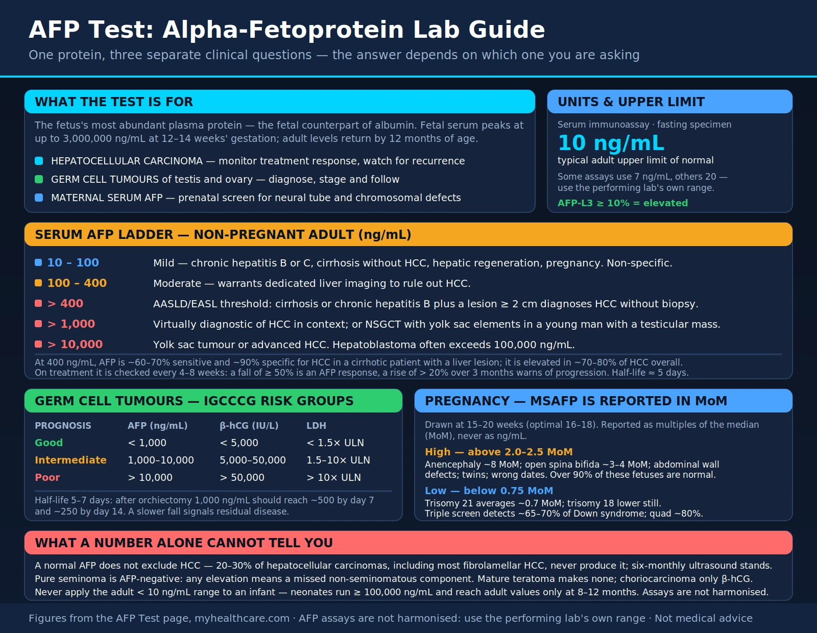
Table of Contents
What is AFP?
Alpha-fetoprotein (AFP) is a serum protein of the fetus. In 1956 Bergstrand and Czar reported a new protein fraction in serum from the human fetus (PubMed PMID: 13351554). In 1963 Abelev and colleagues reported that transplantable mouse hepatomas produced this embryonal alpha-globulin (PubMed PMID: 14010646), and in 1964 Tatarinov reported detecting the embryo-specific alpha-globulin in the blood serum of a patient with primary liver cancer (PubMed PMID: 14207501). Abelev reviewed the experimental and clinical data in 1968 (PubMed PMID: 4174340).
AFP belongs to the albumin gene family. A 1994 paper describing afamin named it the fourth member of the family after albumin, AFP and vitamin D-binding protein, and reported that its gene maps to chromosome 4, as do the other members (PubMed PMID: 7517938).
AFP does not vanish entirely after birth. A 1971 radioimmunoassay study by Ruoslahti and Seppälä demonstrated AFP in the serum of healthy human adults (PubMed PMID: 5002872). In a 1981 study of normal infants, serum AFP fell rapidly after birth, and the normal infant level approached the adult level only at about 8 months of age (PubMed PMID: 6163129).
Measuring serum AFP is used in three main settings covered on this page: liver cancer (hepatocellular carcinoma, HCC), germ cell tumours, and childhood liver tumours, plus maternal serum AFP in prenatal screening. A 2019 review lists testicular germ cell tumours, hepatoblastoma and hepatocellular carcinoma as the cancers in which AFP is useful in infants (PubMed PMID: 30367785).
Biology and Normal Values
AFP is measured by immunoassay on a blood sample. There is no single universal cut-off for "elevated" in the sources on this page; each study states its own. For example, an AFP cut-off of 20 ng/mL is the most common threshold for HCC surveillance, according to the 2023 AASLD practice guidance (PubMed PMID: 37199193). A study of seminoma used an elevated AFP of more than 15 ng/mL (PubMed PMID: 21782217), and the 2021 IGCCCG update modelled risk only for unequivocal AFP elevation, which it defined as more than 30 ng/mL (PubMed PMID: 33822655).
Infants: the 1981 infant study estimated the half-life of AFP breakdown at 5.5 days between birth and 2 weeks, 11 days between 2 weeks and 2 months, and 33 days between 2 and 4 months of age (PubMed PMID: 6163129). A 2019 review found that preterm and term infants up to the third month of life had the highest average AFP concentrations. The data varied too widely for robust reference intervals to be defined. This made it difficult, if not impossible, to use an upper reference limit to rule out a cancer from a single AFP result in the first months of life. The authors proposed judging a retest against an age-specific half-life instead (PubMed PMID: 30367785).
AFP-L3: AFP-L3% is the fraction of AFP that binds the lectin Lens culinaris agglutinin. In a 2007 study of 166 patients with HCC and 106 with benign liver disease, AFP-L3% was not reported when total AFP was below 10 ng/mL, and every patient with total AFP above 200 ng/mL had HCC. Among patients with total AFP of 10–200 ng/mL, an AFP-L3% above 10% had 71% sensitivity and 63% specificity for HCC. An AFP-L3% above 35% had 33% sensitivity and 100% specificity (PubMed PMID: 17368240).
GALAD: a 2014 model combining gender, age, AFP-L3, AFP and des-carboxy-prothrombin (DCP) was developed in 670 patients from a single UK centre and validated in independent data sets. When optimised, it gave sensitivity and specificity above 0.88 regardless of disease stage (PubMed PMID: 24220911). The 2023 AASLD guidance reports that GALAD achieved sensitivities of 60%–80% for early HCC in a multinational case-control study (PubMed PMID: 37199193).
AFP in Hepatocellular Carcinoma
In the GLOBOCAN 2020 estimates, liver cancer was the third leading cause of cancer death worldwide, at 8.3% of cancer deaths (PubMed PMID: 33538338). The 2025 EASL guidelines state that HCC accounts for about 90% of primary liver cancers (PubMed PMID: 39690085). A 2022 review names viral hepatitis and alcohol as important risk factors, with non-alcoholic fatty liver disease rapidly becoming a dominant cause (PubMed PMID: 36084663).
Surveillance
The 2023 AASLD practice guidance recommends HCC surveillance with ultrasound and AFP at roughly six-month intervals in at-risk people, including those with chronic hepatitis B infection or cirrhosis from any cause. It advises diagnostic multiphase CT or MRI when AFP is 20 ng/mL or higher, or rising. In the guidance, an AFP cut-off of 20 ng/mL gives a sensitivity of about 60% and a specificity of about 90% (PubMed PMID: 37199193).
A 2018 meta-analysis of 32 studies (13,367 patients with cirrhosis) found that ultrasound detected any-stage HCC with 84% sensitivity but early-stage HCC with only 47% sensitivity. Ultrasound with AFP detected early-stage HCC with 63% sensitivity, against 45% without AFP. Ultrasound alone had higher specificity than ultrasound plus AFP (PubMed PMID: 29425931).
A 2001 case-control study of 170 patients with HCC and 170 with chronic liver disease found that the best discriminating AFP value was 16 ng/mL. A value of 20 ng/mL gave 60.0% sensitivity and 90.6% specificity. The authors concluded that AFP monitoring misses many HCCs and inappropriately arouses suspicion of malignancy in many patients (PubMed PMID: 11394657).
Diagnosis
The 2023 AASLD guidance states that AFP at 400 ng/mL was previously recommended as a diagnostic criterion for HCC. Over 40% of HCCs have normal AFP levels, and AFP can also be raised in other cancers, including intrahepatic cholangiocarcinoma, gastric cancer and germ cell tumours. Given this insufficient accuracy, the guidance no longer recommends AFP for HCC diagnosis. It advises biopsy for patients whose imaging is not characteristic, whatever the AFP level. It also notes that no clear threshold has been set for a significant AFP elevation: 200 or 400 ng/mL may be considered significant, depending on the clinical context (PubMed PMID: 37199193).
Fibrolamellar HCC is a rare subtype that mainly affects adolescents and young adults without underlying liver disease or cirrhosis. A 2025 review reports that it is less than 5% of all liver tumours and that typical tumour markers such as AFP are usually absent (PubMed PMID: 41178855).
Liver Transplantation
The Metroticket 2.0 model was built from 1,018 patients given a liver transplant for HCC at three Italian centres. It was validated in 341 patients in Shanghai. The sum of tumour number and size, and the log10 AFP level, were associated with HCC-specific death. For a 70% chance of HCC-specific survival at 5 years, the model required an AFP below 200 ng/mL with a tumour number-plus-size sum of no more than 7. An AFP of 200–400 ng/mL required a sum of 5 or less, and an AFP of 400–1000 ng/mL a sum of 4 or less (PubMed PMID: 28989060). The 2023 AASLD guidance states that patients with AFP above 1000 ng/mL must be downstaged to below 500 ng/mL to be considered downstaged. It also reports that AFP levels as low as above 20 ng/mL have been consistently associated with recurrence after transplantation (PubMed PMID: 37199193).
AFP in Drug Trials
The REACH-2 trial enrolled only patients with advanced HCC and AFP of 400 ng/mL or higher who had previously received sorafenib. Its 292 patients were randomised 2:1 to ramucirumab or placebo. Median overall survival was 8.5 months versus 7.3 months (hazard ratio 0.710). The authors describe REACH-2 as, to their knowledge, the first positive phase 3 trial in a biomarker-selected HCC population (PubMed PMID: 30665869). The 2023 AASLD guidance names AFP of 400 ng/mL or higher with ramucirumab as the only biomarker so far linked to treatment-related clinical benefit in HCC, and notes that the absolute survival benefit was modest (PubMed PMID: 37199193).
The RESORCE trial (Bruix 2017) tested regorafenib in 573 patients with HCC that had progressed on sorafenib. Randomisation was stratified by factors including AFP level, and regorafenib improved overall survival (hazard ratio 0.63) (PubMed PMID: 27932229). In an exploratory biomarker analysis of RESORCE samples, plasma AFP was associated with poorer overall survival independently of regorafenib treatment. In other words, it marked prognosis, not benefit from the drug (PubMed PMID: 30738047).
AFP in Germ Cell Tumors
AFP, human chorionic gonadotrophin (hCG) and lactate dehydrogenase (LDH) are the conventional serum markers of testicular germ cell tumours. A 2016 review states that AFP and hCG have limited sensitivity and specificity: AFP indicates the yolk sac tumour subtype, and hCG indicates choriocarcinoma or syncytiotrophoblast. LDH is very nonspecific. As a result, seminomas and nonseminomatous tumours made up of pure embryonal carcinoma are generally negative for these markers (PubMed PMID: 27754472).
Seminoma With a Raised AFP
When a histologically pure seminoma comes with a raised serum AFP, a 1998 study states, it is generally assumed that an undetected focus of yolk sac tumour is present, and the patient is managed as having a nonseminomatous tumour. That study's 10 patients had only minor elevations (10.4–16 ng/mL), and none developed evidence of yolk sac tumour over a mean 6 years of follow-up. The authors suggested that minor elevations may not indicate a hidden yolk sac focus, and called for further studies (PubMed PMID: 9769381). A 2011 series of 22 patients with pure seminoma at orchiectomy and AFP above 15 ng/mL had a median pre-orchiectomy AFP of 248 ng/mL. They were treated as having nonseminomatous tumours, and 67% had nonseminomatous elements in the retroperitoneum (PubMed PMID: 21782217).
IGCCCG Prognostic Classification
The 1997 International Germ Cell Consensus Classification analysed 5,202 patients with nonseminomatous tumours and 660 with seminoma. For nonseminomatous tumours, the independent adverse factors were a mediastinal primary site, the degree of elevation of AFP, hCG and LDH, and nonpulmonary visceral metastases. The good-prognosis group (60% of patients) had 91% 5-year survival. The intermediate group (26%) had 79%, and the poor group (14%, all nonseminomatous) had 48% (PubMed PMID: 9053482). The 2021 IGCCCG update, in 9,728 men treated between 1990 and 2013, found 5-year overall survival of 96%, 89% and 67% in the good, intermediate and poor groups. It added an LDH cut-off of 2.5 times the upper limit of normal, older age and lung metastases as adverse factors (PubMed PMID: 33822655).
Marker Decline During Chemotherapy
In a Swedish-Norwegian population-based protocol (603 patients, 1995–2003), tumour markers were measured weekly during two courses of BEP chemotherapy. A good response was defined as an AFP half-life of 7 days or less and/or an hCG half-life of 3 days or less. Patients with a prolonged half-life received intensified treatment. Ten-year overall survival was 94.7%, 90.0% and 67.4% in the good, intermediate and poor IGCCCG groups (PubMed PMID: 21482994). In the GETUG 13 phase 3 trial of poor-prognosis tumours, hCG and AFP were measured at day 18–21 after one BEP cycle, and 80% of patients had an unfavourable decline. Among those patients, a dose-dense regimen gave a 3-year progression-free survival of 59%, against 48% with continued BEP (hazard ratio 0.66) (PubMed PMID: 25456363).
AFP in Pediatric Cancers
Hepatoblastoma is the most common primary malignant liver tumour of infancy and childhood, occurring mostly in the first two years of life (PubMed PMID: 28126357). A 2024 review reports that monitoring for relapse after treatment has gradually shifted to serial serum AFP alone, because most relapses show a raised AFP. The authors note that little primary data supports this practice and that certain clinical features call for caution (PubMed PMID: 38551915).
A low AFP can itself be a warning sign. In the SIOPEL trials, the 21 children whose serum AFP was below 100 ng/mL at diagnosis formed a high-risk subgroup: they had extensive disease, poor response to chemotherapy and a two-year overall survival of 24% (PubMed PMID: 18166449).
In a review of 194 fetuses and newborns with liver tumours, half of those with hepatoblastoma had an abnormally raised AFP, compared with 14% of those with haemangioma. The review defined "raised" against each institution's normal level for the age group, because laboratory values differed between institutions (PubMed PMID: 18022426). Normal infants have high AFP that falls over the first months of life, so adult ranges do not fit them (PubMed PMID: 6163129) (PubMed PMID: 30367785).
Maternal Serum AFP and Prenatal Screening
Neural Tube Defects
The 1977 UK collaborative study (19 centres) measured maternal serum AFP between 10 and 24 weeks of pregnancy. It included 301 pregnancies with neural-tube defects and more than 18,000 unaffected singleton pregnancies. The best time to detect open spina bifida was 16–18 weeks. Results were expressed as multiples of the median (MoM) for unaffected pregnancies. At 16–18 weeks, 88% of cases of anencephaly, 79% of cases of open spina bifida and 3% of unaffected singleton pregnancies had AFP at or above 2.5 MoM. In the UK, a woman above 2.5 MoM at 16–18 weeks had about a 1-in-20 chance of a fetus with open spina bifida, and about a 1-in-10 chance of any neural-tube defect. The study concluded that the test was an effective way to select women for ultrasonography and amniocentesis (PubMed PMID: 69055).
Other Causes of a Raised Level
- Abdominal wall defects: in a Chinese screening series, median maternal serum AFP was 4.41 MoM with fetal gastroschisis and 2.31 MoM with omphalocele, against 0.98 MoM in controls (PubMed PMID: 29039168).
- Low birthweight: in a 1977 prospective study of 4,224 pregnancies, 10.7% of women with serum AFP above 2.3 times the median delivered infants under 2.5 kg, against 4.2% in the general population (PubMed PMID: 69878). A 1998 study of women having genetic amniocentesis found that those with AFP above 2.0 MoM had more miscarriages (4% against 1%) and more babies under 2,500 g (14.6% against 7.7%) than women with normal or low values (PubMed PMID: 9848697).
Low AFP and Chromosomal Conditions
A 1984 report began with an index case of "undetectable" maternal serum AFP at 16 weeks, in a pregnancy that ended in the birth of an infant with trisomy 18. In 41 pregnancies with fetal autosomal trisomy, maternal serum AFP was significantly lower than in matched controls (PubMed PMID: 6201071). In the same year, a study of 61 pregnancies with Down syndrome found a median maternal serum AFP of 0.72 MoM. Selecting women with AFP at or below 0.5 MoM would have identified 21% of affected pregnancies and 5% of unaffected ones (PubMed PMID: 6201687).
AFP was then combined with other markers. In a 1988 study, hCG in Down syndrome pregnancies was twice the median of unaffected pregnancies. Combining hCG, unconjugated oestriol, AFP and maternal age was projected to detect over 60% of affected pregnancies (PubMed PMID: 2460174). In a 5-year evaluation of the quadruple test in 46,193 pregnancies, 81% of Down syndrome pregnancies screened positive, with a 7% false-positive rate (PubMed PMID: 12642052). A screening result is not a diagnosis. In the 1977 study, the test served to select women for further testing (PubMed PMID: 69055).
Monitoring Treatment Response
In each of these settings, studies have tracked how AFP changes over time, not only a single value.
HCC on systemic therapy: a 2010 study pooled 72 patients from three phase 2 trials of antiangiogenic first-line therapy. It defined an early AFP response as a fall of more than 20% from baseline after 2–4 weeks. The 12 early responders had better response and disease-control rates, longer median progression-free survival (7.5 versus 1.9 months) and longer median overall survival (15.3 versus 4.1 months). AFP response remained an independent predictor in multivariate analysis (PubMed PMID: 20572033).
HCC after resection: a 2013 study of 225 patients, with a validation cohort of 117, defined a prolonged post-surgical AFP half-life as more than 7 days. Within 2 years, 59% of patients with a prolonged half-life had an early recurrence, against 29.3% of those with a normal half-life (PubMed PMID: 23108121). The 2023 AASLD guidance describes surveillance after resection with cross-sectional imaging of the abdomen and chest plus serum AFP every 3–6 months (PubMed PMID: 37199193).
Germ cell tumours: the marker half-life thresholds of the Swedish-Norwegian protocol (PubMed PMID: 21482994) and the day 18–21 marker decline of GETUG 13 (PubMed PMID: 25456363) are described above, under Germ Cell Tumors.
Hepatoblastoma: relapse monitoring in AFP-positive cases has gradually shifted from repeated imaging to serial AFP alone. As noted above, a 2024 review found little primary data behind this practice (PubMed PMID: 38551915).
Interpretation Pitfalls
A normal AFP does not exclude HCC. According to the 2023 AASLD guidance, over 40% of HCCs have normal AFP levels (PubMed PMID: 37199193), and AFP is usually absent in fibrolamellar HCC (PubMed PMID: 41178855).
A raised AFP is not always cancer. Case reports describe patients with cirrhosis and an acute flare of hepatitis B or C whose AFP rose markedly without HCC. In these cases AFP fell gradually after antiviral treatment began (PubMed PMID: 15906959). AFP can also be raised in cancers other than HCC, including intrahepatic cholangiocarcinoma, gastric cancer and germ cell tumours (PubMed PMID: 37199193).
Seminoma with a raised AFP. Studies differ on how to read a raised AFP alongside histologically pure seminoma. In one series, minor elevations of up to 16 ng/mL were not followed by yolk sac tumour (PubMed PMID: 9769381). In another, two-thirds of patients with AFP above 15 ng/mL had nonseminomatous elements in the retroperitoneum (PubMed PMID: 21782217).
Infants. AFP is physiologically high after birth and declines over months (PubMed PMID: 6163129). A 2019 review found that robust reference intervals for the first months of life could not be defined, and judged a single result unsuitable for ruling out cancer (PubMed PMID: 30367785).
Laboratories differ. The 2019 paediatric review excluded studies partly for drawbacks in tracing the AFP assay to higher-order reference materials (PubMed PMID: 30367785). A neonatal tumour review could not assign one standard reference number because AFP values varied between institutions (PubMed PMID: 18022426).
Key Research Papers
- Abelev GI. (1968). Production of embryonal serum alpha-globulin by hepatomas: review of experimental and clinical data. Cancer Res. 28(7):1344-50. — PubMed PMID: 4174340
- Ruoslahti E, Seppälä M. (1971). Studies of carcino-fetal proteins. 3. Development of a radioimmunoassay for α-fetoprotein. Demonstration of α-fetoprotein in serum of healthy human adults. Int J Cancer. 8(3):374-83. — PubMed PMID: 5002872
- Bruix J, Sherman M; Practice Guidelines Committee, American Association for the Study of Liver Diseases. (2005). Management of hepatocellular carcinoma. Hepatology. 42(5):1208-36. — PubMed PMID: 16250051
- Bruix J, Sherman M; American Association for the Study of Liver Diseases. (2011). Management of hepatocellular carcinoma: an update. Hepatology. 53(3):1020-2. — PubMed PMID: 21374666
- Zhu AX, Kang YK, Yen CJ, et al. (2019). Ramucirumab after sorafenib in patients with advanced hepatocellular carcinoma and increased α-fetoprotein concentrations (REACH-2): a randomised, double-blind, placebo-controlled, phase 3 trial. Lancet Oncol. 20(2):282-296. — PubMed PMID: 30665869
- Llovet JM, Ricci S, Mazzaferro V, et al. (2008). Sorafenib in advanced hepatocellular carcinoma. N Engl J Med. 359(4):378-90. — PubMed PMID: 18650514
- International Germ Cell Cancer Collaborative Group. (1997). International Germ Cell Consensus Classification: a prognostic factor-based staging system for metastatic germ cell cancers. J Clin Oncol. 15(2):594-603. — PubMed PMID: 9053482
- Wald NJ, Cuckle HS, Densem JW, et al. (1988). Maternal serum screening for Down's syndrome in early pregnancy. BMJ. 297(6653):883-7. — PubMed PMID: 2460174
- Brock DJ, Barron L, Jelen P, et al. (1977). Maternal serum-alpha-fetoprotein measurements as an early indicator of low birth-weight. Lancet. 2(8032):267-8. — PubMed PMID: 69878
- Vogel A, Meyer T, Sapisochin G, et al. (2022). Hepatocellular carcinoma. Lancet. 400(10360):1345-1362. — PubMed PMID: 36084663
- Bergstrand CG, Czar B. (1956). Demonstration of a new protein fraction in serum from the human fetus. Scand J Clin Lab Invest. 8(2):174. — PubMed PMID: 13351554
- Abelev GI, Perova SD, Khramkova NI, et al. (1963). Production of embryonal alpha-globulin by transplantable mouse hepatomas. Transplantation. 1:174-80. — PubMed PMID: 14010646
- Tatarinov YuS. (1964). [Detection of embryo-specific alpha-globulin in the blood serum of a patient with primary liver cancer]. Vopr Med Khim. 10:90-1. (In Russian.) — PubMed PMID: 14207501
- Lichenstein HS, Lyons DE, Wurfel MM, et al. (1994). Afamin is a new member of the albumin, alpha-fetoprotein, and vitamin D-binding protein gene family. J Biol Chem. 269(27):18149-54. — PubMed PMID: 7517938
- Wu JT, Book L, Sudar K. (1981). Serum alpha fetoprotein (AFP) levels in normal infants. Pediatr Res. 15(1):50-2. — PubMed PMID: 6163129
- Ferraro S, Panzeri A, Braga F, et al. (2019). Serum α-fetoprotein in pediatric oncology: not a children's tale. Clin Chem Lab Med. 57(6):783-797. — PubMed PMID: 30367785
- Leerapun A, Suravarapu SV, Bida JP, et al. (2007). The utility of Lens culinaris agglutinin-reactive alpha-fetoprotein in the diagnosis of hepatocellular carcinoma: evaluation in a United States referral population. Clin Gastroenterol Hepatol. 5(3):394-402; quiz 267. — PubMed PMID: 17368240
- Johnson PJ, Pirrie SJ, Cox TF, et al. (2014). The detection of hepatocellular carcinoma using a prospectively developed and validated model based on serological biomarkers. Cancer Epidemiol Biomarkers Prev. 23(1):144-53. — PubMed PMID: 24220911
- Bae JS, Park SJ, Park KB, et al. (2005). Acute exacerbation of hepatitis in liver cirrhosis with very high levels of alpha-fetoprotein but no occurrence of hepatocellular carcinoma. Korean J Intern Med. 20(1):80-5. — PubMed PMID: 15906959
- Sung H, Ferlay J, Siegel RL, et al. (2021). Global Cancer Statistics 2020: GLOBOCAN Estimates of Incidence and Mortality Worldwide for 36 Cancers in 185 Countries. CA Cancer J Clin. 71(3):209-249. — PubMed PMID: 33538338
- European Association for the Study of the Liver. (2025). EASL Clinical Practice Guidelines on the management of hepatocellular carcinoma. J Hepatol. 82(2):315-374. — PubMed PMID: 39690085
- Singal AG, Llovet JM, Yarchoan M, et al. (2023). AASLD Practice Guidance on prevention, diagnosis, and treatment of hepatocellular carcinoma. Hepatology. 78(6):1922-1965. — PubMed PMID: 37199193
- Tzartzeva K, Obi J, Rich NE, et al. (2018). Surveillance Imaging and Alpha Fetoprotein for Early Detection of Hepatocellular Carcinoma in Patients With Cirrhosis: A Meta-analysis. Gastroenterology. 154(6):1706-1718.e1. — PubMed PMID: 29425931
- Trevisani F, D'Intino PE, Morselli-Labate AM, et al. (2001). Serum alpha-fetoprotein for diagnosis of hepatocellular carcinoma in patients with chronic liver disease: influence of HBsAg and anti-HCV status. J Hepatol. 34(4):570-5. — PubMed PMID: 11394657
- da Fonseca LG, Araujo RLC. (2025). Fibrolamellar hepatocellular carcinoma: Advances, challenges and opportunities in a rare malignancy. World J Gastrointest Surg. 17(10):109107. — PubMed PMID: 41178855
- Mazzaferro V, Sposito C, Zhou J, et al. (2018). Metroticket 2.0 Model for Analysis of Competing Risks of Death After Liver Transplantation for Hepatocellular Carcinoma. Gastroenterology. 154(1):128-139. — PubMed PMID: 28989060
- Bruix J, Qin S, Merle P, et al. (2017). Regorafenib for patients with hepatocellular carcinoma who progressed on sorafenib treatment (RESORCE): a randomised, double-blind, placebo-controlled, phase 3 trial. Lancet. 389(10064):56-66. — PubMed PMID: 27932229
- Teufel M, Seidel H, Köchert K, et al. (2019). Biomarkers Associated With Response to Regorafenib in Patients With Hepatocellular Carcinoma. Gastroenterology. 156(6):1731-1741. — PubMed PMID: 30738047
- Shao YY, Lin ZZ, Hsu C, et al. (2010). Early alpha-fetoprotein response predicts treatment efficacy of antiangiogenic systemic therapy in patients with advanced hepatocellular carcinoma. Cancer. 116(19):4590-6. — PubMed PMID: 20572033
- Shim JH, Han S, Lee YJ, et al. (2013). Half-life of serum alpha-fetoprotein: an early prognostic index of recurrence and survival after hepatic resection for hepatocellular carcinoma. Ann Surg. 257(4):708-17. — PubMed PMID: 23108121
- Murray MJ, Huddart RA, Coleman N. (2016). The present and future of serum diagnostic tests for testicular germ cell tumours. Nat Rev Urol. 13(12):715-725. — PubMed PMID: 27754472
- Nazeer T, Ro JY, Amato RJ, et al. (1998). Histologically pure seminoma with elevated alpha-fetoprotein: a clinicopathologic study of ten cases. Oncol Rep. 5(6):1425-9. — PubMed PMID: 9769381
- Kundu SD, Carver BS, Sheinfeld J. (2011). Retroperitoneal histologic findings of patients with elevated serum alpha-fetoprotein and pure seminoma at orchiectomy. Urology. 78(4):844-7. — PubMed PMID: 21782217
- Gillessen S, Sauvé N, Collette L, et al. (2021). Predicting Outcomes in Men With Metastatic Nonseminomatous Germ Cell Tumors (NSGCT): Results From the IGCCCG Update Consortium. J Clin Oncol. 39(14):1563-1574. — PubMed PMID: 33822655
- Olofsson SE, Tandstad T, Jerkeman M, et al. (2011). Population-based study of treatment guided by tumor marker decline in patients with metastatic nonseminomatous germ cell tumor: a report from the Swedish-Norwegian Testicular Cancer Group. J Clin Oncol. 29(15):2032-9. — PubMed PMID: 21482994
- Fizazi K, Pagliaro L, Laplanche A, et al. (2014). Personalised chemotherapy based on tumour marker decline in poor prognosis germ-cell tumours (GETUG 13): a phase 3, multicentre, randomised trial. Lancet Oncol. 15(13):1442-1450. — PubMed PMID: 25456363
- Sharma D, Subbarao G, Saxena R. (2017). Hepatoblastoma. Semin Diagn Pathol. 34(2):192-200. — PubMed PMID: 28126357
- De Ioris M, Brugieres L, Zimmermann A, et al. (2008). Hepatoblastoma with a low serum alpha-fetoprotein level at diagnosis: the SIOPEL group experience. Eur J Cancer. 44(4):545-50. — PubMed PMID: 18166449
- Isaacs H Jr. (2007). Fetal and neonatal hepatic tumors. J Pediatr Surg. 42(11):1797-803. — PubMed PMID: 18022426
- Davini M, Hastings C, Feusner J. (2024). The Utility of Serum Alpha-fetoprotein for Monitoring for Relapse of Alpha-fetoprotein-Positive Hepatoblastoma. J Pediatr Hematol Oncol. 46(4):206-210. — PubMed PMID: 38551915
- Wald NJ, Cuckle H, Brock JH, et al. (1977). Maternal serum-alpha-fetoprotein measurement in antenatal screening for anencephaly and spina bifida in early pregnancy. Report of U.K. collaborative study on alpha-fetoprotein in relation to neural-tube defects. Lancet. 1(8026):1323-32. — PubMed PMID: 69055
- Chen Y, Zhang W, Lu S, et al. (2017). [Maternal serum alpha fetoprotein and free β-hCG of second trimester for screening of fetal gastroschisis and omphalocele]. Zhejiang Da Xue Xue Bao Yi Xue Ban. 46(3):268-273. — PubMed PMID: 29039168
- Zarzour SJ, Gabert HA, Diket AL, et al. (1998). Abnormal maternal serum alpha fetoprotein and pregnancy outcome. J Matern Fetal Med. 7(6):304-7. — PubMed PMID: 9848697
- Cuckle HS, Wald NJ, Lindenbaum RH. (1984). Maternal serum alpha-fetoprotein measurement: a screening test for Down syndrome. Lancet. 1(8383):926-9. — PubMed PMID: 6201687
- Merkatz IR, Nitowsky HM, Macri JN, et al. (1984). An association between low maternal serum alpha-fetoprotein and fetal chromosomal abnormalities. Am J Obstet Gynecol. 148(7):886-94. — PubMed PMID: 6201071
- Wald NJ, Huttly WJ, Hackshaw AK. (2003). Antenatal screening for Down's syndrome with the quadruple test. Lancet. 361(9360):835-6. — PubMed PMID: 12642052