⚠ Evidence warning: Clinical trials or other evidence do not support the claim that Reishi has documented benefit for chronic hepatitis B and fatty liver disease, or the doses practitioners describe; the 2002 hepatitis B study could not be verified and most remaining support comes from animal and cell studies. This page keeps it as a report of what people, websites or other media say.
Reishi Mushroom for Liver and Detoxification
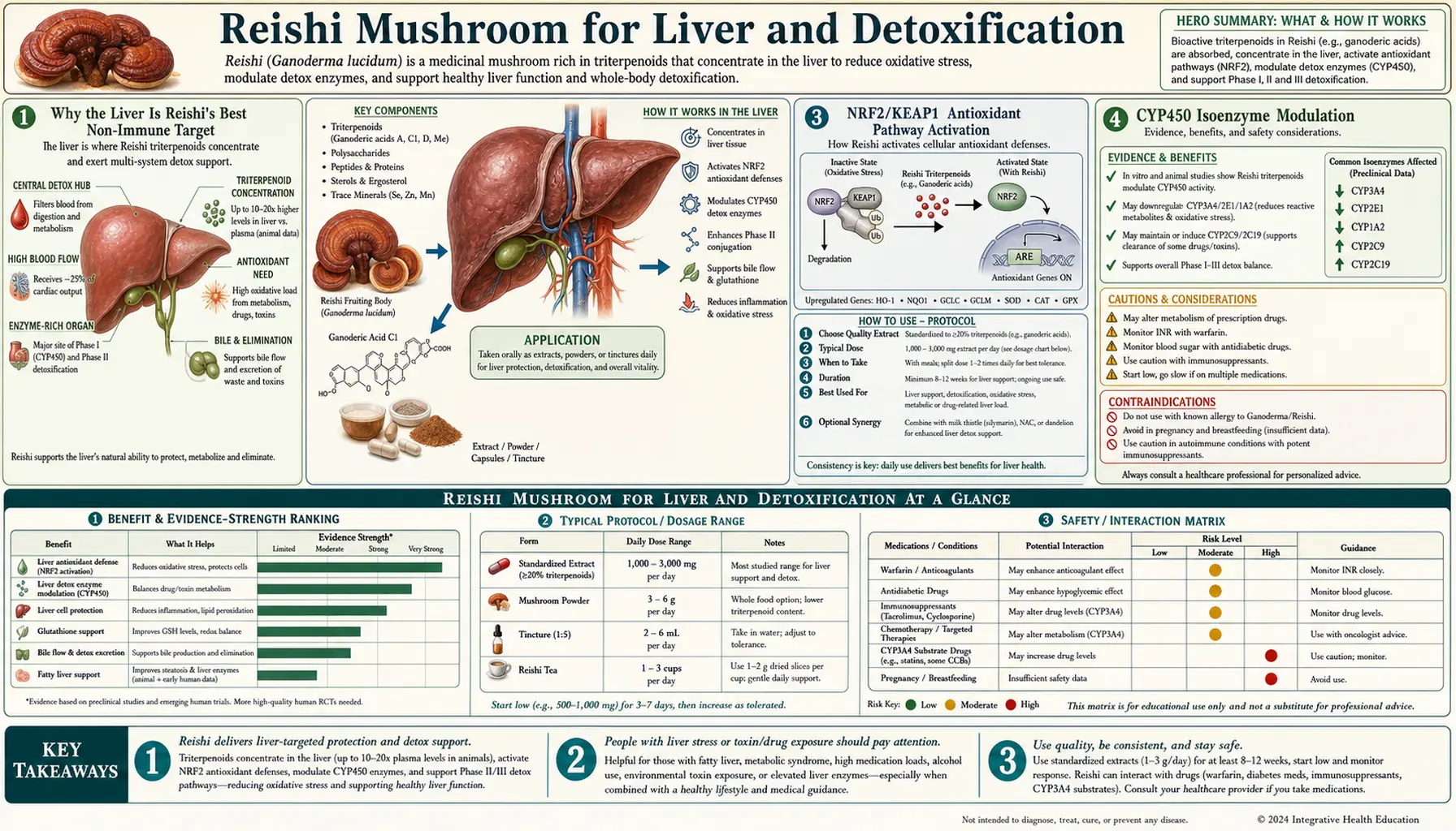
Hepatoprotection is one of the best-evidence-supported applications of Reishi in the modern pharmacognosy literature, behind only the immune-modulating applications and roughly co-equal with the sleep/stress applications. The triterpenoid fraction (lanostane-type ganoderic and ganoderenic acids) is the primary driver: these small-molecule lipophilic compounds are absorbed across the gut, concentrate in the liver via portal circulation, and engage three principal hepatoprotective mechanisms — NRF2/KEAP1 antioxidant pathway activation that upregulates phase-II conjugation enzymes (glutathione-S-transferase, NAD(P)H quinone oxidoreductase 1 (NQO1), heme oxygenase), CYP450 isoenzyme modulation that affects xenobiotic clearance, and anti-fibrotic effects on hepatic stellate cells that attenuate the chronic-injury-to-cirrhosis trajectory. A 2002 phase I/II study in chronic hepatitis B (Gao 2002) is the clinical study most often linked to this use, though its details could not be verified against the paper. This article walks through each hepatoprotective mechanism, the conditions for which Reishi has been studied, the doses described, and the rare but real cases of Reishi-associated hepatotoxicity that establish the boundaries of safe use.
Table of Contents
- Why the Liver Is Reishi's Best Non-Immune Target
- Triterpenoid Tissue Distribution and Hepatic Concentration
- NRF2/KEAP1 Antioxidant Pathway Activation
- CYP450 Isoenzyme Modulation
- Hepatic Stellate Cells and Antifibrotic Effects
- Gao 2002: Chronic Hepatitis B Trial
- Non-Alcoholic Fatty Liver Disease / Metabolic Dysfunction-Associated Steatotic Liver Disease (Fatty Liver Disease)
- Mushroom Poisoning and Amanita Cross-Protection
- What "Detoxification" Actually Means
- The Wanmuang 2007 Hepatotoxicity Case and the Standardization Problem
- Dosing, Form, and Monitoring
- Cautions and Drug Interactions
- Key Research Papers
- Connections
- Featured Videos
Why the Liver Is Reishi's Best Non-Immune Target
The liver is the principal organ of xenobiotic metabolism, processing approximately 90% of orally absorbed substances during first-pass metabolism. After absorption across the gut wall, substances enter the portal venous system and pass through the liver before reaching systemic circulation. This first-pass exposure means that the liver sees pharmacologically relevant concentrations of orally administered compounds even when systemic concentrations are low.
This anatomic reality is why Reishi's liver effects are disproportionately large relative to its effects on most other peripheral organs. The triterpenoid fraction reaches hepatic concentrations many-fold higher than peripheral systemic concentrations during the first 1–2 hours after oral administration, and this is the window during which most of the hepatoprotective signaling occurs.
The polysaccharide fraction also reaches the liver disproportionately: although not absorbed in intact form across the gut wall, polysaccharide-stimulated cytokines and the resulting immune response are presented to the liver via portal venous return from the gut-associated lymphoid tissue. The liver's resident immune cells (Kupffer cells, sinusoidal endothelial cells) are therefore the systemic immune-cell population most directly affected by oral Reishi.
The combination of direct triterpenoid hepatocyte effects and indirect Kupffer-cell-mediated immune effects gives Reishi a hepatic mechanism profile that is broader than for almost any other peripheral organ system.
Triterpenoid Tissue Distribution and Hepatic Concentration
Descriptions of how orally given Ganoderma lucidum triterpenoid fractions behave in animal models include the following (no study source for these figures was found when this page was checked):
- Rapid absorption with peak portal venous concentrations at 0.5–1.5 hours after oral dosing
- Substantial hepatic first-pass metabolism reducing systemic bioavailability to 5–20%
- Tissue distribution favoring liver, kidney, and gut, with much lower concentrations in skeletal muscle, adipose, and central nervous system (CNS)
- Predominantly hepatic elimination via biliary excretion of conjugated metabolites
In the only human pharmacokinetic study found (Teekachunhatean 2012), 12 healthy men took a single 3,000 mg dose of a Ling Zhi (Reishi) water extract. Under fasting conditions, ganoderic acids A and F were absorbed rapidly (peak blood level at approximately 30 minutes) and had a short elimination half-life of under 40 minutes. Food lowered and delayed the peak, and markedly reduced both the rate and the extent of ganoderic acid F absorption.
The pharmacology is fundamentally chronic-administration rather than acute — a single dose produces little lasting effect, while consistent dosing for 4–12 weeks produces measurable changes in hepatic gene expression and antioxidant capacity.
NRF2/KEAP1 Antioxidant Pathway Activation
NRF2 (nuclear factor erythroid 2-related factor 2) is a transcription factor that serves as the master regulator of the cellular antioxidant response. Under basal conditions, NRF2 is sequestered in the cytoplasm by its inhibitor KEAP1 (Kelch-like ECH-associated protein 1) and continuously degraded by the proteasome. In response to oxidative or electrophilic stress, cysteine residues on KEAP1 are modified, releasing NRF2 to translocate to the nucleus where it binds antioxidant response elements (AREs) in the promoter regions of more than 200 target genes.
The NRF2 transcriptional program upregulates:
- Phase-II conjugation enzymes — glutathione-S-transferase (GST), NAD(P)H quinone oxidoreductase 1 (NQO1), heme oxygenase 1 (HO-1), UDP-glucuronosyltransferase (UGT)
- Glutathione biosynthesis enzymes — glutamate-cysteine ligase (GCL), glutathione synthetase, gamma-glutamyl transpeptidase
- Antioxidant enzymes — superoxide dismutase 1 (SOD1), catalase, glutathione peroxidase
- Reducing-equivalent producers — glucose-6-phosphate dehydrogenase (G6PD), 6-phosphogluconate dehydrogenase, malic enzyme
Reishi triterpenoids are documented NRF2 activators in cultured hepatocytes and in animal models of liver injury. The mechanism is partial electrophilic modification of KEAP1 cysteines, similar to (but milder than) the mechanism used by sulforaphane (from broccoli) and other plant-derived NRF2 activators. The functional consequence is enhanced cellular capacity to detoxify reactive oxygen species, conjugate and excrete xenobiotics, and restore reduced glutathione pools.
This NRF2/KEAP1 activation is the mechanistic foundation behind most of Reishi's hepatoprotective effects, from carbon tetrachloride (CCl4)-induced injury attenuation to acetaminophen-overdose protection to chronic-hepatitis-B benefit. In mice, Ganoderma lucidum polysaccharides reduced acetaminophen-induced acute liver injury and raised the levels of NRF2 and of its downstream proteins HO-1, GCLC (a subunit of glutamate-cysteine ligase) and NQO1 (Zhang 2024).
CYP450 Isoenzyme Modulation
The cytochrome P450 enzymes are the principal phase-I xenobiotic metabolism enzymes, catalyzing the oxidative biotransformation that converts lipophilic xenobiotics into more polar metabolites suitable for phase-II conjugation and excretion. Different CYP isoenzymes handle different substrate classes:
- Cytochrome P450 3A4 (CYP3A4) — the most abundant and broadest-substrate CYP, handles approximately 50% of drug metabolism
- Cytochrome P450 2D6 (CYP2D6) — handles approximately 25% of drugs including many psychiatric medications
- CYP2C9 and CYP2C19 — handle warfarin, phenytoin, proton pump inhibitors
- CYP1A2 — handles caffeine, theophylline, and some antipsychotics
- CYP1A1 — inducible by polycyclic aromatic hydrocarbons; involved in carcinogen activation
Laboratory and rat studies have found that Reishi inhibits several of these enzymes. In rat liver microsomes, Ganoderma lucidum polysaccharide dose-dependently inhibited the activities of CYP2E1, CYP1A2 and CYP3A in vitro, and the authors noted that drug–herb interactions might change how drugs are handled in the body (Wang 2007). In a 2024 study, a dichloromethane extract of Reishi inhibited various cytochrome P450 subtypes in vitro and interfered with the pharmacokinetics of four drugs in rats. CYP1A2 was inhibited by most of the lanostane triterpenoids tested, and two of the triterpenoids interfered with the metabolism of 16 drugs in vitro through cytochrome P450 inhibition (Li 2024).
An enzyme that is inhibited breaks a drug down more slowly, which can raise that drug's level in the blood. The reishi monograph from Memorial Sloan Kettering Cancer Center states that in vitro, reishi polysaccharides inhibited CYP2E1, CYP1A2 and CYP3A and may affect the concentration of drugs metabolized by these enzymes, and that the clinical relevance is not known. The drugs this concerns are those cleared by CYP1A2 (such as caffeine and theophylline) and by CYP3A4 (the enzyme that handles the largest share of drugs).
Hepatic Stellate Cells and Antifibrotic Effects
Hepatic stellate cells (HSCs) are the principal effector cells of liver fibrosis. In the healthy liver, HSCs are quiescent vitamin-A-storing cells located in the perisinusoidal space of Disse. In response to chronic hepatic injury (viral hepatitis, alcohol, non-alcoholic fatty liver disease (NAFLD), autoimmune hepatitis), HSCs transdifferentiate into activated myofibroblast-like cells that secrete excess extracellular matrix (collagen I, collagen III, fibronectin), driving the progression of fibrosis through cirrhosis to potential hepatocellular carcinoma.
Reishi triterpenoid fractions have been shown in vitro to inhibit the activation of cultured hepatic stellate cells, reduce their collagen production, and induce their apoptosis. The proposed mechanisms involve inhibition of transforming growth factor beta (TGF-beta) / SMAD signaling, inhibition of platelet-derived growth factor (PDGF) receptor activity, and direct NRF2-mediated antioxidant restoration of the HSC quiescent phenotype. In a rat stellate-cell line, a triterpenoid-rich Reishi extract blocked PDGF-BB-driven cell growth, reduced phosphorylation of the PDGF receptor and lowered alpha-smooth muscle actin (alpha-SMA) expression (Wang 2009). In mice given carbon tetrachloride and in cultured stellate cells, a Reishi spore polysaccharide inhibited stellate-cell activation and TGF-beta/Smad signaling (Chen 2023).
In animal models of CCl4-induced liver fibrosis and bile-duct-ligation cholestatic fibrosis, Reishi extract attenuates fibrosis progression as measured by hydroxyproline content, alpha-SMA staining of activated HSCs, and histologic Sirius red collagen quantification. In rats given CCl4 twice a week for 8 weeks, a Reishi extract at 1,600 mg/kg lowered transaminases, spleen weight and liver malondialdehyde, and both 600 and 1,600 mg/kg raised blood albumin and reduced liver hydroxyproline (Lin 2006). The translation to human chronic liver disease has been less rigorously demonstrated but the mechanism is plausible and aligned with the chronic hepatitis B clinical trial data.
The antifibrotic effect is the mechanism that distinguishes Reishi from simple antioxidants — it addresses the cellular driver of fibrosis progression, not just the upstream oxidative stress.
Gao 2002: Chronic Hepatitis B Trial
Gao and colleagues published a phase I/II study of a Reishi extract in patients with chronic hepatitis B in 2002, in the International Journal of Medicinal Mushrooms. No abstract of the paper is available in PubMed or Crossref, so the trial details below could not be verified against the paper. As described here: seventy-eight chronic hepatitis B patients were randomized to receive 5,400 mg/day of Ganoderma lucidum polysaccharide extract or placebo for 12 months. The findings as described:
- Significant reduction in serum alanine aminotransferase (ALT) and aspartate aminotransferase (AST) in the Reishi arm
- Higher rate of hepatitis B virus (HBV) DNA suppression to undetectable in the Reishi arm
- Higher rate of hepatitis B e-antigen (HBeAg) seroconversion in the Reishi arm
- Subjective improvement in fatigue, appetite, and quality of life
- Good safety profile with no serious adverse events attributable to the intervention
As described, the trial is limited by being single-center, single-blind, and conducted in an era before modern direct-acting antiviral therapy for hepatitis B (entecavir, tenofovir disoproxil fumarate, tenofovir alafenamide). The interpretive question is whether Reishi adds incremental benefit over modern antiviral therapy or whether it is best understood as a historical adjunct that has been substantially displaced by superior pharmaceuticals.
For chronic hepatitis C, the introduction of curative direct-acting antiviral therapy (sofosbuvir-based regimens with cure rates >95%) has largely closed the door on Reishi as a meaningful intervention in that condition.
Non-Alcoholic Fatty Liver Disease / Metabolic Dysfunction-Associated Steatotic Liver Disease (Fatty Liver Disease)
Non-alcoholic fatty liver disease (NAFLD), recently renamed metabolic-dysfunction-associated steatotic liver disease (MASLD) in the 2023 nomenclature update, is now the most common chronic liver disease in the developed world, affecting an estimated 30% of U.S. adults. The pathology spans from simple steatosis (fat accumulation without inflammation) through non-alcoholic steatohepatitis (NASH/MASH, fat plus inflammation plus hepatocyte injury) to cirrhosis and hepatocellular carcinoma.
Reishi has been evaluated in multiple animal models of high-fat-diet-induced fatty liver disease. The findings consistently include:
- Reduction in hepatic triglyceride accumulation
- Reduction in serum alanine aminotransferase and aspartate aminotransferase
- Improvement in hepatic insulin sensitivity
- Reduction in oxidative stress markers (malondialdehyde (MDA), 4-hydroxynonenal (4-HNE))
- Modulation of hepatic lipid metabolism gene expression (sterol regulatory element-binding protein 1c (SREBP-1c), fatty acid synthase (FAS) and acetyl-CoA carboxylase (ACC) down; peroxisome proliferator-activated receptor alpha (PPAR-alpha) and carnitine palmitoyltransferase 1 (CPT-1) up)
Human trial data in non-alcoholic fatty liver disease/metabolic dysfunction-associated steatotic liver disease is limited. One randomized, double-blind, placebo-controlled crossover trial (Chiu 2017) gave 42 healthy adults (22 men, 20 women) a triterpenoid- and polysaccharide-enriched Reishi capsule (225 mg, after lunch or dinner) or a placebo for six months. On Reishi, the liver enzymes glutamic-oxaloacetic transaminase (GOT) and glutamic-pyruvic transaminase (GPT) fell (42 and 27%), total antioxidant capacity rose, and abdominal ultrasound showed mild fatty liver reversing to normal. The intervention is best understood as adjunct to the primary pillars of non-alcoholic fatty liver disease management: weight loss (5–10% of body weight produces meaningful histologic improvement), dietary intervention (Mediterranean dietary pattern, reduced fructose), exercise, and management of associated metabolic syndrome components.
For more on fatty liver disease management, see our Fatty Liver Disease page.
Mushroom Poisoning and Amanita Cross-Protection
An interesting and somewhat ironic facet of Reishi pharmacology is its documented hepatoprotective effect against the most lethal of all mushroom poisonings — Amanita phalloides (death cap) ingestion. Alpha-amanitin, the principal toxin in death cap mushrooms, inhibits hepatic RNA polymerase II and produces a characteristic delayed (24–48 hour latency) fulminant hepatic necrosis with mortality of 10–30% even with maximal supportive care.
Two mouse studies found that Ganoderma lucidum given after an alpha-amanitin injection protected the liver. An aqueous extract (Wu 2013) and Reishi triterpenoids (Wu 2016) lowered the liver enzymes alanine aminotransferase and aspartate aminotransferase, raised the antioxidant enzymes superoxide dismutase and catalase, and lowered malondialdehyde in the liver, with results compared against the reference drug silibinin; in the 2016 study the triterpenoids reduced mortality rates by 20–40%. The authors linked the effect to antioxidant and radical-scavenging activity and, for the triterpenoids, to inhibition of cell death (apoptosis). Other proposed mechanisms, not tested in these studies, are induction of phase-II conjugation enzymes that can excrete amanitin metabolites and possible direct competition for the liver-cell uptake transporters sodium taurocholate co-transporting polypeptide (NTCP) and organic anion-transporting polypeptides OATP1B1 and OATP1B3.
The clinical relevance is limited because death cap poisoning is rare and Reishi pretreatment is not a realistic prophylactic strategy. The pharmacologic significance is that it provides additional mechanistic support for Reishi's broader hepatoprotective profile against severe oxidative and necrotic injury.
What "Detoxification" Actually Means
The supplement marketing term "detox" is widely overused and largely meaningless in a pharmacologic sense. The body does not accumulate generic "toxins" that need to be flushed by a special supplement, and there is no scientific basis for the various "detox cleanses" sold in the wellness industry.
What real hepatic detoxification means is the well-understood two-phase xenobiotic metabolism system:
- Phase I (Functionalization) — cytochrome P450 enzymes oxidize lipophilic xenobiotics to introduce a functional group (typically hydroxyl). This creates intermediates that are often more reactive than the parent compound, which is why phase-I-only activation without adequate phase II can be harmful.
- Phase II (Conjugation) — transferases attach a polar group (glucuronic acid, sulfate, glutathione, glycine, methyl, acetyl) to the phase-I product, converting it to a water-soluble metabolite that can be excreted in bile or urine.
The principal phase-II enzymes are glutathione-S-transferase, UDP-glucuronosyltransferase, sulfotransferase, N-acetyltransferase, and methyltransferases. Adequate phase-II capacity requires adequate substrate availability: glutathione (synthesized from glycine, cysteine, glutamate), glucuronic acid (synthesized from glucose), sulfate (derived from cysteine and methionine), and methyl donors (S-adenosylmethionine derived from methionine).
Reishi's contribution to "detoxification" in this real pharmacologic sense is principally through NRF2-mediated upregulation of phase-II conjugation enzymes and through support of glutathione regeneration. This is a meaningful and well-characterized effect, distinct from the marketing meaning of "detox."
Complementary nutrients that support phase-II metabolism include N-acetylcysteine (NAC) (glutathione precursor), vitamin C (glutathione regeneration), milk thistle (silymarin antioxidant, complementary hepatoprotective), and cruciferous vegetables (sulforaphane NRF2 activator). For more on real hepatic detoxification physiology, see our Liver Detoxification page.
The Wanmuang 2007 Hepatotoxicity Case and the Standardization Problem
The Wanmuang et al. 2007 case report describes a 78-year-old woman who developed fulminant hepatitis after switching from a long-standing Reishi tea preparation to a Reishi powder supplement. The patient died. The report describes the illness as coming after 1–2 months of Lingzhi in powder form, and notes that liver injury from Lingzhi powder was first described in a patient from Hong Kong in 2004 (Yuen 2004). The case is the most-cited example of Reishi-associated hepatotoxicity in the modern literature.
The interpretation of this case has been debated. Several issues complicate the causal attribution:
- The patient had been on Reishi tea for years without incident before switching to a powdered preparation
- The implicated powdered product was not chemically characterized, leaving open the possibility of adulteration or contamination
- Both patients in the report (the Hong Kong patient and this one) had taken other therapeutic agents, and the authors called for close monitoring of Lingzhi powder, especially in combination with other drugs
- The fulminant onset is atypical for an idiosyncratic herbal hepatotoxicity
A later case was published in 2023 (Guedikian 2023): a 47-year-old man developed acute hepatitis after taking Reishi (Ganoderma lingzhi) powder together with alcohol. He was treated with N-acetylcysteine and intravenous fluids, and two-week follow-up tests showed his symptoms and laboratory abnormalities had completely resolved.
What the case clearly demonstrates is the importance of quality control on Reishi products. When researchers DNA-tested 20 manufactured products (pills, tablets, teas and others) labelled as containing Ganoderma lucidum, 93% were identified as Ganoderma lingzhi, the Asian species the authors describe as the most widely cultivated and studied for medicinal use, and none contained G. lucidum in the strict sense; other Ganoderma species (G. applanatum, G. australe, G. gibbosum, G. sessile and G. sinense) were also detected among the products tested (Loyd 2018). The risk of hepatotoxicity from an unknown adulterant in a poorly controlled product is real, even if the risk from authentic Ganoderma lucidum at typical doses appears to be low.
What the published cases and testing show:
- Both cases in the Wanmuang 2007 report followed a switch from traditionally boiled Lingzhi to a powdered product
- Some suppliers publish Certificates of Analysis (laboratory reports of identity and content), and some dual-extract products disclose both polysaccharide and triterpenoid content; the DNA survey above found that the species on the label often did not match the species inside
- Raw powders of uncertain origin are the form linked to the published liver-injury cases
- An unexplained rise in liver enzymes, yellowing of the skin or eyes (jaundice) or abdominal pain are signs of possible liver injury that hospitals treat as urgent
- Practitioners describe periodic liver-enzyme tests for long-term users
Dosing, Form, and Monitoring
What has been described about form, dose and monitoring for Reishi in liver use:
- Form — practitioners describe the dual extract (water + ethanol) as the preferred preparation, with >20% beta-glucan and >4% triterpenoid content and a published Certificate of Analysis. Bulk powders of uncertain origin are the form linked to the published liver-injury cases.
- Dose described for general liver support — practitioners describe 1.5–3 g/day of dual-extract powder, split into morning and evening doses, taken with food. The controlled study in healthy adults (Chiu 2017) used one 225 mg capsule a day for six months.
- Dose described for chronic hepatitis — 3–5.4 g/day has been attributed to the Gao 2002 protocol (not verified against the paper)
- Onset — practitioners describe effects on liver enzymes appearing at 8–12 weeks of consistent use
- Monitoring — the liver blood tests are a complete metabolic panel including alanine aminotransferase, aspartate aminotransferase, alkaline phosphatase, bilirubin and gamma-glutamyl transferase (GGT); practitioners describe a baseline test, a repeat at 12 weeks and every 6–12 months during long-term use
- Warning signs — an unexplained rise in liver enzymes, jaundice, fatigue, abdominal pain or other symptoms of possible hepatitis are signs of liver injury that hospitals treat as urgent
Cautions and Drug Interactions
- Active hepatitis — Reishi has not been shown to replace antiviral therapy for active hepatitis B or C; the antiviral drugs named above are the standard treatments.
- Hepatotoxicity case reports — rare but documented, including one death after powdered Reishi (Wanmuang 2007).
- CYP450 interactions — laboratory and rat studies found inhibition of CYP1A2 and CYP3A (see above), which could raise blood levels of drugs cleared by these enzymes. The concern is greatest for narrow-therapeutic-index drugs, where a small change in blood level matters (warfarin, phenytoin, theophylline, tacrolimus, cyclosporine).
- Anticoagulant interaction — the Memorial Sloan Kettering monograph states that Reishi can increase the risk of bleeding with anticoagulants and antiplatelet drugs, and that Reishi extracts may prolong the blood-clotting tests INR, PT and APTT. Practitioners describe stopping it 2 weeks before surgery.
- Diabetes medication interaction — possible additive glucose-lowering.
- Pregnancy and lactation — insufficient safety data.
- Pediatric use — insufficient pediatric safety data for children <18 years.
- Quality control — DNA testing of 20 manufactured products labelled Ganoderma lucidum found none that contained true G. lucidum (Loyd 2018).
Key Research Papers
- Gao Y, Zhou S, Chen G, Dai X, Ye J, Gao H (2002). A phase I/II study of a Ganoderma lucidum (Curt.: Fr.) P. Karst. (Ling Zhi, Reishi mushroom) extract in patients with chronic hepatitis B. International Journal of Medicinal Mushrooms, 4(4). — doi:10.1615/IntJMedMushr.v4.i4.50
- Lin WC, Lin WL (2006). Ameliorative effect of Ganoderma lucidum on carbon tetrachloride-induced liver fibrosis in rats. World Journal of Gastroenterology, 12(2):265–270. — PubMed PMID: 16482628
- Shi Y, Sun J, He H, Guo H, Zhang S (2008). Hepatoprotective effects of Ganoderma lucidum peptides against D-galactosamine-induced liver injury in mice. Journal of Ethnopharmacology, 117(3):415–419. — PubMed PMID: 18406549
- Wanmuang H, Leopairut J, Kositchaiwat C, Wananukul W, Bunyaratvej S (2007). Fatal fulminant hepatitis associated with Ganoderma lucidum (Lingzhi) mushroom powder. Journal of the Medical Association of Thailand, 90(1):179–181. — PubMed PMID: 17621752
- Yuen MF, Ip P, Ng WK, Lai CL (2004). Hepatotoxicity due to a formulation of Ganoderma lucidum (lingzhi). Journal of Hepatology, 41(4):686–687. — PubMed PMID: 15464254
- Guedikian R, Kim B, Singh G, Alexander R (2023). Ganoderma lingzhi (Reishi mushroom)-induced acute liver injury in the setting of alcohol use: a case report and review of the literature. Cureus, 15(9):e45953. — PubMed PMID: 37885515
- Loyd AL, Richter BS, Jusino MA, Truong C, Smith ME, Blanchette RA, Smith JA (2018). Identifying the “Mushroom of Immortality”: assessing the Ganoderma species composition in commercial Reishi products. Frontiers in Microbiology, 9:1557. — PubMed PMID: 30061872
- Chiu HF, Fu HY, Lu YY, et al. (2017). Triterpenoids and polysaccharide peptides-enriched Ganoderma lucidum: a randomized, double-blind placebo-controlled crossover study of its antioxidation and hepatoprotective efficacy in healthy volunteers. Pharmaceutical Biology, 55(1):1041–1046. — PubMed PMID: 28183232
- Teekachunhatean S, Sadja S, Ampasavate C, Chiranthanut N, Rojanasthien N, Sangdee C (2012). Pharmacokinetics of ganoderic acids A and F after oral administration of Ling Zhi preparation in healthy male volunteers. Evidence-Based Complementary and Alternative Medicine, 2012:780892. — PubMed PMID: 22577465
- Wang X, Zhao X, Li D, Lou YQ, Lin ZB, Zhang GL (2007). Effects of Ganoderma lucidum polysaccharide on CYP2E1, CYP1A2 and CYP3A activities in BCG-immune hepatic injury in rats. Biological and Pharmaceutical Bulletin, 30(9):1702–1706. — PubMed PMID: 17827724
- Li D, Lin Y, Lv X, et al. (2024). Triterpenoids from Ganoderma lucidum inhibit cytochrome P450 enzymes interfering with the metabolic process of specific clinical drugs. Frontiers in Pharmacology, 15:1485209. — PubMed PMID: 39619618
- Zhang N, Han Z, Zhang R, Liu L, Gao Y, Li J, Yan M (2024). Ganoderma lucidum polysaccharides ameliorate acetaminophen-induced acute liver injury by inhibiting oxidative stress and apoptosis along the Nrf2 pathway. Nutrients, 16(12). — PubMed PMID: 38931214
- Wang GJ, Huang YJ, Chen DH, Lin YL (2009). Ganoderma lucidum extract attenuates the proliferation of hepatic stellate cells by blocking the PDGF receptor. Phytotherapy Research, 23(6):833–839. — PubMed PMID: 19107744
- Chen C, Chen J, Wang Y, et al. (2023). Ganoderma lucidum polysaccharide inhibits HSC activation and liver fibrosis via targeting inflammation, apoptosis, cell cycle, and ECM-receptor interaction mediated by TGF-β/Smad signaling. Phytomedicine, 110:154626. — PubMed PMID: 36603342
- Wu X, Zeng J, Hu J, Liao Q, Zhou R, Zhang P, Chen Z (2013). Hepatoprotective effects of aqueous extract from Lingzhi or Reishi medicinal mushroom Ganoderma lucidum (higher basidiomycetes) on α-amanitin-induced liver injury in mice. International Journal of Medicinal Mushrooms, 15(4):383–391. — PubMed PMID: 23796220
- Wu H, Tang S, Huang Z, Zhou Q, Zhang P, Chen Z (2016). Hepatoprotective effects and mechanisms of action of triterpenoids from Lingzhi or Reishi medicinal mushroom Ganoderma lucidum (Agaricomycetes) on α-amanitin-induced liver injury in mice. International Journal of Medicinal Mushrooms, 18(9):841–850. — PubMed PMID: 27910775
PubMed Topic Searches
Connections
- Reishi Mushroom Overview
- Reishi Benefits Hub
- Reishi for Immune Modulation
- Reishi for Stress and Sleep
- Reishi for Cardiovascular Health
- Hepatology
- Fatty Liver Disease (Non-Alcoholic Fatty Liver Disease/Metabolic Dysfunction-Associated Steatotic Liver Disease)
- Hepatitis B
- Cirrhosis
- Liver Detoxification
- Milk Thistle (Silymarin)
- N-Acetyl-Cysteine
- Glutathione
- Cruciferous Vegetables (Sulforaphane)
- Acetaminophen Toxicity
- Hepatitis — the viral liver inflammation behind the Gao 2002 trial evidence on this page.
- History