Varicocele
- 1. Overview
- 2. Anatomy and Pathophysiology
- 3. Epidemiology
- 4. Why 90% Left-Sided
- 5. Clinical Presentation and Grading
- 6. Diagnosis
- 7. Treatment
- 8. Outcomes and Fertility
- Key Research Papers
- 10. Connections
1. Overview
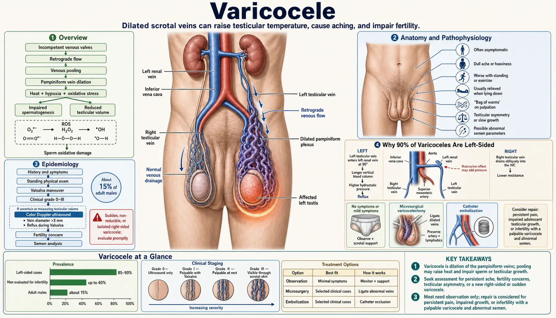
A varicocele is an abnormal dilation and tortuosity of the pampiniform venous plexus — the network of veins that surrounds the testicular artery within the spermatic cord. The analogy is precise: varicoceles are to the scrotum what varicose veins are to the leg, or what hemorrhoids are to the rectum. All three conditions involve valvular incompetence allowing retrograde venous pooling in a low-pressure venous network.
Varicoceles carry outsized clinical importance because they are the single most common correctable cause of male infertility, identified in approximately 35% of men evaluated for primary infertility and in up to 81% of men with secondary infertility — that is, men who fathered a child previously but are now unable to conceive. The World Health Organization has classified varicocele as the most surgically correctable condition in male reproductive medicine.
Despite their prevalence, varicoceles remain frequently underdiagnosed because they are often asymptomatic, require a physical examination performed with the patient standing and performing Valsalva maneuver, and are easily dismissed as an incidental finding when not evaluated in the context of fertility or gonadal function. Progressive Leydig cell dysfunction — leading to declining testosterone over years — makes untreated varicoceles a long-term endocrine concern beyond infertility alone.
2. Anatomy and Pathophysiology
The pampiniform plexus is a network of 8–12 small veins that originate in the testis and epididymis and drain superiorly through the spermatic cord. In healthy anatomy, this plexus wraps tightly around the testicular artery — the arrangement functions as a countercurrent heat exchanger, allowing the arterial blood to be pre-cooled by venous blood before it reaches the testis. This mechanism keeps the testicular temperature approximately 2–3°C below core body temperature, a biological prerequisite for normal spermatogenesis.
Venous valve incompetence is the initiating defect in varicocele formation. The valves in the internal spermatic vein fail to prevent retrograde flow during periods of increased intra-abdominal pressure (standing, exercise, Valsalva). The resulting venous stasis and dilation impair the countercurrent exchange mechanism, allowing testicular temperature to rise by 1–3°C. Even modest sustained temperature elevation above the physiological range disrupts the highly temperature-sensitive stages of spermatogenesis, particularly meiosis and sperm maturation.
Three mechanisms converge to damage testicular function in varicocele:
- Scrotal temperature elevation. Venous stasis disrupts countercurrent cooling. Elevated scrotal temperature impairs DNA integrity of developing spermatocytes, reduces sperm production, and increases apoptosis within the seminiferous tubules.
- Oxidative stress accumulation. Impaired venous drainage leads to accumulation of reactive oxygen species (ROS) within the testicular microenvironment. Oxidative stress directly damages sperm DNA, impairs mitochondrial function (critical for motility), and triggers premature apoptosis of spermatogenic cells. Seminal reactive oxygen species levels are measurably elevated in men with clinical varicocele.
- Reflux of renal and adrenal metabolites. The left testicular vein drains into the left renal vein. Retrograde flow can carry cortisol, catecholamines, and other adrenal or renal metabolites into the testicular venous circulation, where they may suppress testosterone synthesis in Leydig cells and interfere with the local hormonal environment necessary for spermatogenesis.
The cumulative effect is a characteristic deterioration of multiple sperm parameters simultaneously — oligozoospermia (low count), asthenozoospermia (poor motility), and teratozoospermia (abnormal morphology) — together termed the "stress pattern" of the semen analysis. Leydig cell dysfunction adds hypogonadism and declining testosterone to the clinical picture over time.
3. Epidemiology
Varicocele is a common condition with prevalence that escalates sharply in the context of infertility:
- 15% of the general adult male population has a clinical varicocele detectable on physical examination.
- 35% of men presenting with primary infertility (couple has never conceived) have an identifiable varicocele.
- 75–81% of men presenting with secondary infertility (had a prior child but now unable to conceive) have a varicocele — the highest prevalence group, and striking evidence that varicocele-associated testicular damage is progressive rather than static.
These figures from large epidemiological studies and World Health Organization (WHO) data underpin the clinical importance of varicocele screening in all men being evaluated for infertility. The disparity between the 15% population rate and the 75–81% secondary-infertility rate reflects the natural history of untreated varicocele: ongoing oxidative stress, sustained temperature elevation, and progressive Leydig cell dysfunction accumulate over years, so men who were fertile in their 20s may become infertile in their 30s if a varicocele goes unaddressed.
Varicoceles are rare before puberty and increase in prevalence through adolescence as testicular venous flow increases with growth. Pediatric and adolescent varicoceles are monitored for testicular volume asymmetry, which is an early marker of ipsilateral testicular damage and an indication for intervention before fertility is required.
4. Why 90% of Varicoceles Are Left-Sided
The striking left-sided predominance of varicocele — approximately 90% of cases are left-sided or left-dominant — is explained by a fundamental anatomical asymmetry in testicular venous drainage:
- The left testicular vein drains at a near-right angle into the left renal vein. This creates higher hydrostatic pressure in the left spermatic venous column, a longer venous path before reaching the inferior vena cava, and greater susceptibility to retrograde flow when valves are incompetent. The "nutcracker phenomenon" — compression of the left renal vein between the aorta and the superior mesenteric artery — can further impede left renal venous outflow, adding to the hydrostatic burden on the left testicular vein.
- The right testicular vein drains at an oblique angle directly into the inferior vena cava — a shorter, lower-pressure pathway that is inherently more resistant to retrograde flow.
Clinical warning: right-sided or bilateral varicocele demands a secondary-cause workup. An isolated right-sided varicocele, or a right-sided varicocele that does not decompress on lying supine, raises suspicion for an obstructing mass — renal cell carcinoma (RCC), retroperitoneal lymphoma, or other retroperitoneal pathology compressing or invading the right spermatic or renal vein. Because a right-sided varicocele can be a sign of a kidney or abdominal mass, a man with an asymmetric right-sided varicocele is normally sent for computed tomography (CT) of the abdomen and pelvis to look for a tumor before the varicocele itself is treated. Urologists count this among the few truly urgent evaluation pathways in their field.
5. Clinical Presentation and Grading
Symptoms
Many varicoceles are entirely asymptomatic and discovered incidentally during physical examination for infertility evaluation or routine checkup. When symptoms are present, the most common is a dull, dragging ache or heaviness in the left testicle or left groin, often described as a "pulling" sensation. Pain worsens with prolonged standing, physical exertion, and heat exposure; it is typically relieved by lying supine. Sharp acute pain is unusual and raises the possibility of other diagnoses, including torsion.
Physical Examination
The standard examination is performed with the patient standing in a warm room, which promotes scrotal relaxation. The examiner first inspects for visible dilated veins (Grade III), then palpates the spermatic cord above the testis at rest and during Valsalva maneuver (the patient bears down, raising intra-abdominal pressure). Testicular volume is assessed bilaterally using an orchidometer — volume asymmetry greater than 20% is clinically significant and suggests ipsilateral testicular damage.
Grading System (Dubin and Amelar)
- Subclinical: Not palpable or visible; detectable only by scrotal duplex ultrasound. Clinical significance in isolation is debated; most guidelines do not recommend treatment without abnormal semen analysis.
- Grade I: Palpable only during Valsalva maneuver; not palpable at rest; not visible.
- Grade II: Palpable at rest without Valsalva maneuver; not visible through the scrotal skin.
- Grade III: Visible through the scrotal skin — the classic "bag of worms" appearance of tortuous, engorged veins distending the hemiscrotum.
Semen Analysis Findings
Varicocele produces a characteristic "stress pattern" on semen analysis:
- Oligozoospermia: Reduced total sperm count or concentration.
- Asthenozoospermia: Reduced progressive motility — sperm are present but move poorly, reflecting mitochondrial dysfunction from oxidative stress.
- Teratozoospermia: Increased abnormal sperm morphology — elevated percentage of sperm with head, midpiece, or tail defects.
The simultaneous impairment of count, motility, and morphology together is the hallmark pattern. Any single parameter can be selectively impaired in other causes of male factor infertility; the combination across all three is the varicocele fingerprint.
Testosterone and Leydig Cell Dysfunction
Beyond spermatogenesis, varicocele is associated with progressive Leydig cell dysfunction — the testosterone-producing interstitial cells of the testis. Men with varicocele have measurably lower serum testosterone compared to age-matched controls, and this difference increases over time. Studies demonstrate testosterone improvement following varicocelectomy in hypogonadal men with clinical varicocele, making varicocele repair a potentially relevant intervention for symptomatic hypogonadism even in men not pursuing fertility.
6. Diagnosis
Clinical Examination
Physical examination with Valsalva — the cornerstone of varicocele diagnosis — remains the standard first-line evaluation. Experienced examiners detect Grade I–III varicoceles reliably. The examination is performed standing; supine examination misses most varicoceles because venous pooling decompresses in the horizontal position.
Scrotal Duplex Ultrasound
Scrotal duplex ultrasonography with color Doppler is the gold-standard imaging study for varicocele confirmation and grading. Diagnostic criteria:
- Pampiniform plexus veins greater than 3 mm in diameter at rest, or greater than 3.5 mm during Valsalva maneuver.
- Retrograde (reverse) venous flow demonstrated on Doppler during Valsalva maneuver — the definitive functional confirmation of valvular incompetence.
- Subclinical varicocele: Doppler criteria met without palpable findings on examination.
Ultrasound also provides bilateral testicular volume measurement, identifies subclinical contralateral varicoceles, and assesses testicular parenchymal echogenicity for signs of atrophy or prior injury.
Semen Analysis
A complete semen analysis following World Health Organization 2021 reference values is standard in any varicocele evaluation. Two samples collected 48–72 hours apart are preferred for reliability. Parameters assessed: volume, pH, concentration, total motility, progressive motility, morphology (Kruger strict criteria), total motile sperm count.
Hormonal Evaluation
Morning serum total testosterone, follicle-stimulating hormone (FSH), and luteinizing hormone (LH) are appropriate in all infertility evaluations and in men with varicocele who report symptoms of hypogonadism. Elevated follicle-stimulating hormone suggests primary testicular damage to the spermatogenic epithelium. Low testosterone with elevated luteinizing hormone (compensated hypogonadism) is a common pattern in men with larger varicoceles.
Right-Sided Workup
An isolated right-sided varicocele or a right varicocele that does not decompress on lying supine requires computed tomography of the abdomen and pelvis with contrast to exclude retroperitoneal mass, renal cell carcinoma, or venous obstruction before any intervention is planned.
7. Treatment
Indications for Intervention
A 2016 review of the guidelines found that the European Association of Urology (EAU) guidelines give their highest level of evidence (level 1a) for treating varicoceles that combine all three of the following:
- A palpable varicocele on physical examination (Grade I, II, or III).
- Abnormal semen analysis (one or more parameters below reference range).
- Infertility in the couple that is otherwise unexplained.
The same review notes that the American Urological Association (AUA) and the American Society for Reproductive Medicine (ASRM) take the position that there are not enough outcome data to support evidence-based guidelines, and that the evidence behind current recommendations is generally of low quality. The review describes the overall body of evidence on varicocele and its repair as weak and conflicting.
Additional indications include:
- Symptomatic varicocele causing significant scrotal pain or heaviness interfering with daily activity or exercise.
- Adolescent varicocele with progressive testicular volume asymmetry greater than 20%, even before fertility is a concern.
- Hypogonadism (low testosterone) with a palpable varicocele in a man wishing to avoid exogenous testosterone therapy, which suppresses spermatogenesis.
Subclinical varicocele (ultrasound-only, not palpable) is not currently a standard indication for treatment per major guideline bodies including the AUA, EAU, and ASRM.
Microscopic Subinguinal Varicocelectomy — Gold Standard
Microsurgical varicocelectomy via the subinguinal approach, performed under optical magnification (6–25×), is the gold-standard surgical technique with the lowest complication and recurrence rates of all approaches.
Technical advantages of magnification:
- Allows visual identification and preservation of the testicular artery (inadvertent ligation causes testicular atrophy).
- Allows preservation of lymphatic vessels (lymphatic ligation causes postoperative hydrocele, the most common complication of non-microsurgical approaches).
- Allows preservation of the vas deferens.
- All dilated internal spermatic veins, external spermatic veins, and gubernacular veins can be systematically identified and ligated.
Outcomes: A 2009 pooled analysis of 36 studies found the lowest rates with the microsurgical technique: recurrence 1.05% and hydrocele (fluid collection around the testis) 0.44%, compared with recurrence of 4.3% and hydrocele of 2.84% after laparoscopic repair, recurrence of 12.7% after radiological embolization, and hydrocele of 7.3–8.24% after open operations (the Palomo high ligation and the inguinal or subinguinal operations done without a microscope). The 2021 Cochrane update found that the microscopic subinguinal operation probably lowers recurrence compared with other operations (about 0.4–1.1% versus 1.4%, moderate certainty). Return to work typically within 3–5 days for sedentary work.
Laparoscopic Varicocelectomy
Laparoscopic ligation of the internal spermatic vein at the retroperitoneal level is feasible and effective but less commonly performed than microsurgical repair. It requires general anesthesia and carries the standard risks of laparoscopic surgery (port-site injury, bowel, vessel). Recurrence rates are intermediate between open high ligation and microsurgical repair (approximately 3–5%). Hydrocele rates are higher than microsurgery because lymphatics are not routinely preserved.
Percutaneous Embolization (Radiological)
Interventional radiology can occlude the internal spermatic vein via a catheter-based approach using metallic coils, sclerosant, or both, introduced via the femoral or jugular vein. This approach avoids a surgical incision and general anesthesia, making it attractive for men with significant anesthetic risk or those who have had prior surgical failures.
Limitations: Higher technical failure rate than microsurgery when anatomy is complex. Recurrence of 12.7% in the 2009 pooled analysis (versus 1.05% microsurgical). Does not address external spermatic or gubernacular veins. Contrast dye exposure and radiation. Not available at all centers.
Watchful Waiting
For men with subclinical varicocele and normal semen parameters, or for couples who are not actively pursuing fertility and who have no significant symptoms, observation with periodic semen analysis and hormonal monitoring is the usual approach. The progressive nature of varicocele-associated testicular damage argues against indefinite deferral in younger men with Grade II or III varicoceles and borderline semen parameters.
8. Outcomes and Fertility
Semen Parameter Improvement
Meta-analyses and systematic reviews document semen parameter improvement in 60–80% of men following varicocelectomy. All three parameters of the stress pattern — count, motility, and morphology — improve, typically reaching maximum improvement at 3–6 months post-repair as new spermatogenic cycles complete. Men with the most severely impaired preoperative parameters show the greatest relative improvement, though they may not reach normozoospermic values.
Spontaneous Pregnancy Rates
In a randomized controlled trial of 145 infertile men aged 20–39 with palpable varicoceles and at least one abnormal semen value (Abdel-Meguid 2011), spontaneous (unassisted) pregnancy occurred within 12 months in 32.9% of couples after microsurgical subinguinal varicocelectomy, compared with 13.9% with observation alone. In a 2009 meta-analysis of mostly uncontrolled case series, the pooled spontaneous pregnancy rate after microsurgical repair was 41.97%.
The 2012 Cochrane review of varicocele treatment for infertility (Kroese et al., 10 trials, 894 men) found a pregnancy odds ratio of 1.47 favoring treatment, but rated the evidence very low quality: the result was no longer significant with a random-effects model or when only higher-quality studies were analysed, no study reported live births, and the authors called the findings inconclusive.
The 2021 Cochrane update (Persad et al., 48 trials, 5,384 men) replaced it. It found that treatment may improve pregnancy rates (risk ratio 1.55, low certainty): about 21% of couples conceived with no or delayed treatment, compared with 22–48% after surgical or radiological treatment. It is still uncertain whether treatment raises live-birth rates (two trials with results in opposite directions, very low certainty). The microscopic subinguinal operation probably improves pregnancy rates slightly compared with other operations (risk ratio 1.18, moderate certainty).
Assisted Reproductive Technology (ART) Outcomes
For couples proceeding to intrauterine insemination (IUI), in vitro fertilization (IVF), or intracytoplasmic sperm injection (ICSI), varicocelectomy before ART improves outcomes compared to proceeding directly to ART with an untreated varicocele. Improved sperm quality reduces the number of ART cycles needed, lowers the total cost of treatment, and in some cases allows couples to succeed with IUI who would otherwise require IVF. Varicocelectomy before ART is particularly relevant for men with non-obstructive azoospermia — in a subset of these men, surgical repair restores measurable sperm to the ejaculate, converting them from ICSI candidates (requiring surgical sperm retrieval) to IUI or IVF candidates.
Testosterone Improvement
Multiple studies document statistically significant increases in serum testosterone following varicocelectomy in hypogonadal men with clinical varicocele. A 2026 meta-analysis of 18 studies (1,225 men with low testosterone, mostly treated with microsurgical subinguinal repair) found an average rise of about 114 ng/dL (113.87 ng/dL, 95% confidence interval 103–124.75), lasting both before and after 6 months. Erection scores improved, luteinizing hormone fell slightly, and follicle-stimulating hormone did not change. This effect is modest compared to testosterone replacement therapy but occurs without suppression of the hypothalamic-pituitary-gonadal axis, preserving fertility. For this reason varicocelectomy is described as a first-line option for men with hypogonadism who wish to maintain spermatogenesis.
Predictors of Good Outcome
Factors associated with better post-varicocelectomy fertility outcomes include: younger patient age, shorter duration of infertility, Grade II or III (palpable) rather than subclinical varicocele, preoperative total motile sperm count above 5 million, normal partner evaluation, and bilateral repair when bilateral varicoceles are present. Follicle-stimulating hormone below 7.6 mIU/mL predicts preserved spermatogenic potential and better postoperative semen improvement.
Key Research Papers
- Marmar JL, Agarwal A, Prabakaran S, et al. "Reassessing the value of varicocelectomy as a treatment for male subfertility with a new meta-analysis." Fertility and Sterility. 2007;88(3):639–648. — PubMed PMID: 17434508
- Kroese AC, de Lange NM, Collins J, Evers JL. "Surgery or embolization for varicoceles in subfertile men." Cochrane Database of Systematic Reviews. 2012;(10):CD000479. — PubMed PMID: 23076888
- Ficarra V, Cerruto MA, Liguori G, et al. "Treatment of varicocele in subfertile men: the Cochrane Review — a contrary opinion." European Urology. 2006;49(2):258–263. — PubMed PMID: 16426727
- Naughton CK, Nangia AK, Agarwal A. "Pathophysiology of varicoceles in male infertility." Human Reproduction Update. 2001;7(5):473–481. — PubMed PMID: 11556494
- Agarwal A, Sharma R, Harlev A, Esteves SC. "Effect of varicocele on semen characteristics according to the new 2010 World Health Organization criteria: a systematic review and meta-analysis." Asian Journal of Andrology. 2016;18(2):163–170. — PubMed PMID: 26780872
- Cayan S, Shavakhabov S, Kadioglu A. "Treatment of palpable varicocele in infertile men: a meta-analysis to define the best technique." Journal of Andrology. 2009;30(1):33–40. — PubMed PMID: 18772487
- Goldstein M, Gilbert BR, Dicker AP, Dwosh J, Gnecco C. "Microsurgical inguinal varicocelectomy with delivery of the testis: an artery and lymphatic sparing technique." Journal of Urology. 1992;148(6):1808–1811. — PubMed PMID: 1433614
- Cho CL, Esteves SC, Agarwal A. "Novel insights into the pathophysiology of varicocele and its association with reactive oxygen species and sperm DNA fragmentation." Asian Journal of Andrology. 2016;18(2):186–193. — PubMed PMID: 26732105
- Baazeem A, Belzile E, Ciampi A, et al. "Varicocele and male factor infertility treatment: a new meta-analysis and review of the role of varicocele repair." European Urology. 2011;60(4):796–808. — PubMed PMID: 21733620
- Dabaja A, Wosnitzer M, Goldstein M. "Varicocele and hypogonadism." Current Urology Reports. 2013;14(4):309–314. — PubMed PMID: 23754533
- Shridharani A, Owen RC, Elkelany OO, Kim ED. "The significance of clinical practice guidelines on adult varicocele detection and management." Asian Journal of Andrology. 2016;18(2):269–275. — PubMed PMID: 26806081
- Abdel-Meguid TA, Al-Sayyad A, Tayib A, Farsi HM. "Does varicocele repair improve male infertility? An evidence-based perspective from a randomized, controlled trial." European Urology. 2011;59(3):455–461. — PubMed PMID: 21196073
- Persad E, O'Loughlin CA, Kaur S, Wagner G, Matyas N, Hassler-Di Fratta MR, Nussbaumer-Streit B. "Surgical or radiological treatment for varicoceles in subfertile men." Cochrane Database of Systematic Reviews. 2021;4(4):CD000479. — PubMed PMID: 33890288
- Aboukhshaba A, Almajed E, Bin Shunayf M, Albawardy M, AlThunayyan A, Alsuwailim A, Alhumaid T, Alhathal N. "Effect of Varicocelectomy on Testosterone Parameters in Hypogonadism: A Systematic Review and Meta-Analysis." World Journal of Men's Health. 2026. — PubMed PMID: 41946661
PubMed Topic Searches
10. Connections
- Urology — All Conditions
- Testicular Cancer
- Testicular Torsion
- Hydrocele
- Low Testosterone / TRT
- Erectile Dysfunction
- Benign Prostatic Hyperplasia
- Zinc — Supports Leydig Cell Function and Testosterone
- Vitamin C — Antioxidant; Reduces Seminal Oxidative Stress
- Vitamin E — Antioxidant; Semen Quality Studies
- L-Carnitine — Supports Sperm Motility and Energy Metabolism
- Lab Tests — Semen Analysis, Testosterone, follicle-stimulating hormone, luteinizing hormone