Calcium Pyrophosphate Deposition Disease (CPPD, Pseudogout)
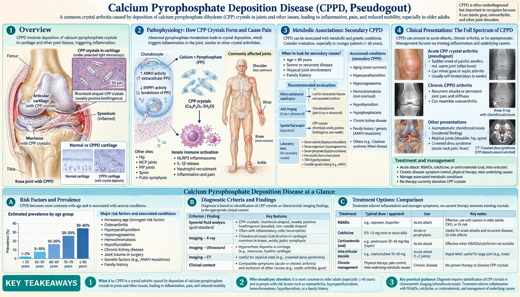
Table of Contents
Overview
Calcium pyrophosphate deposition disease — abbreviated CPPD disease, and historically called pseudogout — is arthritis caused by calcium pyrophosphate (CPP) crystals. A 2016 review in the New England Journal of Medicine (Rosenthal and Ryan) explains the old name: an early description involved patients with an acute gout-like arthritis whose joint-fluid crystals resisted digestion by uricase, so they did not have gout.
In 2011 the European League Against Rheumatism (EULAR) CPPD Task Force agreed new terms. "CPPD" became the umbrella term covering acute CPP crystal arthritis (formerly pseudogout), osteoarthritis with CPPD and chronic CPP crystal inflammatory arthritis. Chondrocalcinosis means cartilage calcification, most commonly due to CPPD, detected by imaging or tissue examination — the Rosenthal and Ryan review notes that the term does not by itself imply clinical arthritis.
CPP crystals and basic calcium phosphate crystals are the two main calcium-containing crystals that can deposit in skeletal tissues (Ea and Lioté, 2014). Gout is a different crystal disease, caused by monosodium urate crystals (Dalbeth, 2021).
How Common It Is
- The 2016 review estimates that CPPD disease affects 4 to 7% of the adult population in Europe and the United States, while noting that these estimates rest largely on X-ray chondrocalcinosis rather than on clinical arthritis.
- In a UK community study of 1,727 people with knee X-rays, the crude prevalence of knee chondrocalcinosis was 7.0%; the age-, sex- and knee-pain-standardised estimate for people over 40 was 4.5%. It rose strongly with age, with no sex predisposition (Neame, 2003).
- Among 100 consecutive admissions to a geriatric unit, chondrocalcinosis rose from 15% at ages 65–74 to 44% over age 84 (Wilkins, 1983).
- A 2024 review in The Lancet Rheumatology (Pascart) states that CPPD disease might be the most common cause of inflammatory arthritis in people over 60, though direct evidence is scarce.
Chondrocalcinosis can be present without any clinical arthritis; the 2016 review's authors regard this as a possible presymptomatic phase, similar to high uric acid before gout, but state that this still needs proof. The 2011 EULAR management recommendations state that asymptomatic CPPD requires no treatment.
Pathophysiology: How CPP Crystals Form and Cause Pain
Crystals Form in Cartilage
According to the 2016 review, the essential first step is the formation of CPP crystals in the matrix around cartilage cells (chondrocytes); CPP crystals rarely form outside cartilage. Chondrocytes release tiny vesicles ("articular cartilage vesicles") that serve as sites of crystal formation, and they produce high levels of extracellular inorganic pyrophosphate (PPi), which is critical for crystal formation. The 2024 review summarises the cause as elevated extracellular pyrophosphate in cartilage.
The Pyrophosphate Pathway
- Source: in cartilage, most PPi is generated from extracellular ATP, by enzymes such as ectonucleotide pyrophosphatase 1 (Rosenthal and Ryan, 2016). Abhishek and Doherty (2011) describe two origins of extracellular PPi: breakdown of nucleotide triphosphates by the enzyme PC-1, and pyrophosphate transport by the ANK protein.
- ANKH: the multipass membrane protein ANKH regulates ATP efflux and so the levels of PPi. Mutations in the ANKH gene cause familial CPPD (Abhishek and Doherty, 2011). The 2016 review notes that the gout drug probenecid antagonises ANKH action in laboratory (in vitro) studies.
- Breakdown: alkaline phosphatase and other pyrophosphatases degrade PPi — which is why the low alkaline phosphatase of hypophosphatasia is linked to CPPD (see below).
- Magnesium: according to the 2016 review, magnesium increases the solubility of CPP crystals and acts as a cofactor for pyrophosphatases.
From Crystals to Inflammation and Damage
Once formed, CPP crystals start inflammation by activating the NLRP3 inflammasome and by creating neutrophil extracellular traps. They also have direct destructive effects on cartilage cells and joint-lining cells, triggering production of matrix metalloproteinases and prostaglandins, and crystal deposits change the mechanical properties of cartilage, which may cause or accelerate joint damage (Rosenthal and Ryan, 2016). The same NLRP3 pathway with release of IL-1β drives the gout flare (Dalbeth, 2021). Colchicine's primary mechanism is tubulin disruption, and newer work describes inhibition of the NLRP3 (NALP3) inflammasome (Leung, 2015).
Familial CPPD
Most CPPD is sporadic. The 2016 review describes two genetic loci in familial CPPD: CCAL2 on chromosome 5p, with autosomal dominant inheritance probably reflecting a gain of function of the ANKH protein, and CCAL1 on chromosome 8, not yet fully characterised; a gain-of-function mutation in TNFRSF11B (osteoprotegerin) has also been described in one family. The review's authors write that CPPD disease before age 60 is a reason to ask about similarly affected relatives. The EULAR 2011 recommendations describe familial forms as rare.
Metabolic Associations: Secondary CPPD
The EULAR 2011 recommendations list the recognised risk factors for CPPD as ageing, osteoarthritis and metabolic conditions such as primary hyperparathyroidism, haemochromatosis and hypomagnesaemia. The 2024 review adds hypophosphatasia and describes all of these metabolic links as uncommon. In the 2023 ACR/EULAR classification criteria, associated metabolic disorders are one of the weighted items.
1. Hyperparathyroidism
In a UK general-practice study of 795 people with pseudogout and 7,770 matched controls, hyperparathyroidism carried an odds ratio of 4.87 (95% CI 2.10–11.3) for pseudogout (Rho, 2012). The 2016 review notes that CPPD disease can persist years after the high calcium has been corrected, which suggests a complex link, and that acute attacks often follow surgery, particularly parathyroidectomy.
2. Haemochromatosis
The 2016 review describes haemochromatosis (iron overload) as strongly associated with CPPD, possibly through iron inhibiting pyrophosphatases or through high levels of parathyroid hormone in cartilage.
3. Hypomagnesaemia
The 2016 review calls low blood magnesium an important risk factor, and links CPPD in the Gitelman variant of Bartter's syndrome to hypomagnesaemia. A UK community study found an association between knee chondrocalcinosis and diuretic use (adjusted OR 2.07); its authors suggested that diuretic-induced low magnesium might theoretically explain it (Neame, 2003).
4. Hypophosphatasia
Hypophosphatasia is a congenital syndrome caused by low functional levels of alkaline phosphatase; CPPD in this condition results from a high ratio of pyrophosphate to phosphate (Rosenthal and Ryan, 2016).
Other Associations
- In the UK general-practice study, pseudogout was also associated with osteoarthritis (OR 2.91), loop diuretic use (OR 1.35) and chronic renal failure (OR 2.29); rheumatoid arthritis, thiazide diuretics and body-mass index were not associated (Rho, 2012).
- Previous joint injury is a strong risk factor: decades after meniscectomy, chondrocalcinosis developed in 20% of operated knees versus 4% of the knees not operated on (cited in Rosenthal and Ryan, 2016).
- As many as 5% of people with gout have CPP crystals in their joint fluid (Rosenthal and Ryan, 2016).
Testing for an Underlying Cause
The authors of the 2016 review write that in people younger than 60 who present with CPPD disease, testing for these associated conditions is indicated because arthritis may be the first sign, and they recommend iron studies (iron, transferrin and ferritin) together with serum calcium, alkaline phosphatase and parathyroid hormone.
Clinical Presentations: The Full Spectrum of CPPD
The 2016 review cites an estimate by McCarty that the chronic degenerative, osteoarthritis-like form accounts for roughly 50% of CPPD disease and acute CPP crystal arthritis for about 25%.
1. Acute CPP Crystal Arthritis ("Pseudogout")
According to the 2016 review, people typically develop sudden arthritis in one or a few joints, with warmth, redness and swelling, and the picture is often indistinguishable from acute gout or septic (infected) arthritis. The knee is the most commonly involved joint, followed by the wrist; an attack at the base of the big toe (podagra) is rare. Fevers, chills and other whole-body symptoms often occur. Gout attacks typically last several days to a week, whereas CPPD attacks may last weeks to months.
In a Japanese series of 50 people with pseudogout (average age 70.8), 25 had fevers averaging 38°C, five with mental confusion; 14 were initially suspected of or misdiagnosed with septic arthritis, and 21 had more than one joint involved (Masuda, 1988). In a 2023 French trial of hospitalised people aged 65 and over (median age 88), the knee was affected in 48%, the wrist in 20% and the ankle in 13% (Pascart, COLCHICORT, 2023).
Triggers described in the 2016 review include acute illness, joint trauma and the period after surgery — particularly parathyroidectomy or hip-fracture repair. Possible drug triggers, some disputed, include injections of hyaluronan into the joint, loop diuretics, granulocyte–macrophage colony-stimulating factor and pamidronate. In the Japanese series, attacks in ten people followed mild trauma, long walks, knee replacement or a glucocorticoid injection into the joint (Masuda, 1988).
2. Chronic CPP Crystal Arthritis
The 2016 review describes several chronic patterns:
- Osteoarthritis-like: the most common chronic form, a polyarticular arthritis resembling osteoarthritis but with inflammatory flares and unusually severe joint damage. Involvement of the shoulder (glenohumeral) joint, wrist and knuckles (metacarpophalangeal joints), which typical osteoarthritis rarely affects, is a clue.
- Rheumatoid-like: a rarer form with persistent inflammatory arthritis of large and small joints; flares often involve joints in sequence and are less symmetric than in rheumatoid arthritis.
- Other forms: CPP crystals in the spine (discs and ligaments), a severely destructive arthritis similar to Charcot's arthropathy, and rare tumour-like soft-tissue deposits that can be mistaken for cancer.
3. Chondrocalcinosis Without Symptoms
In the geriatric-unit study, 7 of the 25 people with knee chondrocalcinosis had no symptoms or signs and no X-ray evidence of osteoarthritis at that knee (Wilkins, 1983). The EULAR 2011 recommendations state that asymptomatic CPPD requires no treatment.
4. Crowned Dens Syndrome
CPP crystals deposited around the C2 vertebra (the dens) cause acute severe neck pain, fever and high inflammatory markers, and the syndrome is often confused with meningitis or sepsis (Rosenthal and Ryan, 2016). A 2025 systematic review of 196 reported patients (mean age 73) found neck pain in 96.8%, a raised CRP in 91.4% and a raised ESR in 80.7%. CT showed abnormalities in 97.3%, compared with 68.4% for MRI and 79.4% for plain X-rays, and patients first misdiagnosed were more likely to undergo lumbar puncture (34.8% versus 13.1%) (Hamdan, 2025). Under the 2023 ACR/EULAR criteria, crowned dens syndrome is on its own sufficient to classify a patient as having CPPD disease.
Diagnosis: How CPPD is Confirmed
The EULAR 2011 recommendations state that definitive diagnosis relies on finding CPP crystals in joint (synovial) fluid; rapid-onset inflammation is suggestive but not definitive. The 2016 review adds that CPPD disease is underdiagnosed: 20% of unselected people examined at knee replacement for osteoarthritis had CPP crystals in their joint fluid.
Synovial Fluid Analysis
- Crystal appearance: CPP crystals are rhomboid-shaped and positively birefringent. Under compensated polarised light microscopy with a red filter they appear blue when parallel to the polariser axis and yellow when perpendicular; monosodium urate crystals of gout show the opposite colours (Rosenthal and Ryan, 2016).
- Hard to see: CPP crystals are small and often weakly birefringent (Rosenthal and Ryan, 2016); only about one in five has enough birefringence for easy detection, and they are easily missed if searched for with a polarising microscope alone (Pascual, 2005).
- Cell counts: cell counts in synovial fluid vary widely (Rosenthal and Ryan, 2016), and the boundaries between non-inflammatory, inflammatory and septic fluid are "very hazy" and have to be read in the clinical setting; microbiological study of the fluid is key to diagnosing infection (Pascual, 2005).
Plain X-Rays
Radiographic chondrocalcinosis is not highly sensitive or specific (EULAR, 2011). The 2016 review estimates that it identifies only about 40% of people with clinically significant CPPD disease, that it is hard to see where cartilage loss is severe, and that knee fibrocartilage calcification can sometimes be a different mineral. The review advises against using chondrocalcinosis as a sole criterion without clinical arthritis, and lists other X-ray clues that help separate CPPD from primary osteoarthritis: hook-like osteophytes, spine involvement, narrowing that predominates at the wrist (radiocarpal) or kneecap (patellofemoral) joint, subchondral cysts, severe joint destruction, and tendon or fascia calcifications.
Musculoskeletal Ultrasound
On ultrasound, chondrocalcinosis appears as linear densities in hyaline cartilage or fibrocartilage (Rosenthal and Ryan, 2016). The EULAR 2011 evidence review found ultrasound more useful than X-ray for peripheral joints (positive likelihood ratio 24.2). In a 2021 OMERACT study using microscopy of knee tissue removed at surgery as the reference, ultrasound had an overall sensitivity of 91% and specificity of 59%; combining the medial meniscus and the medial femoral cartilage gave 88% and 76% (Filippou, 2021). The 2016 review cautions that distinguishing gout from CPPD by ultrasound can be difficult.
CT and Other Imaging
CT detects calcifications accurately and is particularly useful for CPPD in the spine (Rosenthal and Ryan, 2016), such as in crowned dens syndrome (Pascart, 2024). MRI in its present form is insensitive to tissue calcification; dual-energy CT and other newer techniques "hold promise" (Rosenthal and Ryan, 2016).
2023 ACR/EULAR Classification Criteria
The first validated classification criteria for symptomatic CPPD disease were published in 2023 (Abhishek and Tedeschi). Among people with joint pain, swelling or tenderness whose symptoms are not fully explained by another disease, crowned dens syndrome or CPP crystals in synovial fluid are sufficient for classification. Otherwise, a score above 56 points from weighted clinical, metabolic, laboratory and imaging items classifies the disease. Sensitivity was 92.2% and specificity 87.9% in the derivation cohort, and 99.2% and 92.5% in the validation cohort. The authors state that the criteria "will facilitate research in this field."
Treatment: Managing Acute Attacks and Long-Term Disease
No treatment has been shown to dissolve CPP crystals, which is why controlling inflammation is the main focus of therapy (Pascart, 2024). The 2016 review contrasts this with gout, where long-term therapies reduce the urate burden; in gout, long-term urate-lowering therapy leads to dissolution of the urate crystals (Dalbeth, 2021). The EULAR 2011 recommendations state that optimal treatment combines non-drug and drug measures. Doses below are those reported in guidelines, reviews and trials, not instructions.
Acute Attacks
- Local measures and joint injection: EULAR 2011 states that for acute CPP crystal arthritis, cool packs, temporary rest and joint aspiration combined with a steroid injection are often sufficient. The 2016 review describes injected glucocorticoids as typically recommended first-line for joints that can be injected, and its authors use them first-line.
- Colchicine: the 2016 review describes oral colchicine at 0.6–1.2 mg daily in people without significant kidney or liver impairment, with an optional single 1.2 mg loading dose.
- Colchicine versus prednisone (COLCHICORT, 2023): in an open-label trial of 111 hospitalised people aged 65 and over (median age 88), colchicine (1.5 mg on day 1 and 1 mg on day 2) and prednisone (30 mg on days 1 and 2) gave equivalent pain reduction at 24 hours (−36 mm versus −38 mm on a 100 mm scale). Diarrhoea occurred in 22% on colchicine versus 6% on prednisone; high blood pressure in 2% versus 11%, and high blood sugar in none versus 6%.
- NSAIDs: the 2016 review states that anti-inflammatory doses, preferably with stomach protection, may be effective for acute flares.
- Systemic corticosteroids: EULAR 2011 lists oral or injected corticosteroids for people unresponsive to, or unsuited to, other measures; the 2016 review states that most experts consider them moderately effective. The 2024 review states that prednisone might provide the best benefit-risk ratio for acute attacks, while low-dose colchicine is also effective with a risk of mild diarrhoea.
Preventing Recurrent Attacks and Chronic Disease
- Colchicine: EULAR 2011 lists low-dose colchicine 0.5–1.0 mg daily and/or oral NSAIDs with stomach protection for prophylaxis or chronic inflammatory arthritis. The main study behind daily colchicine prophylaxis is small and uncontrolled: ten people with recurrent pseudogout were followed for a year before and a year on colchicine 0.6 mg twice daily, and attacks fell from 32 (3.2 per person per year) to 10 (1 per person per year); 90% of the patients benefited (Alvarellos and Spilberg, 1986 — PubMed PMID: 3772928). The 2016 review calls this "some evidence."
- Methotrexate: an uncontrolled study suggested benefit, but a 2014 randomised crossover trial in 26 people with chronic or recurrent CPPD found no significant difference between weekly 15 mg methotrexate injections and placebo over three months (Finckh, 2014).
- Hydroxychloroquine and low-dose corticosteroids: EULAR 2011 lists low-dose corticosteroid, methotrexate or hydroxychloroquine for chronic inflammatory arthritis with CPPD; the 2016 review states that "some data" support hydroxychloroquine, and the 2024 review describes the evidence for colchicine, low-dose weekly methotrexate and hydroxychloroquine in prophylaxis as limited.
- Real-world use in chronic disease: in a 2024 European chart review of 129 people with chronic CPP crystal inflammatory arthritis, colchicine was the usual first-line drug; colchicine, methotrexate, anakinra and tocilizumab were the most prescribed, and efficacy did not differ significantly between treatments. Adverse events led to 14.1% of colchicine discontinuations (all for diarrhoea) and 31.8% of anakinra discontinuations (Damart, 2024).
IL-1 and IL-6 Blocking Drugs
Anakinra, which blocks interleukin-1, is used off-label. In a 2013 retrospective series of 16 people (mean age 80) who received anakinra 100 mg under the skin daily because conventional drugs had failed or could not be used, 14 responded fully or partly (Ottaviani, 2013). A 2020 systematic review of 76 patients found a response to anakinra in 80.6% with acute and 42.9% with chronic CPPD, with adverse events in 4.1% (Cipolletta, 2020). A meta-analysis of six studies (five observational, one randomised; 84 patients) found a pooled response rate of 76% in acute flares, with injection-site reactions, rash, bacterial pneumonia and low neutrophil counts among the adverse effects (Nakaphan, 2026). The 2024 review states that biologics blocking IL-1 and IL-6 might have a role in refractory disease.
Underlying Conditions and Surgery
The 2016 review notes that CPPD disease can be a presenting sign of hyperparathyroidism, hypophosphatasia, haemochromatosis or hypomagnesaemia, that CPPD can persist years after hypercalcaemia is corrected, and that knee and hip replacement outcomes appear similar in people with osteoarthritis and in those with CPPD disease.
CPPD vs. Gout: Key Differences
Both conditions can present as a sudden, very painful inflammatory arthritis, and the 2016 review notes that acute CPP crystal arthritis is often indistinguishable from acute gout on clinical grounds. The table summarises differences reported in the sources on this page.
| Feature | CPPD | Gout |
|---|---|---|
| Crystal type | Calcium pyrophosphate (CPP) | Monosodium urate |
| Under compensated polarised light | Rhomboid, positively birefringent; blue when parallel, yellow when perpendicular | Yellow when parallel, blue when perpendicular |
| Length of an acute attack | May last weeks to months | Typically several days to 1 week |
| Main risk factors | Ageing, osteoarthritis, prior joint injury; uncommonly hyperparathyroidism, haemochromatosis, hypomagnesaemia, hypophosphatasia | Raised serum urate (hyperuricaemia) is the most important risk factor |
| Inflammation pathway | NLRP3 inflammasome | NLRP3 inflammasome with IL-1β release |
| Long-term treatment | No treatment dissolves CPP crystals; therapy targets inflammation | Long-term urate-lowering therapy dissolves urate crystals |
Sources: Rosenthal and Ryan, 2016; Pascart, 2024; Dalbeth, 2021; EULAR, 2011.
Prognosis and Living with CPPD
Long-Term Outlook
The 2016 review describes chronic CPP crystal arthritis as much more difficult to manage than acute attacks, with few controlled trials of any therapy and no disease-modifying treatment. It states that CPP crystals certainly worsen cartilage damage and likely initiate it, supported by familial cases in which crystals appear before joint degeneration. The 2024 review describes CPPD as strongly associated with cartilage breakdown and osteoarthritis, with the direction of cause unclear. Joint replacement may eventually be needed; the 2016 review reports outcomes similar to those in osteoarthritis.
Diet and Triggers
The 2016 review states that there are no known dietary associations of CPPD disease. Reported triggers of acute attacks are acute illness, joint trauma and surgery (see Clinical Presentations). In the UK general-practice study, loop diuretic use was associated with pseudogout, and its authors suggested that avoiding loop diuretics may help people with recurrent attacks (Rho, 2012).
Fever and a Swollen Joint
Acute CPP crystal arthritis is often clinically indistinguishable from septic arthritis (Rosenthal and Ryan, 2016), fever is common during attacks (Masuda, 1988), and microbiological study of the joint fluid is the key to diagnosing infection (Pascual, 2005). In the Japanese series, 14 of 50 people were first suspected of or misdiagnosed with septic arthritis, and six had surgery under that misdiagnosis (Masuda, 1988).
Key Research Papers
- Zhang W, Doherty M, Bardin T, et al. European League Against Rheumatism recommendations for calcium pyrophosphate deposition. Part I: terminology and diagnosis. Ann Rheum Dis. 2011;70(4):563-570. PubMed PMID: 21216817
- Zhang W, Doherty M, Pascual E, et al. EULAR recommendations for calcium pyrophosphate deposition. Part II: management. Ann Rheum Dis. 2011;70(4):571-575. PubMed PMID: 21257614
- Abhishek A, Tedeschi SK, Pascart T, et al. The 2023 ACR/EULAR classification criteria for calcium pyrophosphate deposition disease. Ann Rheum Dis. 2023;82(10):1248-1257. PubMed PMID: 37495237
- Rosenthal AK, Ryan LM. Calcium pyrophosphate deposition disease. N Engl J Med. 2016;374(26):2575-2584. PubMed PMID: 27355536
- Pascart T, Filippou G, Lioté F, Sirotti S, Jauffret C, Abhishek A. Calcium pyrophosphate deposition disease. Lancet Rheumatol. 2024;6(11):e791-e804. PubMed PMID: 39089298
- Abhishek A, Doherty M. Pathophysiology of articular chondrocalcinosis — role of ANKH. Nat Rev Rheumatol. 2011;7(2):96-104. PubMed PMID: 21102543
- Ea HK, Lioté F. Diagnosis and clinical manifestations of calcium pyrophosphate and basic calcium phosphate crystal deposition diseases. Rheum Dis Clin North Am. 2014;40(2):207-229. PubMed PMID: 24703344
- Neame RL, Carr AJ, Muir K, Doherty M. UK community prevalence of knee chondrocalcinosis: evidence that correlation with osteoarthritis is through a shared association with osteophyte. Ann Rheum Dis. 2003;62(6):513-518. PubMed PMID: 12759286
- Wilkins E, Dieppe P, Maddison P, Evison G. Osteoarthritis and articular chondrocalcinosis in the elderly. Ann Rheum Dis. 1983;42(3):280-284. PubMed PMID: 6859960
- Rho YH, Zhu Y, Zhang Y, Reginato AM, Choi HK. Risk factors for pseudogout in the general population. Rheumatology (Oxford). 2012;51(11):2070-2074. PubMed PMID: 22886340
- Masuda I, Ishikawa K. Clinical features of pseudogout attack. A survey of 50 cases. Clin Orthop Relat Res. 1988;(229):173-181. PubMed PMID: 3349673
- Hamdan O, Alajlouni Y, Mussa Q, et al. Clinical features and diagnostic challenges in crowned dens syndrome: a systematic review and meta-analysis. Rheumatol Int. 2025;45(5):124. PubMed PMID: 40278908
- Pascual E, Jovaní V. Synovial fluid analysis. Best Pract Res Clin Rheumatol. 2005;19(3):371-386. PubMed PMID: 15939364
- Filippou G, Scanu A, Adinolfi A, et al. Criterion validity of ultrasound in the identification of calcium pyrophosphate crystal deposits at the knee: an OMERACT ultrasound study. Ann Rheum Dis. 2021;80(2):261-267. PubMed PMID: 32988839
- Pascart T, Robinet P, Ottaviani S, et al. Evaluating the safety and short-term equivalence of colchicine versus prednisone in older patients with acute calcium pyrophosphate crystal arthritis (COLCHICORT): an open-label, multicentre, randomised trial. Lancet Rheumatol. 2023;5(9):e523-e531. PubMed PMID: 38251496
- Alvarellos A, Spilberg I. Colchicine prophylaxis in pseudogout. J Rheumatol. 1986;13(4):804-805. PubMed PMID: 3772928
- Finckh A, Mc Carthy GM, Madigan A, et al. Methotrexate in chronic-recurrent calcium pyrophosphate deposition disease: no significant effect in a randomized crossover trial. Arthritis Res Ther. 2014;16(5):458. PubMed PMID: 25315665
- Damart J, Filippou G, Andrès M, et al. Retention, safety and efficacy of off-label conventional treatments and biologics for chronic calcium pyrophosphate crystal inflammatory arthritis. Rheumatology (Oxford). 2024;63(2):446-455. PubMed PMID: 37216917
- Ottaviani S, Brunier L, Sibilia J, et al. Efficacy of anakinra in calcium pyrophosphate crystal-induced arthritis: a report of 16 cases and review of the literature. Joint Bone Spine. 2013;80(2):178-182. PubMed PMID: 23022422
- Cipolletta E, Di Matteo A, Scanu A, et al. Biologics in the treatment of calcium pyrophosphate deposition disease: a systematic literature review. Clin Exp Rheumatol. 2020;38(5):1001-1007. PubMed PMID: 32359034
- Nakaphan P, Phutinart S, Pajareya P, et al. Efficacy and safety of anakinra for acute calcium pyrophosphate deposition disease flare: a systematic review and meta-analysis. Mod Rheumatol. 2026;36(3):469-475. PubMed PMID: 41206733
- Leung YY, Yao Hui LL, Kraus VB. Colchicine — update on mechanisms of action and therapeutic uses. Semin Arthritis Rheum. 2015;45(3):341-350. PubMed PMID: 26228647
- Dalbeth N, Gosling AL, Gaffo A, Abhishek A. Gout. Lancet. 2021;397(10287):1843-1855. PubMed PMID: 33798500
PubMed Topic Searches
- Calcium Pyrophosphate Deposition Disease — PubMed
- Pseudogout Treatment — PubMed
- Chondrocalcinosis Pathophysiology — PubMed
- NLRP3 Inflammasome Crystal Arthritis — PubMed
- Colchicine CPPD Prevention — PubMed
Connections
- Rheumatology
- Gout — the most important differential diagnosis; urate crystals vs. CPP crystals
- Osteoarthritis — CPPD can accelerate and mimic OA; both extremely common in the elderly
- Rheumatoid Arthritis — the "pseudo-RA" pattern of chronic CPPD can exactly mimic seronegative RA
- Reactive Arthritis — another cause of acute inflammatory monoarthritis in the differential
- Magnesium — hypomagnesemia is a treatable metabolic risk factor for CPPD; Mg²⁺ inhibits CPP crystal growth
- Lab Tests — joint fluid analysis, serum calcium, PTH, ferritin, and other key tests in CPPD workup
- Calcium — serum calcium is part of every CPPD metabolic workup; hyperparathyroid hypercalcemia is the leading secondary cause