Pulmonary Alveolar Proteinosis
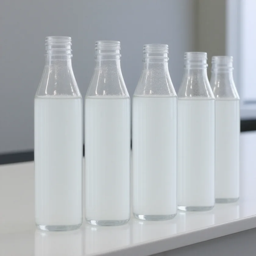
Table of Contents
- How Pulmonary Alveolar Proteinosis Fills the Air Sacs
- The Three Forms of PAP
- Autoimmune PAP: Anti-GM-CSF Antibodies
- Clinical Features and Symptoms
- Imaging and Diagnosis: The Crazy-Paving Pattern
- Whole Lung Lavage: The Historical First-Line Treatment
- GM-CSF Therapy and Emerging Treatments
- Prognosis and Complications
- Key Research Papers
- Featured Videos
How Pulmonary Alveolar Proteinosis Fills the Air Sacs
Pulmonary alveolar proteinosis (PAP) is a rare lung disorder in which the tiny air sacs of the lungs — the alveoli — gradually fill with a thick, milky material made of surfactant proteins and lipids. Surfactant is normally produced by specialized lung cells (type II pneumocytes) and is essential for keeping the alveoli open during breathing. In a healthy lung, alveolar macrophages continuously clear used surfactant to prevent it from accumulating. In PAP, this clearance mechanism breaks down, and the lipoproteinaceous material builds up to a degree that seriously impairs oxygen transfer from air into the bloodstream.
Under the microscope, the alveolar material stains strongly positive with periodic acid-Schiff (PAS) stain, appearing as amorphous globular deposits filling the air spaces. The surrounding lung architecture — the walls, blood vessels, and airways — is typically preserved, distinguishing PAP from fibrotic or inflammatory lung diseases where the structure itself is destroyed. This makes PAP potentially reversible if the underlying cause can be corrected or the material physically removed.
PAP is estimated to affect roughly 6–7 people per million worldwide. Although rare in absolute numbers, it is not vanishingly so, and it appears in clinical practice in pulmonary referral centers with regularity. Delayed diagnosis is common because its symptoms — gradually worsening breathlessness and a dry cough — overlap with many far more common conditions including heart failure, pneumonia, and interstitial lung disease. The average time from first symptoms to diagnosis has historically been one to two years.
Understanding PAP has been transformed over the past three decades. What was once considered a single mysterious disease is now recognized as at least three biologically distinct conditions that share the same final anatomic picture — alveolar filling with surfactant material — but have different causes, different patient populations, and increasingly different treatments.
The Three Forms of PAP
PAP is not one disease but a syndrome — a shared anatomical outcome that can be reached by three different biological routes. Distinguishing between them matters enormously because the treatment strategies diverge completely.
Autoimmune PAP (primary PAP) accounts for about 90% of all adult cases. The cause is the production of autoantibodies — immunoglobulin G (IgG) antibodies directed against the body's own granulocyte-macrophage colony-stimulating factor (GM-CSF). These antibodies circulate in the blood and reach the lungs, where they neutralize GM-CSF signaling. Because GM-CSF is the critical cytokine required for alveolar macrophages to mature and to maintain their surfactant-clearing function, its blockade leads to macrophage dysfunction. Used surfactant accumulates progressively in the alveolar spaces. In the Japanese national registry (Inoue 2008), autoimmune PAP made up 89.9% of all PAP cases and was found in at least 6.2 people per million. Among its 223 patients with autoimmune PAP, the median age at diagnosis was 51 years, men outnumbered women 2.1 to 1, 56% had a history of smoking and 23% had been exposed to dust; the registry authors concluded that the disease was not strongly linked to smoking. A German cohort of 70 patients with PAP (Bonella 2011) found a mean age at diagnosis of 43 years, a male-to-female ratio of 1.3, a smoking history in 79% and dust exposure in 54%, and current smokers needed more whole lung lavages to reach remission.
Secondary PAP comprises 5–10% of cases. Here the macrophage dysfunction is not caused by anti-GM-CSF antibodies but by a reduction in the number or functional capacity of alveolar macrophages due to an underlying systemic condition. The most important causes include:
- Hematological malignancies: Acute myeloid leukemia (AML) is the most common single cause of secondary PAP, but myelodysplastic syndrome (MDS), lymphoma, and chemotherapy-related immunosuppression also feature prominently. Post-bone marrow transplant patients are at particular risk.
- Inhalational exposures: Silica dust inhalation produces a variant called silicoproteinosis, which can be clinically severe and rapid in progression. Other implicated dusts include aluminum, titanium, and indium tin oxide — the latter increasingly recognized in workers in the electronics and solar-cell industries.
- Rare metabolic disorders: Lysinuric protein intolerance (LPI), a disorder of amino acid transport, is associated with secondary PAP through unclear mechanisms involving macrophage function.
Congenital/hereditary PAP is rare, accounting for less than 1% of cases, and presents in neonates or infants rather than adults. It results from mutations in genes encoding either the surfactant proteins themselves (SFTPB, SFTPC, ABCA3 — the gene for a lipid transporter critical for surfactant packaging) or the GM-CSF receptor subunits (CSF2RA encoding the α chain, or CSF2RB encoding the shared β chain). When the GM-CSF receptor is nonfunctional due to mutation, GM-CSF signaling is completely absent even though circulating GM-CSF levels are normal and anti-GM-CSF antibodies are absent. The result is profound alveolar macrophage dysfunction from birth, often presenting as neonatal respiratory distress syndrome that fails to resolve or that recurs after initial stabilization.
Autoimmune PAP: Anti-GM-CSF Antibodies
The discovery that anti-GM-CSF autoantibodies are the cause of primary PAP transformed the understanding of this disease. Before this was established in the 1990s and 2000s, PAP was thought to be an idiopathic disorder of unknown origin. Today, measuring these antibodies in blood or bronchoalveolar lavage (BAL) fluid is both diagnostically definitive and biologically illuminating.
GM-CSF (granulocyte-macrophage colony-stimulating factor) is a cytokine that plays a central role in the maturation and functional maintenance of alveolar macrophages. In the alveolar environment, GM-CSF signals through its receptor on macrophage surfaces, activating transcription factors (particularly PU.1) that drive the expression of proteins required for surfactant catabolism — the breaking down and clearance of the lipid and protein components of used surfactant. Without this signaling, alveolar macrophages remain functionally immature: they are present in the alveolar space but cannot process surfactant effectively. Instead, they accumulate surfactant lipids within their cytoplasm, becoming the characteristic "foamy alveolar macrophages" seen in BAL specimens and biopsies from PAP patients.
In autoimmune PAP, IgG antibodies bind GM-CSF with high affinity and neutralize it before it can engage its receptor. These antibodies are polyclonal, meaning they arise from many different B-cell clones, which is characteristic of an autoimmune rather than a monoclonal (paraneoplastic) origin. Critically, anti-GM-CSF antibodies are present in essentially all patients with autoimmune PAP, are present only at very low levels in healthy people, and are absent in patients with secondary or congenital PAP. Whether the level tracks how sick a person is remains unsettled: in the Japanese registry, disease severity did not correlate with the antibody level (Inoue 2008), while the German cohort found the antibody level related to clinical outcome (Bonella 2011).
How the blood test is read: a standardized blood test (an ELISA) was developed to bring the antibody measurement into routine clinical use (Uchida 2014). With it, the median antibody level was 21.54 IU in 56 people with autoimmune PAP against 0.08 IU in 38 healthy people, so healthy people do carry tiny amounts. A serum level of 5 µg/mL was the best cut-off for telling autoimmune PAP serum apart from normal serum.
The antibodies are also detectable in BAL fluid, where their local concentration in the lung environment contributes directly to macrophage dysfunction at the site of disease. Measuring BAL anti-GM-CSF levels adds sensitivity and can help in cases where serum levels are borderline.
One important consequence of macrophage dysfunction in PAP extends beyond surfactant clearance: alveolar macrophages are the lung's primary innate immune defense against inhaled pathogens. Their dysfunction in PAP explains the elevated risk of opportunistic pulmonary infections — particularly with organisms that are normally controlled by macrophage-mediated immunity, including Nocardia, Cryptococcus, Aspergillus, and Histoplasma. Nocardia in particular appears with striking frequency in PAP case series, which makes it one of the infections looked for when a PAP patient develops fever, productive cough, or worsening infiltrates that do not fit the expected PAP pattern.
Clinical Features and Symptoms
PAP typically announces itself gradually and without drama, which is one reason diagnosis is so often delayed. The disease does not usually cause fever, chills, or the acute systemic upset of pneumonia. Instead, it behaves more like a slowly progressive breathlessness that patients often attribute to deconditioning, weight gain, or aging — until the limitation becomes severe enough to prompt medical evaluation.
Dyspnea on exertion (breathlessness with effort) is the most common presenting symptom. In the Japanese registry it was the presenting symptom in 54.3% of patients, and 31.8% had no symptoms at all and were found through health screening (Inoue 2008). Early in the disease, it appears only with vigorous activity. As alveolar filling progresses, the threshold for breathlessness falls until patients are limited by ordinary activities like climbing stairs or walking short distances. In advanced disease, breathlessness at rest and hypoxemia at rest develop.
Dry cough is present in about half of patients, though it is rarely severe or productive. The cough of PAP is thought to arise from stimulation of irritant receptors in airways partially obstructed by alveolar material, rather than from airway inflammation.
Physical examination findings in PAP are often surprisingly minimal relative to the extent of radiographic disease. Fine bibasilar crackles — similar to the velcro-like crackles of pulmonary fibrosis — are present in about 50% of patients. Clubbing of the fingers is seen but is less common than in fibrotic diseases. Cyanosis indicates severe disease. The absence of fever, wheezing, or pleural rub helps distinguish PAP from more common diagnoses.
Laboratory findings are largely nonspecific. Lactate dehydrogenase (LDH) is elevated in most patients with active PAP, reflecting cellular turnover in the alveoli. Carcinoembryonic antigen (CEA) and cancer antigen 125 (CA-125) can be elevated — these tumor markers are found in high concentrations in surfactant proteins and leak into the bloodstream when surfactant processing is disrupted; they have been used as biomarkers for monitoring disease activity and treatment response. Polycythemia is occasionally present in response to chronic hypoxemia. The definitive laboratory finding is elevated serum anti-GM-CSF antibody titer (above the 5 µg/mL cut-off of the standardized test) in autoimmune PAP.
Pulmonary function tests typically show a restrictive pattern — reduced total lung capacity (TLC) and forced vital capacity (FVC) with a preserved or elevated ratio of forced expiratory volume in 1 second (FEV1) to FVC — along with a reduced diffusing capacity for carbon monoxide (DLCO), reflecting impaired gas exchange across the alveolar-capillary membrane. Arterial blood gas analysis may show hypoxemia with an elevated alveolar-arterial oxygen gradient, often more severe with exercise than at rest early in the disease course.
Imaging and Diagnosis: The Crazy-Paving Pattern
The chest computed tomography (CT) findings of PAP are among the most recognizable in pulmonary radiology. The classic and highly characteristic pattern is called "crazy paving" — a combination of ground-glass opacity (hazy increased attenuation that does not obscure underlying vascular markings) with superimposed thickening of the interlobular septa (the thin walls that divide lung lobules) and intralobular lines. When these two findings coexist, the resulting computed tomography image resembles irregular stone cobblestones or the pattern of crazy-paving stonework, with geometric shapes outlined against a hazy background.
Several features of PAP's computed tomography pattern help distinguish it from other causes of crazy paving (which include pneumocystis pneumonia, pulmonary edema, and mucinous adenocarcinoma):
- Geographic distribution: Areas of ground-glass opacity in PAP are often sharply demarcated from adjacent areas of completely normal lung, creating a patchy or map-like appearance. This sharp boundary between affected and unaffected lung is quite characteristic.
- Bilateral and symmetric: PAP typically affects both lungs and tends to be fairly symmetric, often with a basal predominance, though the distribution can vary.
- Absence of nodules, cavitation, or honeycombing: PAP does not typically cause the nodular densities of hypersensitivity pneumonitis, the cavities of necrotizing pneumonia, or the honeycombing of end-stage fibrosis.
Plain chest radiography shows bilateral alveolar infiltrates — often described as having a "bat-wing" or perihilar distribution — which can closely mimic pulmonary edema. Computed tomography is far more specific, and it is the usual next test when bilateral infiltrates are unexplained.
Bronchoalveolar lavage (BAL) is the gold standard for diagnosis. In PAP, BAL fluid is characteristically milky or opalescent in appearance. When a BAL specimen is left to stand, the white material settles to form a visible white precipitate at the bottom of the collection tube — a finding so distinctive that an experienced bronchoscopist will recognize PAP immediately. Microscopically, BAL cytology shows PAS-positive amorphous globular extracellular material filling the background of the specimen, along with large foamy alveolar macrophages whose cytoplasm is packed with lipid-laden vacuoles and surfactant debris. These findings together are diagnostic of PAP.
Surgical lung biopsy (video-assisted thoracoscopic surgery, VATS) is occasionally performed when BAL is nondiagnostic or when another diagnosis needs to be excluded. Transbronchial biopsy is less reliable for PAP because the specimens may not capture the alveolar filling pattern adequately.
The usual diagnostic work-up after suspicious computed tomography findings includes: serum anti-GM-CSF antibody titer (if positive at diagnostic threshold, confirms autoimmune PAP without need for biopsy in most cases), BAL with cytology and PAS staining, and evaluation for secondary causes (complete blood count with differential, bone marrow evaluation if hematological malignancy is suspected, occupational exposure history).
Whole Lung Lavage: The Historical First-Line Treatment
Whole lung lavage (WLL) is the historical first-line treatment for symptomatic autoimmune PAP and has been in use since the 1960s; a 2025 review states that it remains the preferred treatment for patients in respiratory failure, especially at the time of diagnosis (Jouneau 2025). Despite being a technically demanding procedure performed under general anesthesia, it remains the most effective single intervention for clearing the alveolar material and restoring gas exchange. When performed at experienced centers, WLL produces rapid and often dramatic improvement in symptoms and oxygenation.
How WLL is performed: The patient is placed under general anesthesia and intubated with a double-lumen endotracheal tube — a specialized airway device that has separate channels for the left and right mainstem bronchi, allowing each lung to be independently ventilated or isolated. While one lung is continuously ventilated to maintain oxygenation, the contralateral lung is filled with warmed sterile normal saline, which is then drained by gravity and suction. This cycle of filling and draining is repeated with sequential 1-liter aliquots until the effluent fluid runs clear — typically requiring 10 to 50 liters per lung. The procedure takes several hours. After completing one lung, the same process is performed on the other lung, either in the same session (if the patient tolerates it) or a few days later.
The physical process of repeated lavage dislodges the PAS-positive surfactant material from the alveolar surfaces and washes it out of the lung. In addition to mechanical clearance, WLL may have some immunomodulatory benefit — removing anti-GM-CSF antibodies from the alveolar compartment and allowing partial recovery of macrophage function after the procedure.
Outcomes of WLL: Immediate improvement in oxygenation is seen in 75–85% of patients following bilateral WLL. Gas exchange typically improves within hours to days as the cleared alveoli re-aerate. Functional improvement (better exercise tolerance, reduced dyspnea) follows over the subsequent weeks. A single bilateral WLL induces sustained remission — defined as freedom from PAP symptoms without further treatment — in approximately 40–70% of patients. Those who do not achieve remission or who relapse after initial response can undergo repeat WLL; there is no fixed limit to the number of WLL procedures a patient can have.
Limitations of WLL: The procedure requires general anesthesia, a specialized anesthesia team experienced with double-lumen intubation in an unstable respiratory patient, and a capable bronchoscopy suite. Not all hospitals have this expertise, and patients in some regions must travel significant distances to specialized PAP centers. Patients with severe hypoxemia may not tolerate single-lung ventilation during the procedure. Post-procedure complications including pneumothorax, fluid overload, and saline flooding of the ventilated lung are uncommon but possible.
WLL is used across all three forms of PAP, including secondary PAP (where it provides symptom relief while the underlying cause is addressed) and even congenital PAP (where it is used as a bridge while more definitive treatments are considered). However, in autoimmune PAP specifically, WLL addresses the consequence (alveolar filling) but not the cause (anti-GM-CSF antibodies), which is why disease-modifying therapies targeting the antibody are increasingly important.
GM-CSF Therapy and Emerging Treatments
The recognition that anti-GM-CSF autoantibodies neutralize GM-CSF signaling in autoimmune PAP created an obvious therapeutic hypothesis: if you restore GM-CSF activity in the lungs, alveolar macrophage function might recover, surfactant clearance would resume, and the disease would improve without requiring the heroic physical intervention of WLL. Randomized placebo-controlled trials have since tested it, with mixed results described below: the 2019 PAGE trial of inhaled sargramostim found a modest improvement in blood oxygen and noted no clinical benefits (Tazawa 2019), while the IMPALA trials reported greater improvements in gas transfer with daily inhaled molgramostim than with placebo (Trapnell 2020; Trapnell 2025). The 2024 European Respiratory Society guideline states that several therapies are available to treat PAP, “but supporting evidence is still limited” (McCarthy 2024).
Inhaled GM-CSF is the most carefully studied disease-modifying approach. It has been tested in two laboratory-made forms, sargramostim and molgramostim; the molgramostim trials are the larger ones (IMPALA, 138 patients; IMPALA-2, 164 patients) against 64 patients in the sargramostim PAGE trial. Inhaled recombinant human GM-CSF is delivered directly to the alveolar compartment via inhalation, where it can engage GM-CSF receptors on alveolar macrophages at concentrations sufficient to override the neutralizing effect of anti-GM-CSF antibodies in the local microenvironment — even though the same antibodies in the bloodstream would neutralize systemically administered GM-CSF. This is the key pharmacological rationale for the inhaled route.
The PAGE trial (published in the New England Journal of Medicine in 2019 by Tazawa and colleagues) randomized 64 patients with mild-to-moderate autoimmune PAP to inhaled sargramostim (125 µg twice daily for 7 days, every other week for 24 weeks) or placebo. The alveolar-arterial oxygen gradient — the primary outcome measure of gas exchange — changed by -4.50 mm Hg with sargramostim against +0.17 mm Hg with placebo (P = 0.02), and lung density on computed tomography also improved more. Serious adverse events occurred in 6 patients on sargramostim and 3 on placebo. The authors described a modest effect on arterial oxygen and noted no clinical benefits. The response is gradual, occurring over weeks to months rather than the immediate effect of WLL. A 2025 review states that inhaled GM-CSF, with molgramostim or sargramostim, “is now considered a first-line treatment in the international guidelines” for autoimmune PAP, following the recent randomized placebo-controlled trials; its lead author discloses that he was an IMPALA investigator and a co-author of the guidelines (Jouneau 2025).
Inhaled molgramostim is another inhaled form of recombinant human GM-CSF, taken once a day. In the IMPALA trial (Trapnell 2020), 138 patients with autoimmune PAP received molgramostim 300 µg once daily, either every day or every other week, or placebo for 24 weeks. With daily dosing, the alveolar-arterial oxygen gradient improved by 12.8 mm Hg against 6.6 mm Hg with placebo (P = 0.03), and the St. George's Respiratory Questionnaire total score (a breathing-related quality-of-life score, lower is better) fell by 12.4 points against 5.1. For several outcomes, daily dosing did better than every-other-week dosing. Side effects were similar across the groups except chest pain, which was more common with daily dosing.
The larger phase 3 IMPALA-2 trial (Trapnell 2025) gave 164 patients molgramostim 300 µg or placebo once daily for 48 weeks. Lung gas transfer (the diffusing capacity for carbon monoxide, DLCO, as a percentage of the predicted value) rose 9.8 percentage points with molgramostim against 3.8 with placebo at week 24 (P < 0.001), and 11.6 against 4.7 at week 48. The St. George's total score fell 11.5 points against 4.9 at week 24, while its activity score showed no significant difference between the groups. The share of patients with any side effect, and with a serious one, was similar in both groups.
Subcutaneous GM-CSF has also been studied and shows benefit in some patients, though the systemic route requires higher doses and may be associated with more side effects (including bone pain and flu-like symptoms) compared to the inhaled route.
Rituximab — a monoclonal antibody that depletes B lymphocytes — addresses the autoimmune root cause of autoimmune PAP by reducing the number of B cells capable of producing anti-GM-CSF antibodies. As B cells are depleted, anti-GM-CSF antibody titers gradually fall over months, and alveolar macrophage function may recover. Early case series and small trials have reported favorable responses in refractory autoimmune PAP patients — those who have failed both WLL and GM-CSF therapy. Rituximab is not yet standard of care, and its reported use so far is in treatment-resistant disease.
Spontaneous remission occurs in approximately 15–20% of patients with autoimmune PAP, without any specific treatment. This observation supports a watchful waiting approach for patients with mild disease, acceptable oxygenation, and stable symptoms — monitoring with serial imaging, pulmonary function tests, and symptom assessment every 3–6 months and reserving WLL or GM-CSF therapy for those who progress.
Secondary PAP responds best to treating the underlying cause: cytoreduction for AML/MDS, reduction of immunosuppression where safe after bone marrow transplant, removal from silica exposure in silicoproteinosis. WLL remains useful for symptomatic relief in secondary PAP regardless of cause.
Congenital PAP from GM-CSF receptor mutations does not respond to exogenous GM-CSF (the receptor is absent or nonfunctional) and is managed with serial WLL as a bridge. Lung transplantation has been performed for severe congenital PAP with good outcomes, though the disease can rarely recur in the transplanted lung.
Prognosis and Complications
The prognosis of PAP has improved substantially since effective treatments — particularly WLL and inhaled GM-CSF — became available and better understood. Most patients with autoimmune PAP can expect to maintain a good quality of life and near-normal lung function if the disease is identified and managed appropriately.
Overall survival in modern cohorts of autoimmune PAP is excellent. The 5-year survival rate exceeds 90% in most published series. Death directly attributable to PAP — from progressive respiratory failure — is now uncommon with access to WLL. Historical series from the pre-WLL era reported mortality rates of 30% or higher, underscoring how much the availability of effective treatment has changed the natural history.
Disease course variability is wide. Some patients have a single episode of PAP, respond to one or two WLL procedures, and remain in long-term remission without further therapy. Others have a relapsing-remitting course requiring periodic WLL — sometimes annually or more frequently — to control symptoms. A minority have progressive disease refractory to WLL and GM-CSF therapy, in whom experimental approaches (rituximab, plasmapheresis to remove antibodies) or lung transplantation are considered.
Opportunistic infections represent the most serious and potentially life-threatening complication of PAP. Alveolar macrophage dysfunction impairs the innate immune defense against fungi, Nocardia, and other pathogens that are normally controlled at the alveolar level. Reported rates of opportunistic infection in PAP range from 5–10% in large cohorts. Nocardia species are particularly prevalent in PAP — responsible for pneumonia, brain abscess, and disseminated disease — and they are among the infections looked for in any PAP patient who develops fever or worsening infiltrates. Cryptococcus and Aspergillus are also over-represented compared to the general population. Pneumocystis jirovecii pneumonia (PJP) can occur in PAP even without classic immunosuppression. Reported approaches include heightened vigilance, a low threshold for bronchoscopy in new or changing infiltrates, and preventive (prophylactic) treatment in severely immunocompromised patients, such as those with secondary PAP after a bone marrow transplant (BMT).
Progression to respiratory failure requiring mechanical ventilation is rare with modern management but can occur in patients who present very late, have severe secondary PAP with an underlying malignancy, or develop a superimposed infection on a background of severe PAP.
Lung transplantation has been performed for PAP in cases refractory to all medical and procedural therapy, with outcomes comparable to transplantation for other indications. In autoimmune PAP, there is a theoretical risk of recurrence in the transplanted lung if anti-GM-CSF antibodies persist — this has been reported rarely but represents an important long-term consideration.
Monitoring and follow-up for patients with established PAP usually includes: regular assessment of symptoms and exercise tolerance, serial chest computed tomography (typically every 6–12 months), pulmonary function tests including DLCO, serum anti-GM-CSF antibody titers (although in the Japanese registry disease severity did not correlate with the antibody level), serum lactate dehydrogenase and CEA (surrogate biomarkers of disease burden), and prompt evaluation of any new or worsening respiratory symptoms for opportunistic infection.
Key Research Papers
- Trapnell BC, Whitsett JA, Nakata K. "Pulmonary alveolar proteinosis." N Engl J Med. 2003;349(26):2527–2539. — PubMed PMID: 14695413
- Inoue Y, Trapnell BC, Tazawa R, et al. "Characteristics of a large cohort of patients with autoimmune pulmonary alveolar proteinosis in Japan." Am J Respir Crit Care Med. 2008;177(7):752–762. — PubMed PMID: 18202348
- Trapnell BC, Nakata K, Bonella F, et al. "Pulmonary alveolar proteinosis." Nat Rev Dis Primers. 2019;5(1):16. — PubMed PMID: 30846703
- Seymour JF, Presneill JJ. "Pulmonary alveolar proteinosis: progress in the first 44 years." Am J Respir Crit Care Med. 2002;166(2):215–235. — PubMed PMID: 12119235
- Tazawa R, Ueda T, Abe M, et al. "Inhaled GM-CSF for pulmonary alveolar proteinosis." N Engl J Med. 2019;381(10):923–932. — PubMed PMID: 31483963
- Kavuru MS, Sullivan EJ, Piccin R, Thomassen MJ, Stoller JK. "Exogenous granulocyte-macrophage colony-stimulating factor administration for pulmonary alveolar proteinosis." Am J Respir Crit Care Med. 2000;161(4 Pt 1):1143–1148. — PubMed PMID: 10764303
- Luisetti M, Kadija Z, Mariani F, Rodi G, Campo I, Trapnell BC. "Therapy options in pulmonary alveolar proteinosis." Ther Adv Respir Dis. 2010;4(4):239–248. — PubMed PMID: 20647242
- Bonella F, Bauer PC, Griese M, Ohshimo S, Guzman J, Costabel U. "Pulmonary alveolar proteinosis: new insights from a single-center cohort of 70 patients." Respir Med. 2011;105(12):1908–1916. — PubMed PMID: 21900000
- Shah PL, Hansell D, Lawson PR, Reid KB, Morgan C. "Pulmonary alveolar proteinosis: clinical aspects and current concepts on pathogenesis." Thorax. 2000;55(1):67–77. — PubMed PMID: 10607805
- Uchida K, Beck DC, Yamamoto T, et al. "GM-CSF autoantibodies and neutrophil dysfunction in pulmonary alveolar proteinosis." N Engl J Med. 2007;356(6):567–579. — PubMed PMID: 17287477
- Presneill JJ, Nakata K, Inoue Y, Seymour JF. "Pulmonary alveolar proteinosis." Clin Chest Med. 2004;25(3):593–613. — PubMed PMID: 15331195
- Campo I, Mariani F, Rodi G, et al. "Assessment and management of pulmonary alveolar proteinosis in a reference center." Orphanet J Rare Dis. 2013;8:40. — PubMed PMID: 23497546
- Uchida K, Nakata K, Carey B, et al. "Standardized serum GM-CSF autoantibody testing for the routine clinical diagnosis of autoimmune pulmonary alveolar proteinosis." J Immunol Methods. 2014;402(1-2):57–70. — PubMed PMID: 24275678
- Trapnell BC, Inoue Y, Bonella F, et al. "Inhaled molgramostim therapy in autoimmune pulmonary alveolar proteinosis." N Engl J Med. 2020;383(17):1635–1644. — PubMed PMID: 32897035
- Trapnell BC, Inoue Y, Bonella F, et al. "Phase 3 trial of inhaled molgramostim in autoimmune pulmonary alveolar proteinosis." N Engl J Med. 2025;393(8):764–773. — PubMed PMID: 40834301
- McCarthy C, Bonella F, O'Callaghan M, et al. "European Respiratory Society guidelines for the diagnosis and management of pulmonary alveolar proteinosis." Eur Respir J. 2024;64(5):2400725. — PubMed PMID: 39147411
- Jouneau S, Chauvin P, Lederlin M, Painvin B, Kerjouan M. "Pharmacotherapy for autoimmune pulmonary alveolar proteinosis." Drugs. 2025;85(10):1193–1206. — PubMed PMID: 40866780
Search PubMed for more: Pulmonary alveolar proteinosis Whole lung lavage PAP GM-CSF autoantibodies PAP