Biliary Atresia
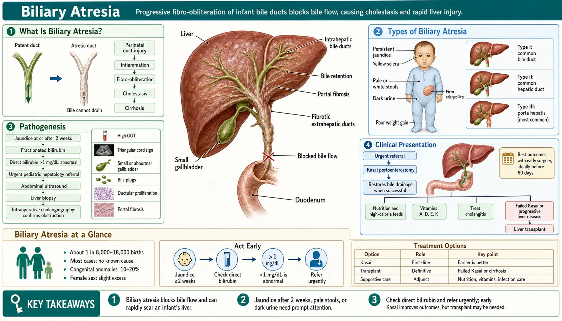
- How Biliary Atresia Damages the Liver
- Types of Biliary Atresia
- Pathogenesis
- Clinical Presentation
- Diagnosis
- Kasai Hepatoportoenterostomy
- Liver Transplantation
- Prognosis and Long-Term Outcomes
- Key Research Papers
- Connections
- Featured Videos
How Biliary Atresia Damages the Liver
Biliary atresia (BA) is a progressive fibro-obliterative cholangiopathy of infancy in which the extrahepatic bile ducts become inflamed, scarred, and obliterated, preventing bile from draining from the liver into the intestine. It is the most common cause of neonatal cholestasis requiring surgical intervention and the leading indication for pediatric liver transplantation worldwide, accounting for approximately 40–50% of all pediatric liver transplants. A 2017 North American and European guideline reports that jaundice from blocked or failed bile flow (cholestatic jaundice) affects about 1 in 2,500 term infants, and that biliary atresia is 25–40% of it in the first months of life.
The disease is not simply a congenital malformation present at birth in a static sense. In the most common form, the bile ducts may be initially patent at birth and only subsequently undergo progressive inflammatory destruction. This dynamic, ongoing process distinguishes biliary atresia from structural ductal anomalies like choledochal cyst and makes early diagnosis and timing of surgery critically important.
Incidence: Biliary atresia affects approximately 1 in 10,000–15,000 live births in Western populations. Incidence is higher in East Asian populations (1:6,000–1:8,000 in Taiwan and Japan). Taiwan's national stool card registry measured 1.51 cases per 10,000 live births from 2004 to 2010: 2.37 in preterm infants and 1.43 in term infants. In the United States, approximately 300–400 new cases are diagnosed each year. There is a slight female predominance (female-to-male ratio approximately 1.1–1.4:1), the reason for which is unknown.
Etiology: The precise cause of biliary atresia remains incompletely understood. Leading hypotheses include:
- Viral triggers: Perinatal viral infections — particularly cytomegalovirus (CMV) and rotavirus — have been most strongly implicated. Cytomegalovirus immunoglobulin M (IgM) seropositivity is found in 30–40% of biliary atresia infants in some series. Animal models using rhesus rotavirus injection in newborn mice reproducibly induce a biliary atresia-like phenotype, suggesting viral-triggered immune activation of bile duct epithelium.
- Immune/inflammatory dysregulation: Even in cases without demonstrable viral trigger, there is evidence of aberrant innate and adaptive immune activation targeting cholangiocytes (bile duct cells). Elevated type 1 helper T cell (Th1) cytokines (interferon gamma (IFN-γ), tumor necrosis factor alpha (TNF-α)), natural killer cell infiltration, and regulatory T cell defects have all been described in biliary atresia liver tissue.
- Developmental abnormality: The embryonic/fetal form of biliary atresia (present from birth with additional structural anomalies) suggests that disrupted morphogenesis of the biliary system during organogenesis plays a role in a subset of patients.
Biliary atresia does not follow Mendelian inheritance patterns, and the risk to siblings is not meaningfully elevated above the population rate. A 2009 review in The Lancet still lists genetic predisposition among the hypotheses and calls the cause "probably multifactorial".
Types of Biliary Atresia
Biliary atresia is not a single uniform entity. The two principal clinical forms differ in their timing of onset, associated anomalies, and (in some evidence) surgical outcomes.
Perinatal Form (Acquired / Isolated biliary atresia)
The perinatal form accounts for 65–90% of all biliary atresia cases. In this form, bile ducts are believed to be initially patent at or shortly after birth. The characteristic clinical course is:
- The normal physiological jaundice of the newborn (indirect hyperbilirubinemia, unconjugated) appears to resolve in the first 1–2 weeks, as expected.
- Then, as the inflammatory and fibrosing process obliterates the extrahepatic ducts, jaundice reappears or persists in weeks 2–4 of life, now with a direct/conjugated hyperbilirubinemia component — a finding that is always pathological and mandates immediate workup.
- No other structural anomalies are present (isolated biliary atresia).
This clinical window of apparent improvement followed by re-emergence is a key reason why biliary atresia can be diagnosed later than optimal: it mimics normal neonatal jaundice resolution in its early phase. The stool color card programs implemented in Taiwan and Japan, where caregivers compare infant stool color with a printed card in the first weeks of life, have shifted diagnosis earlier. In Taiwan the card is built into the child health booklet given to every newborn since 2004, and the national rate of Kasai surgery before 60 days rose from 60% in 2004 to 74.3% in 2005. Japan has screened with a card since 1994.
Embryonic/Fetal Form (Syndromic biliary atresia)
The embryonic/fetal form accounts for 10–35% of cases (estimates vary). In this form:
- Jaundice is persistent from birth — there is no window of apparent resolution, because the ductal abnormality is present from the start of extrauterine life.
- It is associated with additional structural congenital anomalies in a cluster called BASM syndrome (Biliary Atresia Splenic Malformation). At King's College Hospital in London, this was 56 of 548 infants (10.2%) from 1977 to 2004; situs inversus was noted in 37% of them and cardiac abnormalities in 45%, and native liver survival was 46% at 5 years and 32% at 10 years.
BASM syndrome (also called the "splenic malformation syndrome") includes one or more of:
- Polysplenia (multiple small spleens or a bilobed spleen) — most characteristic finding; present in ~90% of syndromic biliary atresia
- Cardiac defects — most commonly interrupted inferior vena cava with azygos continuation, atrial septal defects, or more complex lesions
- Situs inversus (reversal of left-right organ positioning) or situs ambiguus (heterotaxy)
- Intestinal malrotation, preduodenal portal vein, absence of the hepatic artery, or other vascular anomalies
The syndromic form is thought to reflect a defect in laterality determination during early embryogenesis — the same developmental pathway that establishes left-right asymmetry also influences biliary morphogenesis. Genes involved in left-right axis specification (CFC1, CRELD1, and others) have been implicated in some BASM patients. Importantly, some studies suggest that the BASM/syndromic form may have poorer Kasai outcomes than isolated biliary atresia, though results are not uniform across series.
Pathogenesis
The fundamental pathological process in biliary atresia is a progressive inflammatory fibrosis of the extrahepatic bile ducts that extends intrahepatically if untreated. Understanding this sequence explains both why early surgery is so important and why liver transplantation remains necessary for most patients even after successful Kasai surgery.
Step 1 — Bile duct inflammation: A triggering event (viral infection, immune dysregulation, or developmental insult) initiates inflammatory infiltration of the extrahepatic bile duct wall. Cholangiocytes respond by upregulating adhesion molecules and inflammatory cytokines, attracting natural killer (NK) cells, macrophages, and activated CD4+ T cells. The bile duct epithelium begins to sustain injury.
Step 2 — Progressive fibrous obliteration: Ongoing inflammation drives fibrous replacement of the bile duct wall, progressively narrowing and then obliterating the ductal lumen. This process begins in the extrahepatic ducts — the common bile duct, hepatic ducts, and cystic duct — and progresses both distally (toward the duodenum) and proximally (toward the liver hilum). By the time of surgical exploration, in most patients no residual ductal lumen is identifiable; the ducts have been replaced by fibrous tissue.
Step 3 — Biliary obstruction: Complete obliteration of the extrahepatic ducts causes total biliary obstruction. Bile, which is produced continuously by hepatocytes, can no longer drain into the intestine. It backs up into the liver, causing:
- Cholestatic jaundice — conjugated (direct) bilirubin accumulates in blood
- Acholic (pale) stools — absence of bilirubin pigments reaching the intestine
- Dark urine — conjugated bilirubin excreted by the kidney
- Fat malabsorption — absence of bile salts in the intestine impairs fat and fat-soluble vitamin (A, D, E, K) absorption
Step 4 — Progressive biliary cirrhosis: Bile acid retention is directly toxic to hepatocytes. Bile acids accumulate intracellularly, activate apoptotic pathways, and promote reactive oxygen species generation. Simultaneously, portal tract fibrosis expands, bile duct proliferation occurs (a reactive response to obstruction), and the hepatic architecture is progressively distorted. Within weeks to months, biliary cirrhosis develops.
Step 5 — Liver failure: If biliary obstruction is not relieved — either by successful Kasai surgery or liver transplantation — progressive biliary cirrhosis leads to end-stage liver failure with portal hypertension, synthetic dysfunction (coagulopathy, hypoalbuminemia), and ultimately death, typically by age 2 years. This is the universal natural history of untreated biliary atresia.
Clinical Presentation
Biliary atresia presents in the neonatal and early infant period. The baby typically appears well at birth with normal birth weight and early feeding — the disease is not apparent immediately at delivery. The key clinical signs emerge over the first weeks of life.
Prolonged Jaundice
The cardinal presenting feature is persistent or worsening jaundice beyond 14 days of age. In contrast to physiological neonatal jaundice (predominantly unconjugated, peaks at days 3–5, resolves by 2 weeks in term infants), the jaundice of biliary atresia is due to conjugated (direct) hyperbilirubinemia.
The 2017 North American and European guideline states that cholestatic jaundice "is always pathologic" and treats a direct bilirubin above 1.0 mg/dL (17 μmol/L) as the level that warrants timely evaluation and referral; some references also use more than 20% of the total bilirubin. This is the critical lab threshold that triggers the biliary atresia workup. The same guideline recommends measuring total and direct bilirubin in any infant noted to be jaundiced after 2 weeks of age — the color of the baby alone cannot distinguish direct from indirect jaundice.
Acholic (Pale/White) Stools
Pale or acholic stools are the most specific clinical sign of biliary obstruction. Normally, conjugated bilirubin is secreted into bile and converted by intestinal bacteria to urobilinogen and stercobilin, which give stool its brown-yellow color. When bile cannot reach the intestine, stool loses its pigment and becomes pale grey, white, or clay-colored.
Acholic stools are present in up to 80–90% of biliary atresia patients at diagnosis. Parents may notice this change but not report it unless specifically asked. Pediatric practice advice holds that stool color is worth asking about at every well-child visit in the first month of life, and stool color cards (now used in Japan, Taiwan, and some European countries) show parents what normal vs. abnormal stool colors look like.
Dark Urine
Tea-colored or dark-yellow urine reflects conjugated bilirubin excreted by the kidneys (conjugated bilirubin, unlike unconjugated bilirubin bound to albumin, is water-soluble and appears in urine). This finding, combined with pale stools, is a classic combination pointing to obstructive jaundice.
Hepatomegaly
The liver enlarges progressively as biliary obstruction and inflammation cause hepatocyte injury and hepatic fibrosis. A firm or hard liver edge on palpation suggests established fibrosis and carries prognostic significance — excessive fibrosis at the time of Kasai surgery correlates with worse long-term outcome. Splenomegaly develops later as portal hypertension supervenes.
Fat-Soluble Vitamin Deficiency
Since bile acids are absent from the intestinal lumen, fat and fat-soluble vitamins (A, D, E, K) are malabsorbed. Early consequences include:
- Coagulopathy — Vitamin K deficiency → prolonged prothrombin time (PT) and international normalized ratio (INR), which clinicians separate from synthetic liver failure
- Rickets — Vitamin D deficiency
- Growth failure — fat malabsorption reduces caloric intake; patients need supplemental fat-soluble vitamins and high-calorie formula
Infants with biliary atresia often appear otherwise well in the first few weeks despite severe cholestasis — they feed, gain weight modestly, and seem alert. This can falsely reassure caregivers and delay referral. The jaundice and pale stools are the critical clues.
Diagnosis
The diagnosis of biliary atresia requires a systematic workup to exclude other causes of neonatal cholestasis and to confirm the diagnosis before surgical intervention. Time is critical — outcomes deteriorate sharply with delayed surgery.
Initial Laboratory Evaluation
The 2017 guideline recommends a fractionated bilirubin (total and direct) for any infant noted to be jaundiced after 2 weeks of age. If direct bilirubin exceeds 1 mg/dL (or, in some references, 20% of total), the following are obtained:
- Liver function tests (alanine aminotransferase (ALT), aspartate aminotransferase (AST), gamma-glutamyl transferase (GGT), alkaline phosphatase) — typically elevated; gamma-glutamyl transferase is markedly elevated in biliary atresia (often >300 U/L), which helps distinguish biliary atresia from progressive familial intrahepatic cholestasis (PFIC) syndromes where gamma-glutamyl transferase is normal or low
- Coagulation studies (prothrombin time, international normalized ratio, partial thromboplastin time (PTT)) — Vitamin K deficiency or synthetic dysfunction
- Complete blood count, albumin, glucose
- Thyroid function (hypothyroidism causes neonatal cholestasis)
- Cytomegalovirus immunoglobulin M/immunoglobulin G (IgG) serology and urine cytomegalovirus polymerase chain reaction (PCR)
- Alpha-1-antitrypsin level and phenotype (A1AT deficiency is a key mimic)
- Urine organic acids, plasma amino acids, galactose-1-phosphate (metabolic screen)
- Serologies for TORCH infections (toxoplasmosis, other infections, rubella, cytomegalovirus, herpes simplex), hepatitis B surface antigen
Newer Screening and Blood Tests
Newborn blood screening: Fourteen Texas hospitals tested direct or conjugated bilirubin in 124,385 newborns, in two stages. Screening found all 7 known cases, though the confidence range is wide (56.1–100%) and only 5.9% of positive results were true cases. At the region's largest pediatric liver center, the mean age at Kasai surgery was 36 days after screening began, against 56 days before.
Matrix metalloproteinase-7 (MMP-7): A 2025 pooled analysis of 24 studies (7,298 participants) found this blood enzyme had 94% sensitivity and 88% specificity for separating biliary atresia from neonatal hepatitis. The authors call for assay standardization and age-specific reference ranges.
Liver Ultrasound
Abdominal ultrasound is performed early in the workup. Key findings in biliary atresia:
- Absent or abnormal gallbladder — the gallbladder is absent or appears as a small, shrunken, atretic structure in 70–90% of biliary atresia cases. A normal-appearing gallbladder on ultrasound makes biliary atresia less likely but does not exclude it.
- Triangular cord sign — echogenic density at the hepatic hilum representing fibrous tissue obliterating the portal plate; has a positive predictive value of up to 90% when present (though sonographer experience matters)
- Normal intrahepatic bile ducts (the intrahepatic ducts are not dilated in biliary atresia, distinguishing it from choledochal cyst)
- Liver echogenicity may be increased; assess for spleen and portal vein for evidence of portal hypertension
Hepatobiliary Scintigraphy (HIDA Scan)
A technetium-99m iminodiacetic acid (HIDA) scan assesses the hepatic uptake of a radiotracer and its excretion into the biliary system and intestine. In biliary atresia:
- Hepatocyte uptake is intact (the tracer concentrates in the liver normally) — reflecting preserved hepatocellular function early in the disease
- Intestinal excretion is absent — no radiotracer appears in the intestine on delayed imaging at 24 hours
A HIDA scan showing absent intestinal excretion has high sensitivity (~99%) for biliary obstruction, but the specificity for distinguishing biliary atresia from severe intrahepatic cholestasis (which also impairs excretion) is limited. Phenobarbital pretreatment (5 mg/kg/day × 5 days) maximizes specificity by inducing bile flow in intrahepatic causes. The HIDA scan is used as a screening tool; it cannot confirm biliary atresia alone — it is one piece of the diagnostic mosaic.
Liver Biopsy
Percutaneous liver biopsy is the most important non-surgical diagnostic tool. Classic histological features of biliary atresia include:
- Bile duct proliferation — increased number of small bile ductules in the portal tracts (a reactive response to obstruction)
- Bile plugs — inspissated bile in dilated ductules
- Portal tract fibrosis — varying degrees of portal-portal and portal-central fibrosis; the degree of fibrosis at biopsy has prognostic value
- Lobular cholestasis — canalicular and hepatocellular bile stasis
In a multicenter North American study of 227 liver needle biopsies, diagnostic accuracy was 90.1%, sensitivity 88.4% and specificity 92.7% for biliary atresia. It is most useful for excluding other diagnoses (A1AT deficiency has characteristic periodic acid-Schiff (PAS)-positive, diastase-resistant globules; PFIC has specific ultrastructural patterns; neonatal hepatitis shows a distinctive giant-cell hepatitis pattern).
Intraoperative Cholangiography — Gold Standard
The definitive diagnosis of biliary atresia is made at intraoperative cholangiography. Under general anesthesia, the gallbladder (if present) or a duct remnant is cannulated and contrast is injected. In biliary atresia:
- No opacification of the extrahepatic bile ducts or duodenum — contrast is injected but cannot flow due to fibrous obliteration
- The hepatic hilum cannot be traversed by contrast
When intraoperative cholangiography confirms biliary atresia, the surgical team immediately proceeds to Kasai hepatoportoenterostomy — the definitive surgical treatment — without closing and re-operating later. This is why the diagnostic and surgical teams are prepared for immediate intervention at the time of cholangiogram.
Kasai Hepatoportoenterostomy
The Kasai hepatoportoenterostomy — named for Japanese surgeon Morio Kasai, who first described the procedure in 1959 — is the primary surgical treatment for biliary atresia and the only intervention capable of restoring bile flow using the infant's own anatomy, potentially deferring or (in a minority of cases) obviating the need for liver transplantation.
Surgical Technique
The Kasai procedure is a Roux-en-Y hepatoportoenterostomy:
- The atretic extrahepatic bile ducts and gallbladder (en bloc) are dissected and excised up to their junction at the hepatic hilum — the fibrous cone at the porta hepatis.
- The liver hilum is exposed and the fibrous tissue is excised flush with the liver plate, exposing the cut surface of the portal plate where microscopic bile ductules communicate with the intrahepatic biliary system.
- A segment of jejunum is brought up as a Roux limb (typically 40–45 cm) and anastomosed directly to the transected portal plate — the intestine is sewn to the liver hilum, bypassing the absent extrahepatic ducts entirely.
- Bile drains directly from the microscopic ductules at the cut hepatic hilum into the Roux limb and onward to the intestine.
The procedure is typically performed open, though laparoscopic Kasai is performed at specialized centers with comparable short-term outcomes but longer operative times.
Age at Surgery — The Critical Variable
The most powerful predictor of Kasai success is age at time of surgery:
- Under 45 days: Highest success rates — approximately 80% achieve initial bile drainage. Fibrosis is less advanced; bile ductules at the hepatic hilum are more patent and better preserved.
- 45–60 days: Good outcomes; 60–80% achieve bile drainage. Most published guidelines target this window as the acceptable maximum for non-emergency referral.
- 60–90 days: Outcomes decline significantly — approximately 40–50% achieve adequate bile flow. Native liver survival at 5 years falls substantially.
- Over 90 days: Poor surgical outcome; fibrosis at the hilum is typically advanced; less than 20–30% achieve adequate bile drainage. At this stage, primary liver transplantation listing may be preferable at experienced centers, though Kasai is still offered.
The 60-day threshold is a frequently cited clinical rule: every week of delay from 4–12 weeks of age reduces the probability of adequate bile drainage. This drives the urgency of early diagnosis — in countries with stool card screening programs, median age at Kasai is 50–55 days vs. 60–70 days in countries without systematic screening.
A French national study of 743 patients found that at every follow-up from 2 to 15 years, survival with the native liver fell as age at surgery rose across the bands 30 days or less, 31–45, 46–60, 61–75 and 76–90 days, an effect the authors call "progressive and sustained". Median age at surgery there was 60 days. In a North American network of 244 infants, surgery at 75 days or later raised the risk of transplant or death (hazard ratio 1.73).
Outcomes After Kasai Surgery
Success of the Kasai operation is defined as restoration of adequate bile drainage, typically measured as total serum bilirubin falling below 2 mg/dL within 3–6 months of surgery (the "jaundice clearance" endpoint).
- Measured across all infants operated on, jaundice clearance was reached by 45.6% within 3 months in a North American network (244 infants) and by 39% in the French national record (1,340 operated). After Taiwan's stool card program, 60.8% were jaundice-free at 3 months, and a 2009 Lancet review gives up to 60% in experienced centers. Up to 70% has been reported from high-volume Japanese centers with early referral.
- The 2009 Lancet review states that 80% of children who attain satisfactory bile drainage reach adolescence with a good quality of life without a liver transplant. Across all patients after Kasai, the French national record shows 41% with their native liver at 5 years.
- Among patients who do not achieve jaundice clearance after Kasai, most will require liver transplantation within 1–2 years.
- Overall, across all biliary atresia patients regardless of Kasai outcome, approximately 80% will require liver transplantation by adulthood.
Post-Kasai Cholangitis
Ascending cholangitis is the most common serious complication after Kasai, occurring in 40–60% of patients in the first year. The Roux limb lacks normal intestinal flora gating and allows bacterial ascent into the bile drainage pathway. Cholangitis presents as:
- Fever (>38.5°C) in an infant who had achieved bile drainage
- Sudden return of jaundice (rising bilirubin)
- Acholic stools returning
- Elevated inflammatory markers (C-reactive protein (CRP), white blood cell count (WBC))
Each episode of cholangitis can damage the intrahepatic bile ducts and accelerate the progression of hepatic fibrosis, even after successful Kasai. Prophylactic antibiotics (e.g., trimethoprim-sulfamethoxazole) are used at most centers post-Kasai, though evidence for prophylactic benefit is mixed. Cholangitis episodes are treated promptly with intravenous (IV) antibiotics.
Steroids After Surgery
The US START trial randomized 140 infants to high-dose steroids or placebo after Kasai surgery. Good bile drainage at 6 months was seen in 58.6% on steroids and 48.6% on placebo, which was not statistically significant, though the authors could not exclude a small benefit. Survival with the native liver at 24 months of age was 58.7% and 59.4%. Serious side effects were about equally common overall (81.4% and 80.0%), but began earlier on steroids (37.2% vs 19.0% by 30 days after surgery). Growth lagged on steroids for at least 6 months, including length, weight and head growth; head growth recovered during the second 6 months of life.
Liver Transplantation
Liver transplantation is the definitive curative treatment for biliary atresia when the Kasai procedure fails to restore adequate bile drainage, or when progressive cirrhosis and liver failure supervene despite initial Kasai success. Without any treatment, infants with biliary atresia die from liver failure by approximately age 2 years — transplantation is life-saving in this context.
Indications for Transplant After Kasai
- Failed bile drainage — persistent severe jaundice (bilirubin >6 mg/dL) at 3–6 months post-Kasai despite intact surgery
- Decompensated cirrhosis — ascites, variceal bleeding, synthetic failure (international normalized ratio >1.5 despite Vitamin K, albumin <2.5 g/dL) despite adequate bile drainage
- Recurrent severe cholangitis — repeated infections accelerating liver damage
- Hepatopulmonary syndrome or portopulmonary hypertension
- Failure to thrive refractory to nutritional support
- Hepatocellular carcinoma (rare but reported in biliary atresia with cirrhosis)
Timing and Organ Source
Because biliary atresia infants requiring transplant are small (often <10 kg), cadaveric whole-organ transplant requires size-matched donors — supply is severely limited for infants. Techniques that have expanded donor availability include:
- Reduced-size liver transplantation — adult or older child cadaveric donor liver surgically reduced to fit an infant recipient
- Split liver transplantation — one adult cadaveric liver divided to provide a left lateral segment (segments II-III) for a small child and the right lobe for an adult, maximizing one donor organ for two recipients
- Living-donor liver transplantation (LDLT) — a parent or relative donates their left lateral segment; LDLT accounts for up to 80% of pediatric liver transplants in Japan and has expanded to many Western centers; it allows elective timing and avoids wait-list mortality
Outcomes with Transplant
Pediatric liver transplantation for biliary atresia has excellent outcomes at experienced centers:
- 1-year patient survival: 90–95%
- 5-year patient survival: 85–90%
- 10-year patient survival: 80–85%
Children who undergo successful liver transplantation for biliary atresia typically enjoy normal growth, development, and quality of life with chronic immunosuppression (usually tacrolimus ± mycophenolate). The goal is minimizing immunosuppression over time to reduce the risks of opportunistic infections, post-transplant lymphoproliferative disorder (PTLD), and nephrotoxicity while preventing rejection.
Biliary atresia does not recur in the transplanted liver (it is not a systemic disease — it targets only the patient's own bile ducts), and children with a transplant for it have excellent long-term graft survival; in France, patient survival after transplant was 79% at 28 years. The main long-term concerns are chronic kidney disease (tacrolimus nephrotoxicity) and management of adolescent non-adherence to immunosuppression, which is a leading cause of late graft loss in pediatric transplant recipients.
Prognosis and Long-Term Outcomes
The prognosis of biliary atresia has improved dramatically over the past 50 years with the widespread adoption of the Kasai procedure and advances in pediatric liver transplantation. Outcomes are highly variable and depend on several interrelated factors.
Factors Affecting Native Liver Survival After Kasai
- Age at Kasai surgery — the single most important modifiable factor; early surgery (<45–60 days) is the highest-impact intervention
- Jaundice clearance — achieving serum bilirubin <2 mg/dL by 3–6 months post-Kasai is the strongest predictor of long-term native liver survival
- Degree of hepatic fibrosis at time of surgery — more advanced fibrosis (Metavir F3-F4 at Kasai) predicts worse outcomes regardless of surgical success
- Surgical volume and experience — Kasai outcomes are reported as better at high-volume centers (≥5 Kasai procedures per year) than at low-volume centers, and this has been called one of the strongest volume-outcome relationships in pediatric surgery. In the French national record from 1986 to 2015, center caseload had a significant effect in the 1986–1996 cohort only
- Number and severity of post-Kasai cholangitis episodes
- Cytomegalovirus status — some studies show worse outcomes in cytomegalovirus-seropositive infants, though this is not uniformly reproduced
- BASM/syndromic form — some series report worse Kasai outcomes in the syndromic form
Long-Term Native Liver Survival Statistics
Published long-term data from high-volume centers show:
- 5-year native liver survival: 40–55% across all patients (higher in those achieving early jaundice clearance)
- 10-year native liver survival: 25–40%
- 20-year native liver survival: 20–25% (a meaningful minority of patients with excellent early response carry their native liver well into adulthood)
The longest national record is French. Of 1,428 children born from 1986 to 2015, survival with their own liver after Kasai was 41%, 35%, 26% and 22% at 5, 10, 20 and 30 years, and the authors report that 87% of patients now survive overall. Survival with the native liver at 25 years was 38% after surgery in the first month of life, 27% in the second, 22% in the third and 19% later.
Quality of Life and Adult Outcomes
Patients who carry their native liver long-term face ongoing surveillance for:
- Progressive portal hypertension — esophageal varices, hypersplenism, and ascites can develop even in patients with apparent bile drainage clearance if cirrhosis was established before Kasai
- Fat-soluble vitamin deficiency — requires lifelong monitoring and supplementation of vitamins A, D, E, K
- Bone disease (metabolic bone disease from Vitamin D deficiency and cholestasis-related effects on bone metabolism)
- Hepatocellular carcinoma screening — rare but reported in older biliary atresia survivors with cirrhosis; an annual alpha-fetoprotein (AFP) blood test and ultrasound are commonly described
- Transition to adult hepatology care — essential and often challenging; biliary atresia patients reaching adulthood require gastroenterology/hepatology teams experienced with this population
Overall, patients with biliary atresia and liver transplantation lead essentially normal lives with appropriate medical monitoring. Children who received transplants in infancy are today reaching adulthood, attending university, working, and in some cases having children of their own. The psychosocial burden of chronic illness and lifelong immunosuppression is real but manageable.
Key Research Papers
- Bezerra JA, Wells RG, Mack CL, Karpen SJ, Hoofnagle JH, Doo E, Sokol RJ. Biliary Atresia: Clinical and Research Challenges for the Twenty-First Century. Hepatology. 2018;68(3):1163-1173. — Research agenda summary covering immune pathogenesis, genetic factors and future research targets. PubMed PMID: 29604222
- Superina R, Magee JC, Brandt ML, et al. The anatomic pattern of biliary atresia identified at time of Kasai hepatoportoenterostomy and early postoperative clearance of jaundice are significant predictors of transplant-free survival. Ann Surg. 2011;254(4):577-585. — Multicenter North American study (the Childhood Liver Disease Research and Education Network) documenting that anatomic type at Kasai and early jaundice clearance predict transplant-free survival. PubMed PMID: 21869674
- Sokol RJ, Shepherd RW, Superina R, Bezerra JA, Robuck P, Hoofnagle JH. Screening and outcomes in biliary atresia: summary of a National Institutes of Health workshop. Hepatology. 2007;46(2):566-581. — NIH workshop report on screening strategies for early detection, including stool color cards and early conjugated bilirubin, and on outcomes. PubMed PMID: 17661405
- Utterson EC, Shepherd RW, Sokol RJ, et al. Biliary atresia: clinical profiles, risk factors, and outcomes of 755 patients listed for liver transplantation. J Pediatr. 2005;147(2):180-185. — National registry study from the US multicenter experience; survival after transplant was 88% at 3 years. PubMed PMID: 16126046
- Fanna M, Masson G, Capito C, et al. Management of Biliary Atresia in France 1986 to 2015: Long-term Results. J Pediatr Gastroenterol Nutr. 2019;69(4):416-424. — French national cohort with population-based data on survival, Kasai results and transplant outcomes. PubMed PMID: 31335841
- Ng VL, Haber BH, Magee JC, et al. Medical status of 219 children with biliary atresia surviving long-term with their native livers: results from a North American multicenter consortium. J Pediatr. 2014;165(3):539-546. — Children with their native liver 5 or more years after Kasai; over 98% had clinical or biochemical evidence of chronic liver disease. PubMed PMID: 25015575
- Livesey E, Cortina Borja M, Sharif K, et al. Epidemiology of biliary atresia in England and Wales (1999-2006). Arch Dis Child Fetal Neonatal Ed. 2009;94(6):F451-F455. — Population-based incidence study: 0.58 per 10,000 live births, with marked regional differences and no significant seasonal variation. PubMed PMID: 19457876
- Hartley JL, Davenport M, Kelly DA. Biliary atresia. Lancet. 2009;374(9702):1704-1713. — Comprehensive review covering etiology, pathogenesis (including immune mechanisms), surgical management and outcomes. PubMed PMID: 19914515
- Fawaz R, Baumann U, Ekong U, et al. Guideline for the Evaluation of Cholestatic Jaundice in Infants: Joint Recommendations of the North American Society for Pediatric Gastroenterology, Hepatology, and Nutrition and the European Society for Pediatric Gastroenterology, Hepatology, and Nutrition. J Pediatr Gastroenterol Nutr. 2017;64(1):154-168. — Clinical practice guideline on evaluating neonatal cholestasis and the threshold for workup. PubMed PMID: 27429428
- Serinet MO, Wildhaber BE, Broué P, et al. Impact of age at Kasai operation on its results in late childhood and adolescence: a rational basis for biliary atresia screening. Pediatrics. 2009;123(5):1280-1286. — Age-at-surgery outcome analysis in France. PubMed PMID: 19403492
- Hsiao CH, Chang MH, Chen HL, et al. Universal screening for biliary atresia using an infant stool color card in Taiwan. Hepatology. 2008;47(4):1233-1240. PubMed PMID: 18306391
- Lien TH, Chang MH, Wu JF, et al. Effects of the infant stool color card screening program on 5-year outcome of biliary atresia in Taiwan. Hepatology. 2011;53(1):202-208. PubMed PMID: 21140377
- Chiu CY, Chen PH, Chan CF, Chang MH, Wu TC. Biliary atresia in preterm infants in Taiwan: a nationwide survey. J Pediatr. 2013;163(1):100-103. PubMed PMID: 23414661
- Gu YH, Zhao JQ, Kong YY, et al. Repeatability and Reliability of Home-Based Stool Color Card Screening for Biliary Atresia Based on Results in China and Japan. Tohoku J Exp Med. 2020;252(4):365-372. PubMed PMID: 33342972
- Davenport M, Tizzard SA, Underhill J, Mieli-Vergani G, Portmann B, Hadzić N. The biliary atresia splenic malformation syndrome: a 28-year single-center retrospective study. J Pediatr. 2006;149(3):393-400. PubMed PMID: 16939755
- Russo P, Magee JC, Anders RA, et al. Key Histopathologic Features of Liver Biopsies That Distinguish Biliary Atresia From Other Causes of Infantile Cholestasis and Their Correlation With Outcome: A Multicenter Study. Am J Surg Pathol. 2016;40(12):1601-1615. PubMed PMID: 27776008
- Harpavat S, Garcia-Prats JA, Anaya C, et al. Diagnostic Yield of Newborn Screening for Biliary Atresia Using Direct or Conjugated Bilirubin Measurements. JAMA. 2020;323(12):1141-1150. PubMed PMID: 32207797
- Choudhury P, Dhua AK, Jain V, et al. Serum matrix metalloproteinase-7 (MMP-7) as a Biomarker for differentiating biliary atresia from neonatal hepatitis: evidence synthesis through systematic review and meta-analysis. Pediatr Surg Int. 2025;41(1):265. PubMed PMID: 40849391
- Bezerra JA, Spino C, Magee JC, et al. Use of corticosteroids after hepatoportoenterostomy for bile drainage in infants with biliary atresia: the START randomized clinical trial. JAMA. 2014;311(17):1750-1759. PubMed PMID: 24794368
- Alonso EM, Ye W, Hawthorne K, et al. Impact of Steroid Therapy on Early Growth in Infants with Biliary Atresia: The Multicenter Steroids in Biliary Atresia Randomized Trial. J Pediatr. 2018;202:179-185. PubMed PMID: 30244988
- Ge L, Yang J, Li T, et al. Diagnostic and management guidelines for biliary atresia in 2025. Hepatobiliary Surg Nutr. 2026;15(4):103. — International guideline with experts from the United States, Europe and Asia; it makes 23 observations and states that diagnosis and treatment "remains controversial internationally". PubMed PMID: 42569433
Additional PubMed searches:
Biliary atresia Kasai hepatoportoenterostomy outcomes Biliary atresia pediatric liver transplant Neonatal cholestasis biliary atresia diagnosis Overviews of pathogenesis, diagnosis and surgical management
Connections
- Pediatrics
- Neonatal Jaundice
- Congenital Heart Disease
- Gastroenterology
- Liver Disease
- Endocrinology
- Lab Tests
- Vitamin D
- Jaundice — the general symptom; conjugated hyperbilirubinemia persisting past 14 days is what separates biliary atresia from the physiological kind.
- Vitamin K