Spinal Stenosis
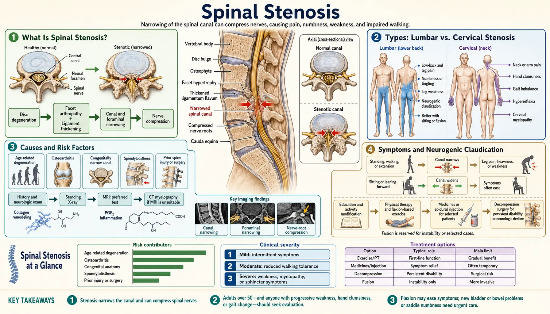
Spinal stenosis is a narrowing of the spaces within the spine that puts pressure on the nerves traveling through the spinal canal. It most commonly affects the lower back (lumbar stenosis) and the neck (cervical stenosis), causing pain, numbness, tingling, and weakness in the arms or legs. Some people with narrowing on imaging have no symptoms at all, while others have significant limits on walking, standing, or daily activities. The condition becomes more common with age, and an analysis of US Medicare claims described the fastest growth in lumbar spine surgery in recent decades as occurring in older patients with spinal stenosis (PubMed PMID: 20371784).
- What Is Spinal Stenosis?
- Types: Lumbar vs. Cervical Stenosis
- Causes and Risk Factors
- Symptoms and Neurogenic Claudication
- Diagnosis (MRI, CT Myelogram, Physical Exam)
- Conventional Treatment (Medications, Epidural Injections, Surgery)
- Physical Therapy and Exercise
- Natural and Complementary Approaches
- Surgical Options (Laminectomy, Spinal Fusion, Percutaneous Decompression)
- Complications and Prognosis
- Living With Spinal Stenosis
- Key Research Papers
- Connections
What Is Spinal Stenosis?
The spinal canal is a tube of bone and ligament that runs the length of the backbone, protecting the spinal cord and the nerve roots that branch off it. In a healthy spine, this canal provides ample room for the nerves. Spinal stenosis occurs when something causes that canal — or the smaller openings where nerve roots exit (foramina) — to become too narrow. When nerves get compressed inside a tight space, they send out distress signals: pain, burning, tingling, numbness, or weakness that can travel far from the actual site of compression.
The word "stenosis" simply means narrowing, and it can happen anywhere along the spine. The lower back and the neck are the most common locations; the thoracic (mid-back) region, whose movement is limited by its connection to the rib cage, is affected less often.
Estimates of how common lumbar stenosis is vary widely with the definition used. A 2020 meta-analysis of 41 papers (55 study samples) put the prevalence based on a clinical diagnosis at 11% in the general population and 25% to 39% among patients seen in primary, secondary or mixed care, and the prevalence based on imaging at 11% in people without symptoms; the authors noted that most included studies carried a high risk of bias (PubMed PMID: 32095908). In a community sample from the Framingham Study examined by CT, acquired narrowing became more common with age: among people aged 60 to 69, relative stenosis was found in 47.2% and absolute stenosis in 19.4% (PubMed PMID: 19398386). Severe cases can lead to cauda equina syndrome (loss of bladder or bowel control), which is handled as a surgical emergency; a meta-analysis of observational studies of cauda equina syndrome caused by disc herniation found that later surgery predicted worse urinary outcomes and supported early surgery (PubMed PMID: 18377315).
Types: Lumbar vs. Cervical Stenosis
Lumbar spinal stenosis involves narrowing in the lower back, specifically in the region between the first lumbar vertebra (L1) and the fifth (L5), or at the L5-S1 junction. This is the most common form and typically causes symptoms in the buttocks, thighs, calves, and feet. The hallmark feature is neurogenic claudication — leg pain and weakness that worsens with walking or standing and is relieved by sitting or bending forward (like leaning over a shopping cart). This positional pattern is one of the features used to tell spinal stenosis apart from a simple herniated disc or peripheral artery disease.
Cervical spinal stenosis affects the neck region (C1 through C7). Because the spinal cord itself passes through this part of the canal — not just individual nerve roots — cervical stenosis can produce a syndrome called cervical myelopathy, which involves coordination problems, difficulty with fine motor tasks (like buttoning a shirt), balance issues, and in severe cases, weakness or spasticity in the arms and legs. Neck pain and radiating arm pain (cervical radiculopathy) are also common. Cervical stenosis with myelopathy is considered more urgent than lumbar stenosis because the spinal cord itself is at risk.
Foraminal stenosis refers specifically to narrowing of the nerve exit holes (foramina) on either side of the vertebrae, rather than the central canal. This type often affects just one nerve root, producing symptoms on one side of the body. It can occur at any spinal level and frequently results from bone spurs growing into the foraminal opening or from disc collapse that reduces the height of the opening.
Tandem stenosis — narrowing at both cervical and lumbar levels simultaneously — occurs in a subset of patients, particularly older adults, and can make diagnosis more complex because symptoms from both regions overlap.
Causes and Risk Factors
The vast majority of spinal stenosis cases are degenerative — meaning they develop gradually over time as a result of normal aging wear on the spine's structures. However, several specific processes contribute to the narrowing:
Osteoarthritis and bone spurs (osteophytes): As the facet joints that link vertebrae together degenerate, extra bone can grow at the joint margins. These bone spurs can project into the spinal canal or foramina and compress nerves.
Ligamentum flavum hypertrophy: The ligamentum flavum is a tough yellow ligament that lines the back wall of the spinal canal. With age, it can thicken and buckle inward when the spine is extended (straightened or bent backward), significantly reducing available space for the nerve roots. Thickened ligamentum flavum is a major contributor to the postural nature of lumbar stenosis symptoms.
Disc degeneration and herniation: Intervertebral discs lose water content and height with age. A flattened disc reduces foraminal height, and disc material that bulges or herniates into the canal adds to compression. Disc collapse also causes the vertebrae above and below to settle closer together, further narrowing the openings.
Spondylolisthesis: This occurs when one vertebra slips forward over the one below it. The mismatch in alignment creates a kink in the canal. Degenerative spondylolisthesis is the form that develops without a fracture.
Congenital factors: Some people are born with a naturally narrower spinal canal. They may be asymptomatic for decades, but any additional age-related narrowing quickly pushes them into symptomatic territory.
Paget's disease of bone: This condition causes abnormal bone remodeling that can lead to enlarged, deformed vertebrae that encroach on the canal. It is an uncommon but recognized cause of stenosis in older adults.
Risk factors commonly listed include older age, female sex (for degenerative spondylolisthesis), a history of spine injury or surgery, obesity (which increases mechanical load on the lumbar spine), occupations involving heavy lifting or prolonged spinal loading, and a family history of spinal problems. In the Framingham CT sample, the prevalence of acquired stenosis rose with age (PubMed PMID: 19398386).
Symptoms and Neurogenic Claudication
Spinal stenosis symptoms range from mild and intermittent to severe and disabling. The pattern of symptoms depends heavily on the location and degree of narrowing.
Neurogenic claudication is the defining symptom of lumbar spinal stenosis. Patients describe a cramping, aching, or burning sensation in the legs — sometimes described as heaviness or a feeling that their legs will give out — that comes on after walking a certain distance or standing for a period of time. The distance or time before symptoms appear varies widely: some people can walk only a block, while others can manage half a mile before discomfort forces them to stop. The key distinguishing feature is that bending forward, sitting down, or leaning over something provides rapid relief. This is because forward flexion opens up the spinal canal slightly, reducing pressure on the compressed nerve roots. Patients often unconsciously adopt a flexed posture and may find they can walk longer distances pushing a grocery cart (the "shopping cart sign").
Lumbar stenosis symptoms also include pain in the low back, buttocks, or thighs; numbness or tingling running down one or both legs; and in more severe cases, weakness in the legs that may cause stumbling or difficulty climbing stairs. The symptoms are often bilateral (both legs), though one side may be worse than the other.
Cervical stenosis symptoms include neck pain; pain, numbness, or tingling radiating into one or both arms (cervical radiculopathy); and — when the spinal cord is compressed — cervical myelopathy. Myelopathy symptoms include clumsy hands (trouble with fine motor tasks), a peculiar "electric shock" sensation down the spine when the neck is flexed (Lhermitte's sign), gait instability (wide-based walking, tendency to fall), urinary urgency or incontinence in advanced cases, and diffuse weakness.
Red flag symptoms include sudden onset of bladder or bowel dysfunction (inability to urinate or loss of bowel control), rapidly progressive leg weakness, or saddle anesthesia (numbness in the groin and inner thighs). These can signal cauda equina syndrome, a neurological emergency; in the meta-analysis of cauda equina syndrome with urinary retention from disc herniation, surgery later than 24 or 72 hours was associated with a significantly higher risk of a fair or poor urinary outcome (PubMed PMID: 18377315).
Diagnosis (MRI, CT Myelogram, Physical Exam)
Diagnosing spinal stenosis combines the patient's history and physical examination with imaging. A 2020 review by the World Federation of Neurosurgical Societies spine committee noted that the severity of narrowing on CT or MRI does not correlate with the severity of symptoms, and that mild-to-moderate stenosis is also found in people without symptoms (PubMed PMID: 32613187).
Medical history and symptom review are central to the diagnostic process. A spine specialist will ask about the specific character of leg or arm symptoms, what makes them better or worse (especially the positional component), how far the patient can walk, and whether symptoms have progressed. The history of neurogenic claudication — worsening with walking, relieved by sitting — is so characteristic that it often points directly to lumbar stenosis before any imaging is ordered.
Physical examination may reveal limited range of motion in the spine, tenderness over the affected vertebral segments, and neurological deficits including reduced reflexes, muscle weakness, or sensory loss in a dermatomal pattern. The "stoop test" — having the patient walk until symptoms appear, then stoop forward — can confirm the postural nature of lumbar stenosis. However, physical examination findings can be subtle or absent in early cases.
MRI (magnetic resonance imaging) was named the best diagnostic test for lumbar stenosis in the 2020 WFNS spine committee review, which also noted that canal diameter measurements have not gained much consensus among radiologists, while qualitative signs such as obliteration of the cerebrospinal fluid space have (PubMed PMID: 32613187). MRI shows the spinal canal, disc tissue, ligaments, nerve roots and spinal cord without radiation. In a 1994 MRI study, a dural-sac cross-sectional area below 100 mm² at more than two of three lumbar levels (L2/3, L3/4, L4/5) was strongly associated with intermittent claudication (PubMed PMID: 7819638).
CT myelography remains useful when MRI is contraindicated (patients with certain pacemakers or metallic implants) or when the degree of bony narrowing needs to be more precisely defined. In this procedure, contrast dye is injected into the spinal fluid space and CT images are obtained, providing excellent detail of the bony anatomy and the outline of the compressed nerve roots.
Electrodiagnostic studies (EMG and nerve conduction tests) can help when peripheral neuropathy, peripheral artery disease, or hip disease could explain the symptoms. The WFNS review found routine electrophysiological tests not helpful for diagnosing lumbar stenosis or predicting its outcome, describing them as complementary to the neurological examination in less clear-cut cases (PubMed PMID: 32613187).
Differential diagnosis includes peripheral artery disease (vascular claudication), hip osteoarthritis, peripheral neuropathy, lumbar disc herniation, sacroiliac joint dysfunction, and — in the cervical spine — amyotrophic lateral sclerosis (ALS) or multiple sclerosis when myelopathy is the presenting feature.
Conventional Treatment (Medications, Epidural Injections, Surgery)
Treatment for spinal stenosis is guided by symptom severity. Mild to moderate stenosis is usually managed non-surgically at first, with more invasive options used for people who do not improve or whose symptoms are severe from the outset. Systematic reviews of non-operative care found the evidence thin: a 2013 Cochrane review of 21 trials found no moderate- or high-quality evidence for any non-operative treatment (PubMed PMID: 23996271), and its 2022 update found moderate-quality evidence only for multimodal programs that include manual therapy and exercise, and against epidural steroids; all other non-operative interventions had evidence of too low a quality to draw conclusions (PubMed PMID: 35046008).
NSAIDs (nonsteroidal anti-inflammatory drugs) such as ibuprofen or naproxen, and acetaminophen (paracetamol), are used for pain relief. No trial of these drugs in spinal stenosis is cited on this page.
Neuropathic pain medications such as gabapentin and pregabalin are prescribed for radiating, burning, or tingling nerve pain. The 2013 Cochrane review rated the evidence that gabapentin improved walking distance as very low quality, from a single small trial (PubMed PMID: 23996271). In a double-blind crossover trial of 29 people with neurogenic claudication, pregabalin was no more effective than an active placebo (diphenhydramine) for time to moderate pain on a treadmill or for any secondary measure of pain or function (PubMed PMID: 25503625).
Muscle relaxants (cyclobenzaprine, methocarbamol) may help when painful muscle spasm accompanies stenosis-related pain, though evidence for their efficacy specifically in stenosis is limited.
Opioid analgesics are sometimes prescribed for severe flares. No trial of opioids in spinal stenosis is cited on this page.
Epidural steroid injections (ESIs) deliver corticosteroid medication into the epidural space around the compressed nerve roots. In the double-blind LESS trial, 400 people with lumbar central stenosis and moderate-to-severe leg pain received epidural injections of a glucocorticoid plus lidocaine or of lidocaine alone; at 6 weeks there was no significant difference in disability (Roland-Morris score) or leg-pain intensity, and the authors concluded that the steroid offered minimal or no short-term benefit over lidocaine alone (PubMed PMID: 24988555). The 2022 systematic review found the steroid-plus-lidocaine improvement statistically, but not clinically, important in the short term and concluded that epidural steroids are not effective for neurogenic claudication from lumbar stenosis (PubMed PMID: 35046008).
Calcitonin (a hormone involved in bone metabolism, available as an injection or nasal spray) has been studied for spinal stenosis. The 2013 Cochrane review found very low-quality evidence from six trials that calcitonin is no better than placebo or paracetamol, regardless of how it was given or which outcome was measured (PubMed PMID: 23996271).
Physical Therapy and Exercise
Physical therapy is one of the most studied non-operative treatments. The 2022 systematic review found moderate-quality evidence that manual therapy plus exercise gave clinically important short-term improvement in symptoms and function compared with medical care or community group exercise, and that manual therapy, education and exercise delivered with a cognitive-behavioural approach improved walking distance compared with self-directed home exercise (PubMed PMID: 35046008). The aims are to reduce pain, improve function, strengthen the muscles that support the spine, and teach postures and movement strategies that reduce nerve compression.
Flexion-based exercise is built around the observation that forward bending opens the lumbar canal; it includes knee-to-chest stretches, posterior pelvic tilts and seated lumbar flexion. In a randomized trial of 58 people with lumbar stenosis, a 6-week program of manual physical therapy, exercise and body-weight-supported treadmill walking led to more patients reporting recovery at 6 weeks than a program of flexion exercises, treadmill walking and subtherapeutic ultrasound (number needed to treat 2.6); at 1 year, 62% and 41% of the two groups still met the threshold for recovery (PubMed PMID: 17047542).
Core stabilization training strengthens the deep abdominal muscles (transversus abdominis), the multifidus muscles and the pelvic floor, with the aim of reducing load on the lumbar spine during daily activities. No trial of core stabilization alone in spinal stenosis is cited on this page.
Aquatic therapy (hydrotherapy) uses the buoyancy of water to unload the spine, so that walking and exercise can be done with less weight on the lower back, and the resistance of water to build strength without heavy impact.
Stationary cycling is another aerobic option. The forward-leaning, slightly flexed position on a bike keeps the lumbar spine flexed, which is the position that relieves neurogenic claudication; recumbent bikes also support the lower back.
Manual therapy — joint mobilization and soft tissue techniques by a physical therapist or chiropractor — is part of the multimodal programs for which the 2022 systematic review found moderate-quality evidence of short-term benefit (PubMed PMID: 35046008). Traction (gently pulling the vertebrae apart) is also used.
Posture and gait training covers walking with the lumbar spine slightly flexed (leaning slightly forward), assistive devices such as walking poles or a walker, changes to daily activities that provoke symptoms, and pacing — stopping to rest before symptoms peak, then resuming.
The SPORT trial enrolled surgical candidates with lumbar stenosis without spondylolisthesis in a randomized cohort (289 patients) and an observational cohort (365 patients). Crossover was heavy: at 2 years, 67% of those assigned to surgery and 43% of those assigned to non-surgical care had had surgery. The intention-to-treat analysis found a significant advantage for surgery on bodily pain but not on physical function or the Oswestry Disability Index, while the as-treated analysis combining both cohorts favored surgery on all primary outcomes (PubMed PMID: 18287602); that as-treated advantage was maintained through 4 years (PubMed PMID: 20453723). In a separate trial of 169 surgical candidates aged 50 or older, surgical decompression and physical therapy produced similar improvements in physical function at 2 years, even though 57% of the physical therapy group crossed over to surgery (PubMed PMID: 25844995).
Natural and Complementary Approaches
Several nutritional and complementary approaches are discussed for spinal stenosis. For most of them, trials in people with spinal stenosis are few or absent; the 2022 systematic review of non-operative care found evidence of too low a quality to draw conclusions for every non-operative intervention other than multimodal programs with manual therapy and exercise, and epidural steroids (PubMed PMID: 35046008).
Anti-inflammatory nutrition: Eating patterns rich in omega-3 fatty acids (from fatty fish such as salmon and sardines), colorful vegetables and whole grains, with few processed foods, refined sugars and trans fats, are discussed for chronic pain in general. No trial of a diet in spinal stenosis is cited on this page.
Magnesium takes part in nerve function and muscle relaxation. No trial of magnesium in spinal stenosis is cited on this page. Foods rich in magnesium include leafy greens, pumpkin seeds, and dark chocolate. See Magnesium for detailed information.
Vitamin D3 is involved in bone health and muscle function. No trial of vitamin D in spinal stenosis is cited on this page. See Vitamin D3.
Collagen and connective tissue: Collagen is a main structural protein of the intervertebral discs and spinal ligaments. No trial of collagen supplements in spinal stenosis is cited on this page. See Collagen.
Curcumin (turmeric): Curcumin has anti-inflammatory effects in animal and laboratory studies and has been tested in human trials for osteoarthritis pain. Its absorption is low; it is often combined with piperine (from black pepper) or formulated with phospholipids. No trial of curcumin in spinal stenosis is cited on this page.
Acupuncture has been studied for lumbar stenosis mainly in China. A 2013 systematic review found six randomized trials (582 participants), all comparing different acupuncture techniques or combinations rather than acupuncture against a sham or no treatment; because of high or uncertain risk of bias and limited generalisability, the authors found no conclusive evidence of its effectiveness or safety for lumbar stenosis (PubMed PMID: 24050593).
TENS (transcutaneous electrical nerve stimulation) uses low-level electrical currents applied to the skin to interrupt pain signals. No trial of TENS in spinal stenosis is cited on this page.
Heat and cold therapy — heating pads for muscle spasm, cold packs for acute inflammation — are simple, low-cost measures used for temporary relief.
Weight management: Excess body weight, particularly central obesity, increases the mechanical load on the lumbar spine. No trial of weight loss in spinal stenosis is cited on this page.
Surgical Options (Laminectomy, Spinal Fusion, Percutaneous Decompression)
Surgery for spinal stenosis is generally considered when non-surgical treatment has not brought adequate relief, when symptoms significantly impair daily function or quality of life, or when neurological deficits (weakness, loss of bladder/bowel control) are progressing; the SPORT trial enrolled surgical candidates who had had symptoms for at least 12 weeks (PubMed PMID: 18287602). The choice of procedure depends on the location and extent of stenosis, the presence of spinal instability or deformity, and factors such as age, bone density, and overall health.
Laminectomy (decompressive laminectomy) is the standard decompression operation for lumbar spinal stenosis. The surgeon removes all or part of the lamina — the bony arch that forms the back wall of the spinal canal — along with thickened ligamentum flavum and bone spurs, creating more space for the nerve roots. At 8 years in SPORT, the intention-to-treat analysis found no differences between the randomized groups; in the as-treated analysis of the randomized cohort, the early benefit of surgery seen out to 4 years converged over time, with no significant treatment effect in years 6 to 8, while the observational cohort kept a stable advantage for surgery. Data at 8 years were obtained for 55% of the randomized and 52% of the observational participants (PubMed PMID: 25569524).
Spinal fusion joins two or more vertebrae permanently, using bone graft material along with metal screws and rods, and has often been added to decompression when one vertebra slips on another (spondylolisthesis) or when decompression could leave the spine unstable. The Swedish Spinal Stenosis Study randomized 247 patients aged 50 to 80 with stenosis at one or two levels, 135 of whom had degenerative spondylolisthesis, to decompression plus fusion or decompression alone. At 2 years there was no significant difference in the Oswestry Disability Index (27 vs 24) or the 6-minute walk test (397 m vs 405 m), with similar results in patients with and without spondylolisthesis and no significant differences at 5 years; fusion meant longer operations, more bleeding, higher costs and longer hospital stays (7.4 vs 4.1 days) (PubMed PMID: 27074066). In US Medicare data for 2007, life-threatening complications rose with surgical invasiveness, from 2.3% with decompression alone to 5.6% with complex fusion (PubMed PMID: 20371784).
Minimally invasive spine surgery (MISS) and other limited decompression techniques (unilateral or bilateral laminotomy, split-spinous-process laminotomy) aim to decompress the canal while preserving the midline structures of the spine. A 2015 Cochrane review of 10 trials (733 participants) found low- or very low-quality evidence that these techniques and conventional laminectomy have similar effects on functional disability and leg pain (PubMed PMID: 25760812).
Laminoplasty is used for cervical stenosis. Instead of removing the lamina entirely, the surgeon cuts it open on one side and props it open like a hinge, enlarging the canal while preserving more of the posterior spinal structures.
Percutaneous lumbar decompression is an outpatient procedure for lumbar stenosis caused mainly by a thickened ligamentum flavum: instruments passed through the skin under imaging guidance remove small portions of ligament and lamina from the back of the canal. A 2-year report on 45 patients treated at 11 US sites, with no control group, described significant reductions in pain and improvements in function (PubMed PMID: 23446067). A randomized trial compared the procedure (143 patients) with epidural steroid injections (131 patients) up to 1 year, with 2-year follow-up of the procedure group only; at 2 years the Oswestry Disability Index in that group had improved by 22.7 points and pain scores by 3.6 points (PubMed PMID: 30199512). The 2016 Cochrane review rated the evidence from a smaller comparison with epidural steroids (38 participants) as low quality (PubMed PMID: 26824399).
Interspinous spacer devices are implants placed between the spinous processes — the bony projections at the back of each vertebra — to hold the spine in slight flexion at the treated level. In a double-blind Dutch trial of 159 patients with neurogenic claudication, the spacer was not superior to conventional decompression at 8 weeks, outcomes did not differ during the first year, and repeat surgery was much more common after the spacer (29% vs 8%) (PubMed PMID: 24231273).
Surgical outcomes compared with non-surgical care: A 2016 Cochrane review of five randomized trials (643 participants) rated the evidence as low quality. Pooling two trials, it found no significant difference in Oswestry Disability Index between decompression (with or without fusion) and multimodal non-operative care at 6 months or 1 year, and a difference favoring decompression at 24 months; in one included trial, complications occurred in 10% and 24% of participants (PubMed PMID: 26824399).
Complications and Prognosis
The natural history of lumbar stenosis is variable, and it does not inevitably worsen. In a Swedish study of 32 untreated patients followed for a mean of 49 months, symptoms on a visual analog scale were unchanged in 70%, improved in 15% and worsened in 15%, and no severe deterioration was found after four years (PubMed PMID: 1534726).
Spontaneous improvement does occur: in that same follow-up study, the same number of patients still had claudication at the end, but their symptoms were milder (PubMed PMID: 1534726).
Severe symptoms: The trials of surgery cited on this page enrolled surgical candidates with persistent symptoms, and the long-term comparisons between surgery and non-surgical care are summarized in the surgical section above.
Surgical complications include infection, bleeding, dural tear (inadvertent puncture of the membrane surrounding the spinal fluid), nerve injury, persistent pain after technically successful surgery, and adjacent segment disease (accelerated degeneration above or below a fusion). In US Medicare patients operated on for stenosis in 2007, life-threatening complications occurred in 2.3% after decompression alone and 5.6% after complex fusion, and rehospitalization within 30 days in 7.8% and 13.0% (PubMed PMID: 20371784).
Reoperation rates: In the Swedish fusion trial, additional lumbar spine surgery was performed during a mean follow-up of 6.5 years in 22% of the fusion group and 21% of the decompression-alone group (PubMed PMID: 27074066). In the Dutch interspinous spacer trial, repeat surgery was needed in 29% after the spacer and 8% after conventional decompression (PubMed PMID: 24231273).
Cervical myelopathy carries a more guarded outlook without surgery. A 2013 systematic review found moderate-strength evidence that 20% to 60% of patients with cervical spondylotic myelopathy deteriorate neurologically over time without surgery, and low-strength evidence linking circumferential cord compression to deterioration (PubMed PMID: 23963004).
Living With Spinal Stenosis
Many people with spinal stenosis manage the condition for years without surgery; in the untreated Swedish cohort, most patients' symptoms were unchanged after about four years (PubMed PMID: 1534726). The positional nature of the pain — bending forward helps and straightening up hurts — shapes many day-to-day adaptations.
Adaptive strategies for daily life that people with stenosis describe include leaning on a shopping cart or using walking poles for longer walks; "interval walking", sitting to rest briefly before symptoms peak; chairs or car seats that allow a slightly reclined position; and sleeping with a pillow between the knees on the side, or under the knees on the back, to keep the lumbar spine slightly flexed.
Ergonomic modifications include raised work surfaces to reduce prolonged bending, recliners or adjustable chairs in place of hard upright chairs, grab bars in the bathroom when balance is affected, and raised toilet seats when getting up from low positions is painful. Bending forward tends to relieve stenosis symptoms, while prolonged standing in extension tends to provoke them.
Activity: Deconditioning — the muscle weakness and cardiovascular decline that come with inactivity — adds to the functional impact of stenosis. Aquatic exercise and stationary cycling allow aerobic activity even when walking is limited.
Mental health and pain management: Depression and anxiety are common alongside chronic musculoskeletal pain. In the 2022 systematic review, a program of manual therapy, education and exercise delivered with a cognitive-behavioural approach improved walking distance compared with self-directed home exercise, with moderate-quality evidence (PubMed PMID: 35046008). Mindfulness meditation and stress-reduction techniques are also used for chronic pain.
Flares: Stenosis symptoms often fluctuate. A symptom diary is one way people track specific triggers (long car rides, cold weather, certain activities) and changes over time.
Tracking change: Because the severity of narrowing on imaging does not match the severity of symptoms (PubMed PMID: 32613187), the changes clinicians ask about are mainly reported symptoms — shorter walking distance, weaker hands, new bladder urgency — which are the signs used to judge whether the condition is stable or worsening. Decisions about treatment belong with a clinician.
Key Research Papers
- Weinstein JN, Tosteson TD, Lurie JD, et al. (2008). Surgical versus nonsurgical therapy for lumbar spinal stenosis. N Engl J Med — PubMed PMID: 18287602
- Lurie JD, Tosteson TD, Tosteson A, et al. (2015). Long-term outcomes of lumbar spinal stenosis: eight-year results of the Spine Patient Outcomes Research Trial (SPORT). Spine — PubMed PMID: 25569524
- Försth P, Ólafsson G, Carlsson T, et al. (2016). A randomized, controlled trial of fusion surgery for lumbar spinal stenosis (Swedish Spinal Stenosis Study). N Engl J Med — PubMed PMID: 27074066
- Ammendolia C, Stuber KJ, Rok E, et al. (2013). Nonoperative treatment for lumbar spinal stenosis with neurogenic claudication. Cochrane Database Syst Rev — PubMed PMID: 23996271
- Kreiner DS, Shaffer WO, Baisden JL, et al.; North American Spine Society (2013). An evidence-based clinical guideline for the diagnosis and treatment of degenerative lumbar spinal stenosis (update). Spine J — PubMed PMID: 23830297
- Overdevest GM, Jacobs W, Vleggeert-Lankamp C, et al. (2015). Effectiveness of posterior decompression techniques compared with conventional laminectomy for lumbar stenosis. Cochrane Database Syst Rev — PubMed PMID: 25760812
- Deyo RA, Mirza SK, Martin BI, et al. (2010). Trends, major medical complications, and charges associated with surgery for lumbar spinal stenosis in older adults. JAMA — PubMed PMID: 20371784
- Jensen RK, Jensen TS, Koes B, Hartvigsen J. (2020). Prevalence of lumbar spinal stenosis in general and clinical populations: a systematic review and meta-analysis. Eur Spine J — PubMed PMID: 32095908
- Chopko BW. (2013). Long-term results of percutaneous lumbar decompression for LSS: two-year outcomes. Clin J Pain — PubMed PMID: 23446067
- Zaina F, Tomkins-Lane C, Carragee E, Negrini S. (2016). Surgical versus non-surgical treatment for lumbar spinal stenosis. Cochrane Database Syst Rev — PubMed PMID: 26824399
- Delitto A, Piva SR, Moore CG, et al. (2015). Surgery versus nonsurgical treatment of lumbar spinal stenosis: a randomized trial. Ann Intern Med — PubMed PMID: 25844995
- Whitman JM, Flynn TW, Childs JD, et al. (2006). A comparison between two physical therapy treatment programs for patients with lumbar spinal stenosis: a randomized clinical trial. Spine — PubMed PMID: 17047542
- Weinstein JN, Tosteson TD, Lurie JD, et al. (2010). Surgical versus nonoperative treatment for lumbar spinal stenosis four-year results of the Spine Patient Outcomes Research Trial. Spine — PubMed PMID: 20453723
- Friedly JL, Comstock BA, Turner JA, et al. (2014). A randomized trial of epidural glucocorticoid injections for spinal stenosis (LESS trial). N Engl J Med — PubMed PMID: 24988555
- Ammendolia C, Hofkirchner C, Plener J, et al. (2022). Non-operative treatment for lumbar spinal stenosis with neurogenic claudication: an updated systematic review. BMJ Open — PubMed PMID: 35046008
- Markman JD, Frazer ME, Rast SA, et al. (2015). Double-blind, randomized, controlled, crossover trial of pregabalin for neurogenic claudication. Neurology — PubMed PMID: 25503625
- Moojen WA, Arts MP, Jacobs WC, et al. (2013). Interspinous process device versus standard conventional surgical decompression for lumbar spinal stenosis: randomized controlled trial. BMJ — PubMed PMID: 24231273
- Staats PS, Chafin TB, Golovac S, et al. (2018). Long-term safety and efficacy of minimally invasive lumbar decompression procedure for the treatment of lumbar spinal stenosis with neurogenic claudication: 2-year results of MiDAS ENCORE. Reg Anesth Pain Med — PubMed PMID: 30199512
- Johnsson KE, Rosén I, Udén A. (1992). The natural course of lumbar spinal stenosis. Clin Orthop Relat Res — PubMed PMID: 1534726
- Kalichman L, Cole R, Kim DH, et al. (2009). Spinal stenosis prevalence and association with symptoms: the Framingham Study. Spine J — PubMed PMID: 19398386
- Hamanishi C, Matukura N, Fujita M, et al. (1994). Cross-sectional area of the stenotic lumbar dural tube measured from the transverse views of magnetic resonance imaging. J Spinal Disord — PubMed PMID: 7819638
- Zileli M, Crostelli M, Grimaldi M, et al. (2020). Natural course and diagnosis of lumbar spinal stenosis: WFNS Spine Committee recommendations. World Neurosurg X — PubMed PMID: 32613187
- Kim KH, Kim TH, Lee BR, et al. (2013). Acupuncture for lumbar spinal stenosis: a systematic review and meta-analysis. Complement Ther Med — PubMed PMID: 24050593
- DeLong WB, Polissar N, Neradilek B. (2008). Timing of surgery in cauda equina syndrome with urinary retention: meta-analysis of observational studies. J Neurosurg Spine — PubMed PMID: 18377315
- Karadimas SK, Erwin WM, Ely CG, et al. (2013). Pathophysiology and natural history of cervical spondylotic myelopathy. Spine — PubMed PMID: 23963004