Vulvar Cancer
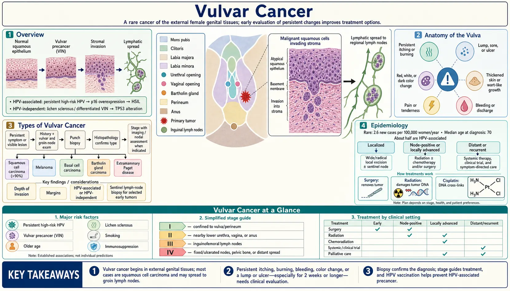
Table of Contents
1. Overview
Vulvar cancer is a cancer of the external female genitalia, the vulva. The U.S. National Cancer Institute’s SEER program estimates 7,130 new cases and 1,750 deaths in the United States in 2026, about 0.3% of all new cancer cases (SEER Cancer Stat Facts: Vulvar Cancer). The 2021 FIGO cancer report describes it as uncommon, accounting for about 4% of gynecological malignancies (PubMed PMID: 34669204); the 2024 NCCN guideline summary gives a range of 5% to 8% (PubMed PMID: 38503056). A 2018 systematic review of patient-reported outcomes found that its treatment is associated with considerable morbidity that lowers quality of life (PubMed PMID: 29420364).
Squamous cell carcinoma (SCC) makes up more than 80% of vulvar cancers, according to the FIGO report, which describes two main pathological pathways to vulvar SCC: one caused by persistent HPV infection and one that is HPV-independent and often associated with lichen sclerosus. Both pathways are described in Section 3.
SEER reports that 58% of vulvar cancers are diagnosed at the localized stage, where the 5-year relative survival is 85.5% (SEER Cancer Stat Facts: Vulvar Cancer). Survival by stage is set out in Section 10.
2. Anatomy of the Vulva
The term vulva refers to the external female genitalia. It is distinct from the vagina, the internal canal leading to the uterus. The FIGO report classifies a tumor as vulvar when the vulva is its primary site, including tumors that involve both the vulva and the vagina (PubMed PMID: 34669204).
The structures that make up the vulva include:
- Labia majora — the outer, larger folds of skin on either side.
- Labia minora — the inner, thinner folds of skin just inside the labia majora.
- Clitoris — the erectile organ at the top of the labia minora.
- Vaginal vestibule — the space between the labia minora that contains the vaginal opening and the urethral opening.
- Bartholin glands — the greater vestibular glands, one on each side of the vaginal opening. These can give rise to a rare, distinct type of vulvar cancer.
- Skene glands — the lesser vestibular glands.
Why anatomy matters for staging and surgery. The FIGO report notes that most vulvar cancers arise in the skin of the labia, while cancers of the clitoris and vestibular glands are extremely rare. Lymph from the vulva drains primarily to the inguinofemoral (groin) nodes and secondarily to the external and internal iliac nodes. Depending on the tumor’s location, its size and its closeness to the midline, drainage can be one-sided or two-sided, and a lesion on or close to the clitoris can drain directly to the iliac region (PubMed PMID: 34669204).
3. Types of Vulvar Cancer
The type of vulvar cancer is linked to who develops it, how it is staged and how it is treated.
Squamous Cell Carcinoma — Two Distinct Pathways
The FIGO report describes two main pathological pathways to vulvar SCC (PubMed PMID: 34669204):
HPV-associated pathway: Warty/basaloid SCC generally occurs in younger women, is caused by persistent infection with oncogenic HPV types (particularly HPV 16, 18, 31 and 33), and has a high-grade squamous intraepithelial lesion (HSIL, formerly called usual-type vulvar intraepithelial neoplasia) as its precursor. These lesions are frequently multifocal and may be associated with squamous intraepithelial lesions elsewhere in the lower genital tract (cervix, vagina, anus); HIV infection and cigarette smoking are common predisposing factors. The 2015 ISSVD terminology names three vulvar precursor categories: vulvar LSIL, vulvar HSIL and differentiated VIN (PubMed PMID: 26704327).
HPV-independent pathway: Keratinizing SCC usually occurs in older women and is often associated with lichen sclerosus and/or differentiated VIN (dVIN). The FIGO report states that dVIN accounts for less than 5% of preneoplastic vulvar lesions but has a higher rate of progression to cancer, a shorter interval to progression and a higher recurrence rate than HSIL, and is rarely associated with persistent HPV infection (less than 2%) (PubMed PMID: 34669204). A 2014 review by Reyes and Cooper describes keratinizing SCC as the most common type (60% of cases), with basaloid or warty SCC making up 40%, and reports that dVIN is consistently negative for p16 while most cases carry TP53 mutations (PubMed PMID: 24399036).
How often HPV is found: In a 2023 meta-analysis of 91 studies (8,200 cases), HPV DNA was detected in 39.1% of vulvar cancers and in 76.1% of vulvar intraepithelial neoplasia; HPV16 was the most common type in vulvar cancer (78.1%), followed by HPV33 (7.5%) (PubMed PMID: 36933562).
Other, Less Common Types
- Melanoma — the FIGO report describes vulvar melanoma as the second most common vulvar malignancy, though very rare (an incidence of 0.1 per 100,000). Mucosal melanomas of the vulva and vagina have a 15% 5-year survival, and their cause and genetics differ from those of skin melanomas; most vulvar melanomas involve the clitoris or labia minora. They are staged with the AJCC system (which includes Breslow thickness) rather than FIGO staging. The report describes radical wide local excision with margins of at least 1 cm, and testing for c-kit and BRAF mutations because immunotherapies such as nivolumab may have a role (PubMed PMID: 34669204).
- Bartholin gland carcinoma — about 5% of vulvar cancers, according to the FIGO report, which notes that the diagnosis is often made after removal of a persistent or recurrent Bartholin cyst and lists transitional cell, squamous, adenocarcinoma, adenoid cystic and adenosquamous forms (PubMed PMID: 34669204). In a single-institution series of 429 invasive vulvar carcinomas, 33 (7.7%) arose in the Bartholin gland (87.9% squamous, 12.1% adenocarcinoma). These patients were younger at diagnosis (median 57 vs 63 years) and more often had stage III/IV disease (60.6% vs 35.8%), yet recurrence-free and overall survival did not differ significantly from other vulvar carcinomas (PubMed PMID: 26844611).
- Paget disease of the vulva — the FIGO report describes extramammary Paget disease as rare. A primary form begins as an intraepithelial lesion; a secondary form comes from an underlying adenocarcinoma, which may be anorectal, urothelial or of the genital tract. It occurs mainly in postmenopausal women, who present with itching and pain, often with an eczema-like weeping lesion. Its histological changes often extend far beyond what is visible, and recurrence rates are high even with adequate margins (PubMed PMID: 34669204).
- Verrucous carcinoma, basal cell carcinoma and sarcoma — listed by the FIGO report and the 2024 NCCN guideline summary among the rarer histological types (PubMed PMID: 34669204; PubMed PMID: 38503056).
4. Epidemiology
SEER estimates 7,130 new cases and 1,750 deaths in the United States in 2026. The age-adjusted rate of new cases is 2.6 per 100,000 women per year (2019–2023) and the death rate 0.6 per 100,000 women per year (2020–2024); about 0.3% of women will be diagnosed with vulvar cancer during their lifetime (SEER Cancer Stat Facts: Vulvar Cancer).
The median age at diagnosis is 70, and vulvar cancer is most frequently diagnosed among women aged 65–74 (SEER Cancer Stat Facts: Vulvar Cancer). The FIGO report states that the mean age at diagnosis has fallen in recent years owing to the increase in HPV infections, and cites an epidemiological study of 13 high-income countries that found a significant 14% overall increase in incidence: 38% in women younger than 60, with no significant increase in women older than 60 (PubMed PMID: 34669204).
SEER notes that vulvar cancer is more common among women with a history of vulvar intraepithelial neoplasia, HPV infection or genital warts (SEER Cancer Stat Facts: Vulvar Cancer). The NCCN guideline summary lists increasing age, HPV infection, cigarette smoking, inflammatory conditions of the vulva and immunodeficiency as known risk factors (PubMed PMID: 38503056).
5. Risk Factors
- HPV infection — the FIGO report states that persistent HPV infection, particularly with HPV 16, has been associated with the long-term development of vulvar HSIL and SCC (PubMed PMID: 34669204).
- Lichen sclerosus — in a Dutch registry cohort of 3,038 women diagnosed with lichen sclerosus between 1991 and 2011, the cumulative incidence of vulvar SCC was 6.7%. The 10-year incidence was 18.8% in women with concurrent VIN and 2.8% in women without it, and rose with age at lichen sclerosus diagnosis (1.8% under 50, 3% at 50–70, 5.9% at 70 or older). The incidence of lichen sclerosus itself rose from 7.4 to 14.6 per 100,000 woman-years over the period (PubMed PMID: 27257093).
- Cigarette smoking — listed as a risk factor by the NCCN guideline summary and as a common predisposing factor for HPV-associated disease by the FIGO report (PubMed PMID: 38503056; PubMed PMID: 34669204).
- Immunodeficiency, including HIV infection — listed by the same two sources.
- Precancerous lesions of the lower genital tract — SEER notes higher rates in women with a history of vulvar intraepithelial neoplasia, and the FIGO report links HPV-associated vulvar lesions with squamous intraepithelial lesions of the cervix, vagina and anus (SEER Cancer Stat Facts: Vulvar Cancer; PubMed PMID: 34669204).
- Age — the median age at diagnosis is 70 (SEER Cancer Stat Facts: Vulvar Cancer).
6. Staging
Vulvar cancer is staged with the FIGO 2021 system. It was the first FIGO staging derived from data analyses, built from 12,063 cases in the U.S. National Cancer Database treated between 2010 and 2017. The revision introduced a new definition of depth of invasion, adopted the lymph node definitions used in cervical cancer (micrometastasis and macrometastasis; isolated tumor cells do not count), and allows cross-sectional imaging findings to be used. It applies to all vulvar cancer types except melanoma, and documentation of HPV status (HPV-associated or HPV-independent) is strongly recommended (PubMed PMID: 34520062).
Stage I — Tumor confined to the vulva:
- Stage IA: Tumor 2 cm or smaller and stromal invasion of 1 mm or less.
- Stage IB: Tumor larger than 2 cm, or stromal invasion of more than 1 mm.
Stage II: Tumor of any size extending to the lower third of the urethra, the lower third of the vagina or the lower third of the anus, with negative nodes.
Stage III — Extension to the upper part of adjacent perineal structures, or any number of non-fixed, non-ulcerated lymph nodes:
- Stage IIIA: Tumor of any size extending to the upper two-thirds of the urethra, the upper two-thirds of the vagina, the bladder mucosa or the rectal mucosa, or regional lymph node metastases of 5 mm or less.
- Stage IIIB: Regional lymph node metastases larger than 5 mm.
- Stage IIIC: Regional lymph node metastases with extracapsular spread.
Stage IV — Tumor fixed to bone, fixed or ulcerated lymph node metastases, or distant metastases:
- Stage IVA: Disease fixed to pelvic bone, or fixed or ulcerated regional lymph node metastases.
- Stage IVB: Distant metastases.
“Regional” refers to the inguinal and femoral nodes. Depth of invasion is measured from the basement membrane of the deepest adjacent dysplastic, tumor-free rete ridge to the deepest point of invasion (PubMed PMID: 34520062). The FIGO report calls appropriate management of the groin lymph nodes the most important factor in reducing mortality from early vulvar cancer, because recurrences in the groin carry poorer survival despite multimodal rescue treatment (PubMed PMID: 34669204).
7. Diagnosis
According to the FIGO report, most women present with vulvar itching or pain, or have noticed a lump or ulcer; some have abnormal bleeding or discharge, many have a history of vulvar symptoms from underlying lichen sclerosus or HSIL, and advanced disease may present as a lump in the groin. The report states that any suspicious vulvar lesion should be biopsied to exclude invasion. This can be done under local anesthetic with a 3 or 4 mm Keyes biopsy instrument, or as an incisional or wedge biopsy; the report states that, even for a small lesion, it is better not to excise the whole lesion at biopsy, as this makes the definitive surgery harder to plan (PubMed PMID: 34669204).
The report also describes early evaluation of patients with symptoms such as chronic vulvar itching, or signs such as pigmented lesions or irregular ulcers, as candidates for skin biopsy (PubMed PMID: 34669204).
Investigations listed in the FIGO report (PubMed PMID: 34669204):
- Cervical cytology and colposcopy of the cervix and vagina, because HPV-related cancers are associated with other squamous intraepithelial lesions.
- Blood tests (full blood count, biochemical and liver profiles) and HIV testing; chest X-ray.
- CT or MRI of the pelvis and groins, especially for locally advanced tumors, to detect enlarged lymph nodes, erosion into bone or other metastases, and for treatment planning.
- PET-CT, which detects inguinofemoral lymph node involvement more effectively than CT and may be used for larger tumors when metastatic disease is suspected or at recurrence.
Tumor marker: The FIGO report states that elevated pretreatment serum squamous cell carcinoma antigen (SCC-Ag) may be an independent prognostic factor for disease-free and overall survival, and that for a select group of patients SCC-Ag may be used as a serum marker for surveillance (PubMed PMID: 34669204).
8. Treatment
The FIGO report states that treatment depends primarily on histology and stage, and also on age, other illnesses and performance status. Treatment is predominantly surgical, particularly for SCC, although concurrent chemoradiation is an effective alternative, particularly for advanced tumors and those where exenteration would be needed to achieve adequate surgical margins. The report describes management as individualized and carried out by a multidisciplinary team in an experienced cancer center, with treatment of the primary lesion and of the groin nodes considered independently (PubMed PMID: 34669204).
Early-Stage Disease (Stages I–II)
Radical wide local excision. The FIGO report describes radical wide local excision as the gold standard for early vulvar cancer: as effective as radical vulvectomy in preventing local recurrence, with substantially less psychosexual morbidity. Surgeons aim for surgical margins of 2 cm to achieve pathological margins of at least 8 mm (allowing for shrinkage of the fixed tissue), although the report adds that many “recurrent” vulvar cancers are probably new tumors arising in the surrounding abnormal tissue (PubMed PMID: 34669204). The 8 mm figure is debated: in an AGO-CaRE-1 subset analysis of 289 node-negative, surgically treated patients, vulvar recurrence occurred in 12.6% of patients with a margin under 8 mm and 10.2% with a margin of 8 mm or more, margin distance had no statistically significant effect on local recurrence, and the authors concluded that the need for an 8 mm minimum could not be confirmed (PubMed PMID: 27837710).
Stage IA (2 cm or smaller, invasion 1 mm or less). The FIGO report states that these lesions are managed with radical wide local excision and that groin node dissection is not necessary. When the initial biopsy shows a lesion of 2 cm or less with invasion of 1 mm or less, a radical wide local excision is usually done to assess the maximum depth of invasion; if no part invades more than 1 mm, that excision is adequate definitive treatment (PubMed PMID: 34669204).
Groin node assessment. The FIGO report recommends inguinofemoral lymphadenectomy for all women with stage IB or resectable stage II cancers, and describes the sentinel node procedure (below) as increasingly used in early disease to omit full lymphadenectomy when the sentinel node is negative. Fewer than 1% of patients with small lateral lesions (under 4 cm and at least 2 cm from the midline) and negative nodes on that side have metastases in the opposite groin, so a one-sided dissection is described as adequate for them; tumors closer than 2 cm to the midline or crossing it, very large lateral tumors (over 4 cm), and positive nodes on the tumor side are managed with dissection of both groins (PubMed PMID: 34669204). In a GOG 173 analysis of 234 patients, preoperative lymphoscintigraphy showed two-sided drainage in 22% of lateral, 58% of near-midline and 70% of midline tumors; among patients with midline tumors whose scan showed one-sided drainage, 4 of 32 who had the opposite groin dissected had metastases there, and none of 27 with near-midline tumors did (PubMed PMID: 23201592).
Sentinel lymph node biopsy (SLNB). The FIGO report lists the GROINSS-V indications for a sentinel node procedure: a unifocal tumor confined to the vulva, less than 4 cm in diameter, with stromal invasion of more than 1 mm, and clinically and radiologically negative groin nodes. Sentinel nodes are identified with radio-labelled technetium and blue dye; indocyanine green with near-infrared fluorescence has become an option. The report states that when a sentinel node is not detected on the tumor side, a complete lymphadenectomy of that groin is done, and that a positive sentinel node leads to a recommendation of lymphadenectomy of both groins (PubMed PMID: 34669204).
GROINSS-V I. In this multicenter observational study (Van der Zee et al., 2008; PubMed PMID: 18281661), the sentinel node procedure was performed in 623 groins of 403 patients with T1/2 (under 4 cm) squamous cell cancer of the vulva between 2000 and 2006. Among 259 patients with unifocal disease and a negative sentinel node, in whom lymphadenectomy was omitted, six groin recurrences were diagnosed (2.3%) over a median follow-up of 35 months, and the 3-year survival rate was 97%. Compared with sentinel-node-positive patients who had an inguinofemoral lymphadenectomy, patients who had only the sentinel node removed had less groin wound breakdown (11.7% vs 34.0%), cellulitis (4.5% vs 21.3%), recurrent erysipelas (0.4% vs 16.2%) and lymphedema of the legs (1.9% vs 25.2%). The authors suggested that sentinel node dissection, performed by a quality-controlled multidisciplinary team, should be part of standard treatment in selected patients. Long-term follow-up (median 105 months) found an isolated groin recurrence rate at 5 years of 2.5% in sentinel-node-negative and 8.0% in sentinel-node-positive patients (PubMed PMID: 26428940).
GOG 173. In this Gynecologic Oncology Group study (Levenback et al., 2012; PubMed PMID: 22753905), 452 women with SCC (at least 1 mm invasion, tumors 2–6 cm) had both a sentinel node biopsy and a full inguinofemoral lymphadenectomy. The sentinel node biopsy had a sensitivity of 91.7% and a false-negative predictive value of 3.7%, which was 2.0% in women with tumors under 4 cm.
GROINSS-V II. This phase II single-arm trial (Oonk et al., 2021; PubMed PMID: 34432481) gave inguinofemoral radiotherapy (50 Gy) instead of lymphadenectomy to patients with early-stage vulvar cancer (under 4 cm) whose sentinel node contained metastasis. Of 1,535 eligible patients registered between 2005 and 2016, 322 (21.0%) had sentinel node metastasis. In June 2010 a stopping rule was triggered because isolated groin recurrences rose above the predefined threshold; nine of the ten patients with an isolated groin recurrence had sentinel node metastases larger than 2 mm and/or extracapsular spread. The protocol was amended so that macrometastases (over 2 mm) were treated with lymphadenectomy, while micrometastases (2 mm or less) continued to receive radiotherapy. Among 126 patients with micrometastases who received radiotherapy, the isolated groin recurrence rate on that side at 2 years was 1.6%; among patients with macrometastases it was 22% after radiotherapy and 6.9% after lymphadenectomy. Treatment-related morbidity was less frequent after radiotherapy. The authors concluded that inguinofemoral radiotherapy is a safe alternative to lymphadenectomy for sentinel node micrometastases.
Adjuvant radiation. The FIGO report lists extracapsular spread and two or more positive groin nodes as indications for pelvic and groin irradiation in patients with positive groin nodes (PubMed PMID: 34669204).
Advanced Disease (Stages III–IVA)
The FIGO report defines advanced vulvar cancer as tumors that extend beyond the vulva and/or bulky positive groin nodes, and describes determining groin node status, ideally before treatment is planned, by fine needle aspiration or biopsy of suspicious nodes and by CT, MRI or PET-CT. For clinically positive nodes, it describes removing enlarged groin and pelvic nodes if possible followed by groin and pelvic radiation, and states that full lymphadenectomy is not performed because full groin dissection followed by groin irradiation may result in severe lymphedema. Ulcerated or fixed groin nodes are biopsied and then treated with primary radiotherapy, with or without chemosensitization (PubMed PMID: 34669204).
For the primary tumor, the report describes surgical excision with clear margins and without sphincter damage, whenever possible, as the optimum treatment. If adequate excision could only be achieved by exenteration with a bowel or urinary stoma, radiotherapy with or without concurrent chemotherapy may be the preferred alternative, and concurrent chemoradiation is a well-described option for large tumors where surgery would damage the anus or urethra (PubMed PMID: 34669204).
Primary chemoradiation — GOG 205. In this Gynecologic Oncology Group phase II trial (Moore et al., 2012; PubMed PMID: 22079361), 58 evaluable patients with locally advanced T3 or T4 tumors not amenable to radical vulvectomy received radiation (1.8 Gy daily for 32 fractions, 57.6 Gy) with weekly cisplatin (40 mg/m²), followed by surgical resection of residual tumor or a biopsy. Thirty-seven (64%) had a complete clinical response; of the 34 of these who had a surgical biopsy, 29 (78%) also had a complete pathological response. Forty (69%) completed study treatment; nine stopped because of toxicity. Common adverse effects included leukopenia, pain, radiation dermatitis and metabolic changes. In a later National Cancer Database analysis of 1,352 patients who did not have surgery (2003–2014), 5-year overall survival was 49.9% after chemoradiation and 27.4% after radiation alone (adjusted hazard ratio 0.76); this was an observational comparison, and the authors wrote that concurrent chemoradiation may be beneficial for select patients (PubMed PMID: 28662775).
Recurrent and Metastatic Disease
The FIGO report states that the management of recurrent disease depends on the site(s) of recurrence, the patient’s performance status, previous treatment and re-staging findings, with options including surgery, (chemo)radiation, neoadjuvant or palliative chemotherapy, a targeted agent where available, or best supportive care. Local recurrences most often occur in the first 2 years. In an analysis the report cites from Sydney, recurrences at the primary site occurred after a median disease-free interval of 21 months and were associated with a histological margin of 8 mm or less, while “recurrences” at remote vulvar sites occurred later (median 69 months) and were more often associated with lichen sclerosus (PubMed PMID: 34669204).
- Immunotherapy: In the vulvar SCC cohort of the phase 2 KEYNOTE-158 study, 101 patients with previously treated advanced disease received the PD-1 inhibitor pembrolizumab. The objective response rate was 10.9% overall, 9.5% among 84 patients with PD-L1-positive tumors and 28.6% among 7 with PD-L1-negative tumors; the median duration of response was 20.4 months, and median progression-free and overall survival were 2.1 and 6.2 months. Treatment-related adverse events occurred in 50.5% (grade 3–5 in 11.9%), with two treatment-related deaths from hepatitis (PubMed PMID: 35361487).
- Targeted therapy: The FIGO report states that EGFR inhibition has been used in vulvar cancer, and that in a phase 2 study the EGFR inhibitor erlotinib showed a clinical benefit rate of 67% in women with metastatic vulvar cancer. It names PI3K, CDKN2A and PTEN mutations in non-HPV-associated cancers as possible targets (PubMed PMID: 34669204).
- Neoadjuvant chemotherapy with cisplatin and 5-fluorouracil, or other combinations, has been reported to help preserve the anal sphincter and/or urethra in advanced disease; the FIGO report describes this as the subject of ongoing clinical research (PubMed PMID: 34669204).
9. Complications
A 2018 systematic review of 11 studies (535 women) concluded that vulvar cancer treatment is associated with considerable morbidity that lowers quality of life; sexuality, lymphedema, body image, urinary and bowel function, and vulva-specific symptoms were reported as potentially important issues (PubMed PMID: 29420364).
Surgical Complications
- Wound breakdown and infection — in GROINSS-V I, groin wound breakdown occurred in 34.0% and cellulitis in 21.3% of patients who had an inguinofemoral lymphadenectomy, compared with 11.7% and 4.5% after sentinel node removal alone (PubMed PMID: 18281661). The FIGO report states that resecting the primary tumor and the lymph nodes through separate incisions allows better healing than en bloc resection (PubMed PMID: 34669204).
- Lymphedema — leg lymphedema was reported in 25.2% after lymphadenectomy and 1.9% after sentinel node removal alone in GROINSS-V I (PubMed PMID: 18281661). The FIGO report notes that full groin dissection followed by groin irradiation may result in severe lymphedema (PubMed PMID: 34669204).
- Urinary function — the FIGO report states that the distal 1 cm of the urethra can be removed to achieve an adequate margin without compromising urinary continence, and that reconstructive flaps may be used to close large defects and maintain vaginal function (PubMed PMID: 34669204).
Radiation Complications
- Acute effects — in GOG 205, common adverse effects of radiation with weekly cisplatin included leukopenia, pain, radiation dermatitis and metabolic changes, and 9 of 58 patients stopped treatment because of toxicity (PubMed PMID: 22079361). The FIGO report states that intensity-modulated radiation therapy decreases acute skin and soft-tissue effects but is complex to plan, with a substantial risk of underdosing the target (PubMed PMID: 34669204).
- Groin radiotherapy vs lymphadenectomy — in GROINSS-V II, treatment-related morbidity was less frequent after groin radiotherapy than after lymphadenectomy (PubMed PMID: 34432481).
Psychological Impact
The FIGO report states that psychosexual counselling services should be available to all women with preinvasive and invasive vulvar disease from diagnosis, throughout treatment and afterwards, and notes that excision of HSIL can cause psychosexual morbidity, particularly in younger women (PubMed PMID: 34669204). The 2018 systematic review identified sexuality and body image among the issues patients report (PubMed PMID: 29420364).
10. Prognosis
SEER reports a 5-year relative survival of 69.7% for vulvar cancer overall (2016–2022), and the following by stage at diagnosis (SEER Cancer Stat Facts: Vulvar Cancer):
- Localized (confined to the primary site; 58% of cases): 5-year relative survival 85.5%.
- Regional (spread to regional lymph nodes; 27% of cases): 51.4%.
- Distant (metastasized; 8% of cases): 20.8%.
- Unstaged (7% of cases): 61.0%.
Lymph node status. In the long-term follow-up of GROINSS-V I, 10-year disease-specific survival was 91% for sentinel-node-negative patients and 65% for sentinel-node-positive patients (PubMed PMID: 26428940). The FIGO report cites several studies showing the prognostic importance of the number and size of groin node metastases and of extracapsular spread (PubMed PMID: 34669204). In a Polish series of 104 patients treated between 1990 and 2003, 5-year overall survival was 44.4%, and age, clinical inguinal lymph node status and stage were independent prognostic factors (PubMed PMID: 18592791).
HPV status. A 2018 meta-analysis of 18 studies (1,638 women) found better 5-year overall survival in HPV-positive than in HPV-negative vulvar cancer (pooled hazard ratio 0.61, 95% CI 0.48–0.77), with moderate to high heterogeneity between studies (PubMed PMID: 29090456).
Recurrence. In GROINSS-V I, local recurrence occurred in 27.2% of patients at 5 years and 39.5% at 10 years, and 10-year disease-specific survival was 90% in patients without and 69% in patients with a local recurrence (PubMed PMID: 26428940). The FIGO report states that groin recurrences are associated with poorer survival despite multimodal rescue treatment (PubMed PMID: 34669204).
SEER notes that survival statistics are based on large groups of people and cannot be used to predict exactly what will happen to an individual patient (SEER Cancer Stat Facts: Vulvar Cancer).
11. Prevention
HPV Vaccination
The U.S. Advisory Committee on Immunization Practices (ACIP) routinely recommends HPV vaccination at age 11 or 12, with vaccination possible from age 9. In June 2019 it recommended catch-up vaccination for all persons through age 26, and shared clinical decision-making for adults aged 27 through 45 rather than routine catch-up vaccination (PubMed PMID: 31415491). The FIGO report states that HPV vaccination has been shown to reduce noncervical premalignant lesions among vaccinated women who were not initially infected, with vaccine efficacies of more than 90%, and that long-term analyses by the Norwegian Cancer Registry show promising estimates of fewer HPV-associated vulvar cancers in vaccinated communities in future years (PubMed PMID: 34669204). The authors of the 2023 HPV meta-analysis wrote that the high prevalence of HPV16 and HPV33 in vulvar neoplasia emphasised the importance of nine-valent HPV vaccination (PubMed PMID: 36933562).
Lichen Sclerosus Treatment and Cancer Risk
The FIGO report states that there is no definitive treatment for lichen sclerosus; cornerstone measures are avoiding precipitating factors (such as local irritants and an occlusive, moist environment) and potent or ultrapotent topical corticosteroids, with topical calcineurin inhibitors, retinoids and photodynamic therapy as alternatives in selected or resistant cases (PubMed PMID: 34669204). In a prospective single-center cohort of 507 women with biopsy-proven vulvar lichen sclerosus in Sydney, followed for a mean of 4.7 years, biopsy-proven SCC or VIN occurred in none of the 357 patients who adhered to long-term preventive topical corticosteroid treatment, compared with 7 (4.7%) of the 150 who were partially compliant. Symptoms were suppressed in 93.3% vs 58.0%, scarring developed in 3.4% vs 40.0%, and reversible steroid-induced skin atrophy occurred in 1.1% vs 2.0%. The authors concluded that such individualized regimens, with regular long-term follow-up, may modify the course of the disease (PubMed PMID: 26070005).
Treatment of Precancerous Lesions
The FIGO report describes the treatment of predisposing and preneoplastic lesions as an effective strategy to reduce vulvar cancer incidence. Excision with 0.5–1 cm margins is described as the treatment of choice for dVIN. For HSIL, simple excision with 5 mm margins and 4 mm depth is the most common approach; carbon dioxide laser and topical imiquimod are alternatives that preserve anatomy, and moderate-quality evidence shows response rates with imiquimod and cidofovir similar to surgery or laser at 6 months. Whatever the treatment, HSIL recurs in up to 30–40% of cases (PubMed PMID: 34669204).
Vulvar Self-Examination
The FIGO report states that there is no evidence for specific screening for vulvar cancer, that self-examination by women with lichen sclerosus should be encouraged, and that women known to have squamous intraepithelial lesions of the cervix, vagina or anus should have the vulva inspected as part of their follow-up colposcopy visits (PubMed PMID: 34669204).
Key Research Papers
Historical Background
The FIGO report states that vulvar cancer has been surgically staged since 1988, that the staging was revised in 2009, and that the 2021 revision was the first FIGO staging to be derived from data analyses (PubMed PMID: 34669204; PubMed PMID: 34520062). Radical wide local excision replaced radical vulvectomy as the gold standard for early disease, being as effective in preventing local recurrence with substantially less psychosexual morbidity (PubMed PMID: 34669204). GROINSS-V I, published in 2008, showed that the sentinel node procedure can be applied safely in selected early-stage patients (PubMed PMID: 18281661), and GROINSS-V II, published in 2021, extended groin radiotherapy in place of lymphadenectomy to patients with sentinel node micrometastases (PubMed PMID: 34432481). The precursor lesions of the two SCC pathways were renamed in the 2015 ISSVD terminology as vulvar HSIL and differentiated VIN (PubMed PMID: 26704327).
- Van der Zee AG, Oonk MH, De Hullu JA, et al. Sentinel node dissection is safe in the treatment of early-stage vulvar cancer. J Clin Oncol. 2008;26(6):884–889. — PubMed PMID: 18281661 · doi:10.1200/JCO.2007.14.0566
- Oonk MHM, Slomovitz B, Baldwin PJW, et al. Radiotherapy versus inguinofemoral lymphadenectomy as treatment for vulvar cancer patients with micrometastases in the sentinel node: results of GROINSS-V II. J Clin Oncol. 2021;39(32):3623–3632. — PubMed PMID: 34432481 · doi:10.1200/JCO.21.00006
- Moore DH, Ali S, Koh WJ, et al. A phase II trial of radiation therapy and weekly cisplatin chemotherapy for the treatment of locally-advanced squamous cell carcinoma of the vulva: a gynecologic oncology group study. Gynecol Oncol. 2012;124(3):529–533. (GOG 205.) — PubMed PMID: 22079361 · doi:10.1016/j.ygyno.2011.11.003
- Rao YJ, Chin RI, Hui C, et al. Improved survival with definitive chemoradiation compared to definitive radiation alone in squamous cell carcinoma of the vulva: a review of the National Cancer Database. Gynecol Oncol. 2017;146(3):572–579. — PubMed PMID: 28662775 · doi:10.1016/j.ygyno.2017.06.022
- Bornstein J, Bogliatto F, Haefner HK, et al. The 2015 International Society for the Study of Vulvovaginal Disease (ISSVD) terminology of vulvar squamous intraepithelial lesions. J Low Genit Tract Dis. 2016;20(1):11–14. — PubMed PMID: 26704327 · doi:10.1097/LGT.0000000000000169
- Reyes MC, Cooper K. An update on vulvar intraepithelial neoplasia: terminology and a practical approach to diagnosis. J Clin Pathol. 2014;67(4):290–294. — PubMed PMID: 24399036 · doi:10.1136/jclinpath-2013-202117
- Te Grootenhuis NC, van der Zee AGJ, van Doorn HC, et al. Sentinel nodes in vulvar cancer: long-term follow-up of the Groningen International Study on Sentinel Nodes in Vulvar Cancer (GROINSS-V) I. Gynecol Oncol. 2016;140(1):8–14. — PubMed PMID: 26428940 · doi:10.1016/j.ygyno.2015.09.077
- Woelber L, Griebel LF, Eulenburg C, et al. Role of tumour-free margin distance for loco-regional control in vulvar cancer — a subset analysis of the Arbeitsgemeinschaft Gynäkologische Onkologie CaRE-1 multicenter study. Eur J Cancer. 2016;69:180–188. — PubMed PMID: 27837710 · doi:10.1016/j.ejca.2016.09.038
- Bhalwal AB, Nick AM, Dos Reis R, et al. Carcinoma of the Bartholin gland: a review of 33 cases. Int J Gynecol Cancer. 2016;26(4):785–789. — PubMed PMID: 26844611 · doi:10.1097/IGC.0000000000000656
- Blecharz P, Karolewski K, Bieda T, et al. Prognostic factors in patients with carcinoma of the vulva — our own experience and literature review. Eur J Gynaecol Oncol. 2008;29(3):260–263. — PubMed PMID: 18592791
- Lewin SN, Herzog TJ, Barrena Medel NI, et al. Comparative performance of the 2009 International Federation of Gynecology and Obstetrics’ staging system for uterine corpus cancer. Obstet Gynecol. 2010;116(5):1141–1149. — PubMed PMID: 20966700 · doi:10.1097/AOG.0b013e3181f39849 (Editorial note: this paper concerns uterine corpus cancer staging, not vulvar cancer.)
- Olawaiye AB, Cuello MA, Rogers LJ. Cancer of the vulva: 2021 update (FIGO Cancer Report 2021). Int J Gynaecol Obstet. 2021;155 Suppl 1:7–18. — PubMed PMID: 34669204 · doi:10.1002/ijgo.13881
- Olawaiye AB, Cotler J, Cuello MA, et al. FIGO staging for carcinoma of the vulva: 2021 revision. Int J Gynaecol Obstet. 2021;155(1):43–47. — PubMed PMID: 34520062 · doi:10.1002/ijgo.13880
- Levenback CF, Ali S, Coleman RL, et al. Lymphatic mapping and sentinel lymph node biopsy in women with squamous cell carcinoma of the vulva: a gynecologic oncology group study. J Clin Oncol. 2012;30(31):3786–3791. — PubMed PMID: 22753905 · doi:10.1200/JCO.2011.41.2528
- Coleman RL, Ali S, Levenback CF, et al. Is bilateral lymphadenectomy for midline squamous carcinoma of the vulva always necessary? An analysis from Gynecologic Oncology Group (GOG) 173. Gynecol Oncol. 2013;128(2):155–159. — PubMed PMID: 23201592 · doi:10.1016/j.ygyno.2012.11.034
- Shapira-Frommer R, Mileshkin L, Manzyuk L, et al. Efficacy and safety of pembrolizumab for patients with previously treated advanced vulvar squamous cell carcinoma: results from the phase 2 KEYNOTE-158 study. Gynecol Oncol. 2022;166(2):211–218. — PubMed PMID: 35361487 · doi:10.1016/j.ygyno.2022.01.029
- Li Z, Liu P, Wang Z, et al. Prevalence of human papillomavirus DNA and p16(INK4a) positivity in vulvar cancer and vulvar intraepithelial neoplasia: a systematic review and meta-analysis. Lancet Oncol. 2023;24(4):403–414. — PubMed PMID: 36933562 · doi:10.1016/S1470-2045(23)00066-9
- Bleeker MC, Visser PJ, Overbeek LI, et al. Lichen sclerosus: incidence and risk of vulvar squamous cell carcinoma. Cancer Epidemiol Biomarkers Prev. 2016;25(8):1224–1230. — PubMed PMID: 27257093 · doi:10.1158/1055-9965.EPI-16-0019
- Rasmussen CL, Sand FL, Hoffmann Frederiksen M, et al. Does HPV status influence survival after vulvar cancer? Int J Cancer. 2018;142(6):1158–1165. — PubMed PMID: 29090456 · doi:10.1002/ijc.31139
- Lee A, Bradford J, Fischer G. Long-term management of adult vulvar lichen sclerosus: a prospective cohort study of 507 women. JAMA Dermatol. 2015;151(10):1061–1067. — PubMed PMID: 26070005 · doi:10.1001/jamadermatol.2015.0643
- Froeding LP, Greimel E, Lanceley A, et al. Assessing patient-reported quality of life outcomes in vulva cancer patients: a systematic literature review. Int J Gynecol Cancer. 2018;28(4):808–817. — PubMed PMID: 29420364 · doi:10.1097/IGC.0000000000001211
- Meites E, Szilagyi PG, Chesson HW, et al. Human papillomavirus vaccination for adults: updated recommendations of the Advisory Committee on Immunization Practices. MMWR Morb Mortal Wkly Rep. 2019;68(32):698–702. — PubMed PMID: 31415491 · doi:10.15585/mmwr.mm6832a3
- Abu-Rustum NR, Yashar CM, Arend R, et al. Vulvar cancer, version 3.2024, NCCN Clinical Practice Guidelines in Oncology. J Natl Compr Canc Netw. 2024;22(2):117–135. — PubMed PMID: 38503056 · doi:10.6004/jnccn.2024.0013
PubMed Topic Searches
The links below run live searches on PubMed, the U.S. National Library of Medicine's database of peer-reviewed biomedical literature. Results update as new studies are published.
- PubMed: Vulvar cancer sentinel lymph node GROINSS-V
- PubMed: Vulvar squamous cell carcinoma HPV lichen sclerosus
- PubMed: Vulvar intraepithelial neoplasia (VIN) treatment
- PubMed: Chemoradiation locally advanced vulvar cancer cisplatin
- PubMed: Vulvar cancer FIGO staging prognosis
- PubMed: Vulvar cancer inguinofemoral lymphadenectomy lymphedema
- PubMed: Pembrolizumab squamous cell carcinoma vulva immunotherapy
- PubMed: Vulvar cancer wide local excision surgical margins
- PubMed: Bartholin gland carcinoma treatment
- PubMed: Lichen sclerosus vulvar squamous cell carcinoma risk
Connections
- Oncology
- Cancer (Overview)
- Cervical Cancer
- Endometrial Cancer
- Ovarian Cancer
- Anal Cancer
- Metastatic Cancers
- Lichen Sclerosus — the chronic vulvar skin condition behind the HPV-negative dVIN pathway to vulvar SCC.
- Reproductive Medicine