Gallbladder Cancer
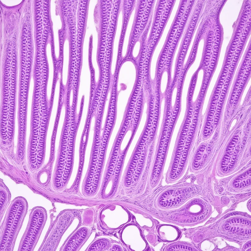

Gallbladder cancer is the most common malignancy of the biliary tract — a deceptively hidden organ that rarely announces its transformation until the disease is advanced. With most patients diagnosed at a stage when surgery can no longer remove it, and low five-year survival (see Epidemiology), it stands among the most lethal gastrointestinal cancers. Yet early-stage gallbladder cancers discovered incidentally at cholecystectomy carry an excellent prognosis when managed correctly — making pathological examination of every removed gallbladder a life-saving practice. The approval of durvalumab plus gemcitabine and cisplatin in 2022 opened a new first-line option for advanced disease, and molecular profiling is identifying additional targets including HER2 and FGFR.
Table of Contents
- What Gallbladder Cancer Is
- Epidemiology and Global Incidence
- Risk Factors and Carcinogenesis
- Molecular Pathology: HER2, FGFR, and Beyond
- Clinical Presentation
- Staging and Resectability Assessment
- Surgical Treatment: From Incidental to Radical Resection
- Systemic Therapy: Chemotherapy and Immunotherapy
- Targeted Therapy and Biomarker-Driven Treatment
- Biliary Drainage and Supportive Care
- Key Research Papers
- PubMed Topic Searches
- Connections
- Featured Videos
1. What Gallbladder Cancer Is
The gallbladder is a small pear-shaped sac tucked beneath the right lobe of the liver, functioning as a reservoir for bile produced by the liver. Its inner surface is lined with a single layer of columnar epithelial cells. Gallbladder cancer (GBC) arises when these epithelial cells undergo malignant transformation, with adenocarcinoma accounting for more than 90% of cases. Less common histological variants include papillary adenocarcinoma (better prognosis), mucinous adenocarcinoma, squamous cell carcinoma, and rarely neuroendocrine carcinoma.
The gallbladder's anatomical position — nestled directly against hepatic segments IVb and V, with immediate proximity to the cystic artery, hepatic artery, and portal vein — means that even modest local tumor growth can involve critical vascular structures or the liver parenchyma, rapidly rendering the disease unresectable. The lymphatic drainage flows to the cystic duct node (Calot's triangle), then to the periportal, hepatoduodenal ligament, and celiac nodes, providing multiple routes of regional spread.
A unique and clinically important characteristic of gallbladder cancer is the frequency of incidental discovery. Because gallstones cause identical symptoms to early GBC (right upper quadrant pain, nausea after fatty meals), many patients undergo cholecystectomy for presumed benign gallstone disease and have GBC found only on pathological examination of the removed specimen. This incidental discovery pattern accounts for 40–50% of all GBC diagnoses — which is why, in standard practice, every gallbladder removed at surgery is sent to pathology, however straightforward the clinical picture appeared.
2. Epidemiology and Global Incidence
Gallbladder cancer affects approximately 11,000 Americans annually, making it relatively rare in the United States. Globally, however, incidence varies dramatically by geography — a variation so striking that it has driven important etiological research.
High-Incidence Regions
The highest age-standardized incidence rates in the world occur in:
- South America: Chile (highest worldwide; 27 per 100,000 women in some regions), Bolivia, Ecuador, Peru. The South American GBC belt corresponds closely with indigenous populations who have high gallstone prevalence and specific dietary carcinogen exposures.
- South Asia: Northern India (Gangetic plain), Pakistan, Bangladesh. India contributes the largest absolute number of GBC cases globally.
- East Asia: Japan, Korea, and parts of China have intermediate-high incidence, with specific risk tied to anomalous pancreaticobiliary junction prevalence.
- Eastern Europe: Poland, Czech Republic, and Slovakia have higher rates than Western Europe.
Demographics
Gallbladder cancer shows a pronounced female predominance (3:1 female-to-male ratio in most populations), mirroring the higher prevalence of gallstone disease in women. In the United States, Native Americans and Hispanic women have substantially higher GBC incidence than non-Hispanic white women, again tracking with gallstone disease prevalence. The median age at diagnosis is 65–70 years, with most patients presenting in the sixth and seventh decades.
In absolute numbers, the United States sees approximately 11,000 new cases and 4,000 deaths annually from GBC. While these numbers are small relative to colorectal or breast cancer, the lethality — fewer than 15% of patients surviving five years — gives GBC an outsized death-to-incidence ratio.
3. Risk Factors and Carcinogenesis
The dominant risk factor for gallbladder cancer is gallstone disease, and the carcinogenic pathway likely involves chronic mucosal inflammation from stone-related trauma, bile acid dysregulation, and bacterial colonization. However, not all stones carry equal risk, and several additional factors substantially modify individual risk.
Gallstones and Chronic Inflammation
Gallstones are present in 70–90% of GBC patients, making them the most prevalent associated condition. Risk features that particularly elevate GBC risk include:
- Stone size: Stones larger than 3 cm carry a 10-fold higher GBC risk than stones smaller than 1 cm. Large stones cause more extensive mucosal trauma and inflammation.
- Duration: More than 20 years of known gallstone disease correlates with significantly higher GBC risk.
- Number: Multiple stones and chronic cholecystitis with repeated inflammatory bouts increase cumulative mucosal injury.
Gallbladder Polyps
Gallbladder polyps detected on ultrasound are sorted by risk, mainly by size. The thresholds below are those long used in surgical practice; guidelines differ on them:
- Polyps >1 cm: High malignant potential; removal of the gallbladder (cholecystectomy) has been the usual recommendation for polyps exceeding 1 cm in maximum diameter, regardless of symptom status.
- Polyps 0.6–0.9 cm: Intermediate risk; surveillance ultrasound at 6-month intervals; cholecystectomy if growth occurs.
- Polyps <0.5 cm: Very low malignant potential (almost certainly cholesterol polyps); annual surveillance acceptable.
- Sessile polyps and those associated with gallstones or primary sclerosing cholangitis carry higher malignant risk than pedunculated polyps at any size.
A 2022 consensus of the Society of Radiologists in Ultrasound states that the vast majority of gallbladder polyps smaller than 10 mm are not true neoplastic polyps but benign cholesterol polyps with no inherent risk of malignancy, and that recent studies have found the overall risk of gallbladder cancer is not increased in people with small polyps — calling into question frequent and prolonged follow-up of these common lesions. Reported follow-up schedules for small polyps therefore vary between guidelines.
Porcelain Gallbladder
Calcification of the gallbladder wall (porcelain gallbladder) was historically considered a strong GBC risk factor, triggering prophylactic cholecystectomy. Contemporary evidence is more nuanced: complete, diffuse wall calcification does not increase GBC risk, while selective mucosal (incomplete) calcification may carry modestly elevated risk. Patients with porcelain gallbladder discovered incidentally on imaging are typically evaluated by a hepatobiliary surgeon, particularly if calcification is incomplete or focal.
Anomalous Pancreaticobiliary Junction (APBJ)
APBJ — a congenital malunion of the pancreatic and common bile ducts creating a long common channel outside the duodenal wall — is the strongest relative risk factor for GBC, conferring a 20-fold or higher increased risk. The abnormal union allows pancreatic secretions to reflux chronically into the biliary tree, causing repeated chemical inflammation of the gallbladder epithelium. APBJ is particularly prevalent in Japan and East Asia and accounts for a disproportionate share of GBC in younger patients and women in these regions. When APBJ is detected on MRCP (magnetic resonance cholangiopancreatography), it is generally managed with preventive removal of the gallbladder (prophylactic cholecystectomy), even in the absence of symptoms or stones.
Additional Risk Factors
- Primary sclerosing cholangitis (PSC): The chronic inflammatory biliopathy associated with inflammatory bowel disease carries substantially elevated GBC risk alongside cholangiocarcinoma risk.
- Salmonella typhi chronic carriage: Chronic typhoid carriers who harbor S. typhi in the biliary tree have elevated GBC risk, explaining higher incidence in regions with endemic typhoid fever.
- Xanthogranulomatous cholecystitis (XGC): A severe inflammatory variant of cholecystitis characterized by lipid-laden macrophages within the gallbladder wall; associated with a 5–10% concurrent GBC rate and frequently mimics GBC on imaging.
- Obesity: Independently increases GBC risk beyond its effect on gallstone disease, likely through adipokine-mediated inflammation.
- Heavy metal and carcinogen exposure: Aflatoxin, radon, and nitrosamine exposure have been associated with elevated GBC risk in specific geographic and occupational contexts.
4. Molecular Pathology: HER2, FGFR, and Beyond
Gallbladder cancer has a heterogeneous molecular landscape. Unlike pancreatic cancer (where KRAS dominates) or colorectal cancer (with its relatively homogeneous MMR/KRAS/BRAF landscape), GBC harbors a broader distribution of oncogenic alterations — a feature that makes comprehensive molecular profiling particularly important to identify actionable targets.
ERBB2/HER2 Amplification or Overexpression
HER2 amplification or overexpression is the most clinically actionable alteration in GBC, present in approximately 15–20% of cases. This is a substantially higher rate than in biliary tract cancers overall (where HER2 alterations are less common in intrahepatic cholangiocarcinoma). The MyPathway basket trial and subsequent studies have demonstrated responses to trastuzumab-based regimens in HER2-positive GBC, and several dedicated clinical trials are now evaluating anti-HER2 agents in this population. HER2 testing by immunohistochemistry (IHC, a tissue stain) and fluorescence in situ hybridization (FISH, a gene-copy test) is used to find these patients among those with advanced GBC.
FGFR Alterations
FGFR2 fusion genes (which drive approximately 15% of intrahepatic cholangiocarcinoma and are the target of pemigatinib, approved by the U.S. Food and Drug Administration, and of infigratinib, whose approval was withdrawn in 2024 at the manufacturer's voluntary request, per a Federal Register notice of 16 May 2024) are uncommon in GBC. However, FGFR1 amplifications and FGFR3 mutations occur in a small subset of GBC, and FGFR inhibitors may represent a treatment option in patients whose tumors harbor these alterations on next-generation sequencing (NGS).
KRAS and TP53
KRAS mutations occur in approximately 10–25% of GBC and, as in other gastrointestinal (GI) cancers, have long been considered untargetable. TP53 mutations are among the most frequent alterations in GBC (40–60% of cases), reflecting its role in late-stage carcinogenesis after chronic inflammatory mucosal injury. TP53 mutations correlate with more aggressive histological grade and worse prognosis but currently lack specific targeted interventions.
PIK3CA, CDKN2A, and IDH
PIK3CA mutations (activating PI3K/AKT/mTOR pathway) occur in 10–20% of GBC and have led to early-phase trials of PI3K pathway inhibitors. CDKN2A (p16) deletion and IDH1/2 mutations are present in smaller subsets; IDH inhibitors (ivosidenib for IDH1) have shown efficacy in cholangiocarcinoma and may benefit the small fraction of IDH-mutated GBC patients.
Mismatch Repair Deficiency and TMB
Microsatellite instability-high (MSI-H) or mismatch repair-deficient (dMMR) status occurs in 5–8% of GBC. These tumors may respond to pembrolizumab per the tissue-agnostic U.S. Food and Drug Administration (FDA) approval. Additionally, high tumor mutational burden (TMB-High, ≥10 mut/Mb) occurs in a subset of GBC and may predict benefit from pembrolizumab regardless of MSI status.
Because of this molecular variety, comprehensive tumor profiling (NGS panel, MMR IHC, HER2 IHC/FISH) is used in patients with advanced GBC at the time of diagnosis to inform second-line and subsequent therapy options.
5. Clinical Presentation
The most challenging aspect of gallbladder cancer is its tendency to remain clinically silent until locally advanced or metastatic. The symptoms of early-stage GBC — right upper quadrant discomfort, nausea, and intolerance to fatty foods — are indistinguishable from symptomatic gallstone disease, which is far more common and in which clinicians' initial reflex is to attribute symptoms to benign cholelithiasis.
Incidental Discovery (40–50% of Cases)
The largest single mode of GBC detection is incidental discovery at laparoscopic cholecystectomy performed for presumed symptomatic cholelithiasis or acute cholecystitis. The gallbladder is removed, the patient recovers, and the pathology report returns days later with a diagnosis of carcinoma. This scenario is handled with a structured management algorithm (see Surgical Treatment section) and is the reason every gallbladder is sent for formal pathological examination — including gross sectioning with attention to the mucosa and wall thickness.
Symptomatic Presentation
When GBC presents symptomatically, the clinical picture varies by stage:
- Right upper quadrant (RUQ) pain: The most common presenting symptom. Early disease produces colicky or dull right-sided pain similar to biliary colic. As the tumor invades the liver bed, the pain becomes persistent and continuous rather than episodic, an important qualitative change that typically leads to cross-sectional imaging rather than empirical stone management.
- Jaundice: Obstructive jaundice in GBC indicates involvement of the common hepatic or common bile duct, either by direct tumor extension or compression by metastatic periportal lymph nodes. Painless progressive jaundice in an older patient with no prior biliary history raises concern for biliary malignancy. When GBC presents with jaundice, it is almost invariably at an unresectable stage.
- Weight loss and anorexia: Constitutional symptoms of malignancy, typically appearing when the disease is locoregionally advanced or metastatic.
- Palpable right upper quadrant mass: A late finding indicating a large primary tumor or hepatic involvement; present in fewer than 20% of patients at diagnosis but signals advanced disease.
- Acute cholecystitis presentation: A small subset of GBC presents acutely, mimicking bacterial acute cholecystitis with fever, RUQ tenderness, and leukocytosis. This presentation is a further reason every cholecystectomy specimen is examined by a pathologist.
Imaging Red Flags
Certain findings on routine abdominal ultrasound (the most common first-line investigation for biliary symptoms) raise concern for malignancy rather than benign gallbladder disease: gallbladder wall thickening greater than 3–4 mm (particularly focal or asymmetric), a polypoid intraluminal mass, discontinuity of the mucosal line, hepatic invasion at the gallbladder fossa, and biliary ductal dilation. Any of these findings is usually followed by cross-sectional imaging with contrast-enhanced computed tomography (CT) or MRI/MRCP before cholecystectomy.
6. Staging and Resectability Assessment
Gallbladder cancer is staged using the AJCC (American Joint Committee on Cancer) eighth edition TNM system. T stage reflects the depth of tumor invasion through the gallbladder wall layers, which is the most critical determinant of surgical approach for early disease:
- T1a: Tumor confined to the lamina propria (innermost mucosal layer). These are the most favorable tumors, typically found incidentally; simple cholecystectomy is curative.
- T1b: Tumor invades the muscularis (muscle layer of the gallbladder wall). The optimal treatment for T1b remains debated — cholecystectomy alone may be curative for T1b, but some centers advocate re-resection with hepatic bed resection given a 15–25% nodal positivity rate.
- T2: Tumor invades the perimuscular connective tissue on the peritoneal surface (T2a) or on the hepatic side (T2b). T2 requires radical resection (hepatic bed + lymphadenectomy). Hepatic-side T2b may have a worse prognosis due to proximity to liver vasculature.
- T3: Tumor perforates the visceral peritoneum or directly invades liver, gallbladder fossa, or adjacent organs (stomach, duodenum, colon, pancreas, bile ducts, omentum). Selected T3 tumors are resectable with major hepatic resection.
- T4: Tumor invades the main portal vein, hepatic arteries, or two or more extrahepatic organs/structures. Generally unresectable.
Staging Workup
For newly diagnosed or suspected GBC, the staging workup includes:
- Multiphasic computed tomography abdomen/pelvis with contrast: Defines local extent, hepatic invasion, vascular involvement, regional lymph node enlargement, and distant metastases. The arterial and portal venous phases are essential to assess hepatic artery and portal vein involvement.
- Magnetic resonance imaging (MRI) with MRCP: Superior to computed tomography for characterizing biliary anatomy, delineating hepatic extension, and assessing biliary ductal involvement. Essential when APBJ is suspected or when the relationship of the tumor to the biliary confluence must be defined before surgery.
- Positron emission tomography (PET)/computed tomography: Useful for detecting distant metastases not apparent on cross-sectional imaging. Detects peritoneal implants poorly (positron emission tomography (PET)/computed tomography sensitivity for peritoneal disease is low), which is why staging laparoscopy is commonly performed before planned resection.
- Staging laparoscopy: Performed by most hepatobiliary centers before any planned radical resection. Peritoneal deposits are found in 20–30% of patients thought to be resectable on computed tomography/magnetic resonance imaging. Port-site metastases from prior laparoscopic cholecystectomy may also be detected and excised.
- Serum CA 19-9 and carcinoembryonic antigen (CEA): Elevated CA 19-9 (>100 U/mL) occurs in 60–70% of GBC and serves as a baseline tumor marker for monitoring treatment response. Neither marker is specific enough for diagnosis, but elevation in the context of a suspicious mass strongly supports malignancy.
Factors Rendering Disease Unresectable
The majority of GBC patients (70–80%) present with unresectable disease; a 2015 review puts the share presenting at a stage amenable to surgical resection at only 10%. Key contraindications to resection include: bilateral hepatic involvement beyond what can be removed with adequate functional reserve, main portal vein or bilateral hepatic artery encasement, extensive peritoneal carcinomatosis, and distant metastases (liver, lung, or bone parenchymal metastases).
7. Surgical Treatment: From Incidental to Radical Resection
Surgery remains the only potentially curative treatment for gallbladder cancer. The appropriate surgical approach depends critically on T stage and the circumstances of diagnosis (incidental vs. planned resection for suspected GBC).
Management of Incidentally Discovered GBC
When pathology unexpectedly reveals GBC in a cholecystectomy specimen, the management algorithm is guided by T stage and the adequacy of prior resection:
- T1a (lamina propria only): Simple cholecystectomy is adequate; disease-specific survival exceeds 95%. No re-operation needed if margins are negative and bile duct is uninvolved.
- T1b (muscularis): Cholecystectomy alone may be sufficient; some guidelines and centers advocate re-resection (hepatic bed + portal lymphadenectomy) given the 15–25% reported rate of lymph node positivity. An individualized multidisciplinary tumor board decision is appropriate. Negative margins on the initial cholecystectomy specimen are a prerequisite for observation vs. re-resection.
- T2 (perimuscular connective tissue): Re-operation is the usual approach: radical cholecystectomy including resection of the liver segments IVb and V (hepatic bed directly underlying the gallbladder fossa) plus formal lymphadenectomy of the hepatoduodenal ligament (portal triad) nodes. The biliary duct does not need to be resected unless directly involved, as routine bile duct excision does not improve survival and adds morbidity.
- T3 (visceral peritoneal perforation or liver/adjacent organ invasion): Radical resection with major hepatic resection (segments IVb/V or more) + regional lymphadenectomy ± adjacent organ resection if feasible. In patients who underwent prior laparoscopic cholecystectomy, port-site excision (excision of all trocar insertion sites) has been practised because of the risk of tumor seeding; a 2017 multi-institution study found that routine port-site excision did not improve survival (see below).
Port-Site Resection After Laparoscopic Cholecystectomy
When GBC is discovered after laparoscopic cholecystectomy, there is a well-documented risk of port-site implantation — tumor seeding along the abdominal wall at the trocar insertion sites. This occurs because laparoscopic surgery disrupts the fascial barrier and creates opportunities for tumor cell implantation in the pneumoperitoneum environment, particularly when bile spillage occurred during surgery. Excision of the trocar port sites at re-operation has long been practised. The clinical significance of port-site metastases is debated (whether they represent a truly isolated local recurrence or simply reflect systemic spread). A 2017 analysis from the US Extrahepatic Biliary Malignancy Consortium (10 institutions, 2000–2015) studied 193 patients who had curative re-resection for incidentally found gallbladder cancer: 47 had port-site excision and 146 did not. Three-year survival was similar (65% vs 43%, P = 0.07, not a significant difference), port-site excision did not remain linked to survival after adjustment (only tumor grade and T stage did), and distant recurrence was identical (80% vs 81%). The authors concluded that port-site excision is not associated with improved overall survival and that routine port-site excision is not recommended.
Radical Cholecystectomy for Planned GBC Resection
For patients with GBC diagnosed preoperatively (by imaging with or without biopsy), the surgical approach consists of:
- Staging laparoscopy first to exclude peritoneal disease.
- Hepatic bed resection: Segments IVb and V are resected en bloc with the gallbladder fossa. For T3 tumors with deeper hepatic invasion, an extended hepatectomy may be required.
- Regional lymphadenectomy: Resection of the cystic duct node, hepatoduodenal ligament nodes, portal lymph nodes, and periportal tissue to achieve N1 staging. Examination of at least six nodes is the standard described for accurate staging.
- Bile duct resection: Performed only if the cystic duct margin is positive or if the common hepatic or common bile duct is directly involved. Routine bile duct excision is not performed.
8. Systemic Therapy: Chemotherapy and Immunotherapy
Adjuvant Therapy After Curative Resection
The BILCAP trial (Primrose et al., 2019) established capecitabine as the standard adjuvant therapy for resected biliary tract cancers, including gallbladder cancer. In this randomized phase III trial, patients with resected cholangiocarcinoma or GBC were randomized to capecitabine (1,250 mg/m² twice daily, days 1–14, every 21 days for 8 cycles) versus observation. The primary endpoint of overall survival in the intention-to-treat population narrowly missed statistical significance (median OS 51.1 months vs. 36.4 months; hazard ratio (HR) 0.81; p=0.097), but the pre-specified per-protocol analysis showed significant benefit (hazard ratio 0.75; p=0.028). Most international guidelines now recommend capecitabine adjuvant therapy for 6 months after R0 or R1 resection of biliary tract cancers.
First-Line Palliative Chemotherapy: Gemcitabine + Cisplatin
For patients with unresectable, locally advanced, or metastatic GBC, gemcitabine plus cisplatin (GemCis) became the reference first-line regimen following the ABC-02 trial (Valle et al., 2010). This landmark randomized phase III trial enrolled 410 patients with advanced biliary tract cancers (cholangiocarcinoma, GBC, and ampullary carcinoma) and demonstrated superior overall survival with GemCis versus gemcitabine alone (11.7 months vs. 8.1 months; hazard ratio 0.64; p<0.001). Progression-free survival and response rate also favored the combination. GemCis remained standard of care for a decade, until the addition of immunotherapy.
First-Line Immunotherapy: Gemcitabine + Cisplatin + Durvalumab (TOPAZ-1)
The TOPAZ-1 trial (Oh et al., 2022) evaluated the addition of durvalumab (a PD-L1 inhibitor, 1500 mg flat dose every 3 weeks) to gemcitabine plus cisplatin versus chemotherapy alone in patients with previously untreated advanced biliary tract cancer, including GBC. The trial met its primary endpoint: durvalumab plus GemCis improved overall survival compared to placebo plus GemCis (median OS 12.8 months vs. 11.5 months; hazard ratio 0.80; p=0.021), with 24-month OS rates of 24.9% vs. 10.4%. The benefit was consistent across biliary tract cancer subtypes, including GBC. Based on these results, the U.S. Food and Drug Administration approved durvalumab in combination with gemcitabine and cisplatin in September 2022 as first-line treatment for adult patients with locally advanced or metastatic biliary tract cancers. The 3-year update of TOPAZ-1 (2025; 685 patients, median follow-up 41.3 months) reported median overall survival of 12.9 vs. 11.3 months (hazard ratio 0.74) and 36-month survival of 14.6% vs. 6.9%; the authors state these long-term results support durvalumab plus gemcitabine and cisplatin as standard of care.
First-Line Immunotherapy: Gemcitabine + Cisplatin + Pembrolizumab (KEYNOTE-966)
The KEYNOTE-966 trial (Kelley et al., 2023) randomly assigned 1,069 people with previously untreated, unresectable or metastatic biliary tract cancer at 175 centres to pembrolizumab (200 mg every 3 weeks) or placebo, each with gemcitabine and cisplatin. Median overall survival was 12.7 months with pembrolizumab versus 10.9 months with placebo (hazard ratio 0.83). Severe (grade 3–4) side effects were similar in the two groups (79% vs 75%). The authors concluded that pembrolizumab with gemcitabine and cisplatin could be a new treatment option, making it a second immunotherapy-chemotherapy first-line combination alongside durvalumab.
Alternative First-Line Regimens
The NALIRIFOX regimen (liposomal irinotecan, fluorouracil, leucovorin, and oxaliplatin) is being evaluated in biliary tract cancers. FOLFOX (fluorouracil, leucovorin, oxaliplatin) is used in some centers for cisplatin-ineligible patients. Gemcitabine plus oxaliplatin (GEMOX) represents another cisplatin-sparing alternative for patients with impaired renal function.
Adding nab-paclitaxel to gemcitabine and cisplatin (the GAP regimen) looked promising in a 2019 phase II trial (45% partial response, median survival 19.2 months against historical controls), but the phase III SWOG S1815 trial (441 analysable patients, 16% with gallbladder cancer) found no survival gain: median overall survival 14.0 months with the triplet versus 13.6 months with gemcitabine and cisplatin (hazard ratio 0.91, P = .41), with more side effects from the triplet. A greater improvement in progression-free survival was seen in the gallbladder cancer subgroup, but not in overall survival.
Second-Line Therapy
No randomized phase III trial has definitively established a standard second-line regimen for GBC. Options used in practice include FOLFOX (as demonstrated in the ABC-06 trial for biliary tract cancers generally), pembrolizumab for MSI-H/dMMR tumors (tissue-agnostic approval), and clinical trial participation. FOLFIRI and mFOLFOX6 are also used. The relatively poor performance status of many GBC patients at disease progression often limits second-line treatment options.
9. Targeted Therapy and Biomarker-Driven Treatment
The molecular heterogeneity of GBC has motivated a comprehensive biomarker-testing strategy at diagnosis, particularly for patients with advanced disease. Several actionable alterations can guide treatment selection beyond standard chemotherapy.
HER2-Directed Therapy
HER2 overexpression or amplification occurs in 15–20% of GBC — the highest rate among biliary tract cancer subtypes. The MyPathway multi-basket trial evaluated trastuzumab (8 mg/kg loading, then 6 mg/kg every 3 weeks) plus pertuzumab (840 mg loading, then 420 mg every 3 weeks) in HER2-positive biliary tract cancers and demonstrated an objective response rate of 23% with a median duration of response of 10 months. Several ongoing trials are evaluating trastuzumab deruxtecan (T-DXd), a HER2-targeted antibody-drug conjugate with demonstrated efficacy across multiple HER2-expressing solid tumors, in advanced GBC. HER2 testing (IHC 3+ or FISH amplification) identifies the patients with unresectable or metastatic GBC who are candidates for these drugs.
Zanidatamab, an antibody that binds two different sites on HER2, was tested in the HERIZON-BTC-01 trial (2023) in people with HER2-amplified biliary tract cancer that had progressed after gemcitabine-based treatment. Among the 80 patients with HER2-positive tumours (IHC 2+ or 3+), 41.3% had a confirmed tumour response; 18% of all patients had grade 3 treatment-related side effects, most often diarrhoea (5%) and reduced heart pumping strength (decreased ejection fraction, 3%), with no grade 4 events or treatment-related deaths.
MSI-H/dMMR Tumors
Pembrolizumab is U.S. Food and Drug Administration approved (tissue-agnostic) for all MSI-H or dMMR solid tumors, including GBC. MSI-H/dMMR status occurs in 5–8% of GBC. In the KEYNOTE-158 basket trial, pembrolizumab demonstrated an objective response rate of approximately 40% in MSI-H biliary tract cancers, substantially higher than in microsatellite-stable tumors (<5% response). MMR IHC testing (staining for MLH1, MSH2, MSH6, PMS2) of advanced GBC specimens identifies these tumors.
IDH1 Inhibition
IDH1 mutations are present in approximately 5% of GBC (more common in intrahepatic cholangiocarcinoma at 20%). Ivosidenib (an IDH1 inhibitor, 500 mg daily) received U.S. Food and Drug Administration approval in 2021 for previously treated IDH1-mutant cholangiocarcinoma based on the ClarIDHy trial. While most evidence derives from cholangiocarcinoma cohorts, GBC patients with IDH1 mutations are eligible for ivosidenib and may derive clinical benefit.
FGFR Inhibitors
FGFR2 fusions (the primary target of pemigatinib, approved by the U.S. Food and Drug Administration; infigratinib's approval was withdrawn in 2024 at the manufacturer's voluntary request, per the Federal Register) are rare in GBC (<1%). However, FGFR1 amplifications and FGFR3 mutations occur in a small fraction of GBC, and patients with these alterations may be candidates for clinical trials of FGFR inhibitors or off-label use.
TMB-High Tumors
Pembrolizumab is U.S. Food and Drug Administration approved for TMB-high (≥10 mut/Mb) solid tumors regardless of histology, per the KEYNOTE-158 data. Approximately 5–10% of GBC tumors are TMB-high, making this another avenue for checkpoint inhibitor therapy in patients without MSI-H or other actionable mutations.
A molecular testing protocol described for advanced GBC at diagnosis includes: comprehensive NGS panel (covering HER2 amplification, IDH1/2, FGFR1-4, KRAS, TP53, PIK3CA, TMB), MMR IHC, and HER2 IHC/FISH as a parallel rapid test given the high actionability rate of HER2 alterations.
10. Biliary Drainage and Supportive Care
Obstructive jaundice is a debilitating complication of advanced GBC that significantly impairs quality of life, interferes with systemic treatment (chemotherapy must be dose-modified or withheld for elevated bilirubin), and can rapidly progress to cholangitis and biliary sepsis. Effective biliary drainage is a critical component of GBC palliative care.
Endoscopic Biliary Drainage (ERCP)
Endoscopic retrograde cholangiopancreatography (ERCP) with self-expanding metal stent (SEMS) placement is the preferred first-line drainage approach for most patients with GBC-related biliary obstruction involving the distal common bile duct or common hepatic duct. Metal stents are preferred over plastic stents for malignant biliary obstruction because they remain patent significantly longer (median 7–12 months vs. 3 months), requiring fewer repeat procedures. Covered metal stents reduce tumor ingrowth but have a higher migration rate; uncovered stents are most commonly used for malignant strictures.
Percutaneous Transhepatic Cholangiography (PTC)
When ERCP fails (most commonly due to duodenal invasion preventing endoscope positioning or failure to cannulate) or when the anatomy of the biliary stricture makes endoscopic access impossible (high hilar strictures), percutaneous transhepatic biliary drainage (PTBD) provides an alternative route. PTBD can be internalized to internal-external drainage or converted to an internal stent once the access track is matured. It is more invasive than ERCP and carries risks including pneumothorax, arteriovenous fistula, and bile leak, but is effective and often necessary in patients with GBC invading the hepatic hilum.
Nutritional Support and Palliative Care Integration
GBC patients frequently experience significant weight loss, anorexia, and cachexia. Nutritional assessment and support — including dietitian consultation and oral supplementation — are commonly part of care from early in the treatment course. Early palliative care consultation (as recommended by the American Society of Clinical Oncology (ASCO) and the European Society for Medical Oncology (ESMO) for advanced cancers generally) improves quality of life, symptom control, and in some studies survival. Goals-of-care conversations are especially important in GBC given its rapid progression in many patients.
Key Research Papers
- Valle J, et al. Cisplatin plus gemcitabine versus gemcitabine for biliary tract cancer. N Engl J Med. 2010;362(14):1273-1281. — PubMed PMID: 20375404 — The ABC-02 trial establishing gemcitabine plus cisplatin as standard first-line chemotherapy for advanced biliary tract cancers, including gallbladder cancer (OS 11.7m vs 8.1m, HR 0.64).
- Primrose JN, et al. Capecitabine compared with observation in resected biliary tract cancer (BILCAP): a randomised, controlled, multicentre, phase 3 study. Lancet Oncol. 2019;20(5):663-673. — PubMed PMID: 30922733 — Landmark BILCAP trial of adjuvant capecitabine after resection of biliary tract cancers; intention-to-treat OS 51.1m vs 36.4m (HR 0.81, p=0.097, primary endpoint not met); per-protocol OS 53m vs 36m (HR 0.75), now the reference adjuvant regimen.
- Oh DY, Ruth He A, Qin S, et al. Durvalumab plus Gemcitabine and Cisplatin in Advanced Biliary Tract Cancer. NEJM Evid. 2022;1(8):EVIDoa2200015. — PubMed PMID: 38319896 — TOPAZ-1 trial: addition of durvalumab to GemCis improved OS (HR 0.80; 24-month OS 24.9% vs 10.4%) in 685 patients with advanced biliary tract cancers.
- Shroff RT, Javle MM, Xiao L, et al. Gemcitabine, Cisplatin, and nab-Paclitaxel for the Treatment of Advanced Biliary Tract Cancers: A Phase 2 Clinical Trial. JAMA Oncol. 2019;5(6):824-830. — PubMed PMID: 30998813 — Single-arm phase II triplet chemotherapy (GAP regimen) in 60 treated patients: partial response 45% and median OS 19.2m against historical controls; the phase III SWOG S1815 trial later found no OS benefit.
- Lamarca A, et al. Second-line FOLFOX chemotherapy versus active symptom control for advanced biliary tract cancer (ABC-06): a phase 3, open-label, randomised, controlled trial. Lancet Oncol. 2021;22(5):690-701. — PubMed PMID: 33798493 — ABC-06 trial demonstrating modest but statistically significant OS benefit of FOLFOX over active symptom control in second-line biliary tract cancer (OS 6.2m vs 5.3m, HR 0.69).
- Javle M, Borad MJ, Azad NS, et al. Pertuzumab and trastuzumab for HER2-positive, metastatic biliary tract cancer (MyPathway): a multicentre, open-label, phase 2a, multiple basket study. Lancet Oncol. 2021;22(9):1290-1300. — PubMed PMID: 34339623 — MyPathway HER2-positive biliary tract cancer cohort: 9 of 39 patients had a partial response (ORR 23%), supporting HER2 as an actionable target.
- Abou-Alfa GK, et al. Ivosidenib in IDH1-mutant, chemotherapy-refractory cholangiocarcinoma (ClarIDHy): a multicentre, randomised, double-blind, placebo-controlled, phase 3 study. Lancet Oncol. 2020;21(6):796-807. — PubMed PMID: 32416072 — ClarIDHy trial of ivosidenib in IDH1-mutant cholangiocarcinoma (PFS 2.7m vs 1.4m), establishing IDH1 inhibition as a target relevant to IDH1-mutant GBC as well.
- Tewari M, Mishra RR, Shukla HS. Salmonella typhi and gallbladder cancer: report from an endemic region. Hepatobiliary Pancreat Dis Int. 2010;9(5):524-530. — PubMed PMID: 20943463 — Case-control study from northern India (54 gallbladder cancer cases, 54 gallstone controls): S. typhi DNA found in tumour tissue in 33% of cases vs 0% of controls.
- Kanthan R, Senger JL, Ahmed S, Kanthan SC. Gallbladder Cancer in the 21st Century. J Oncol. 2015;2015:967472. — PubMed PMID: 26421012 — Review of GBC epidemiology, risk factors, pathology, management and prognosis; only 10% of patients present at a resectable stage.
- Vogel A, Bridgewater J, Edeline J, et al. Biliary tract cancer: ESMO Clinical Practice Guideline for diagnosis, treatment and follow-up. Ann Oncol. 2023;34(2):127-140. — PubMed PMID: 36372281 — Current ESMO guidelines for biliary tract cancers including GBC, incorporating molecular testing recommendations and updated treatment algorithms.
- Kelley RK, Ueno M, Yoo C, et al. Pembrolizumab in combination with gemcitabine and cisplatin compared with gemcitabine and cisplatin alone for patients with advanced biliary tract cancer (KEYNOTE-966): a randomised, double-blind, placebo-controlled, phase 3 trial. Lancet. 2023;401(10391):1853-1865. — PubMed PMID: 37075781
- Oh DY, He AR, Qin S, et al. Durvalumab plus chemotherapy in advanced biliary tract cancer: 3-year overall survival update from the phase III TOPAZ-1 study. J Hepatol. 2025;83(5):1092-1101. — PubMed PMID: 40381735
- Shroff RT, King G, Colby S, et al. SWOG S1815: A Phase III Randomized Trial of Gemcitabine, Cisplatin, and Nab-Paclitaxel Versus Gemcitabine and Cisplatin in Newly Diagnosed, Advanced Biliary Tract Cancers. J Clin Oncol. 2025;43(5):536-544. — PubMed PMID: 39671534
- Harding JJ, Fan J, Oh DY, et al. Zanidatamab for HER2-amplified, unresectable, locally advanced or metastatic biliary tract cancer (HERIZON-BTC-01): a multicentre, single-arm, phase 2b study. Lancet Oncol. 2023;24(7):772-782. — PubMed PMID: 37276871
- Ethun CG, Postlewait LM, Le N, et al. Routine port-site excision in incidentally discovered gallbladder cancer is not associated with improved survival: A multi-institution analysis from the US Extrahepatic Biliary Malignancy Consortium. J Surg Oncol. 2017;115(7):805-811. — PubMed PMID: 28230242
- Kamaya A, Fung C, Szpakowski JL, et al. Management of Incidentally Detected Gallbladder Polyps: Society of Radiologists in Ultrasound Consensus Conference Recommendations. Radiology. 2022;305(2):277-289. — PubMed PMID: 35787200
- Food and Drug Administration. Withdrawal of Approval of New Drug Application for Infigratinib Phosphate Capsules, 25 Milligrams and 100 Milligrams. Federal Register, 16 May 2024, document 2024-10714. — Federal Register 2024-10714
12. PubMed Topic Searches
- Gallbladder cancer surgery radical cholecystectomy
- Incidental gallbladder carcinoma cholecystectomy management
- Gallbladder cancer gemcitabine cisplatin durvalumab TOPAZ-1
- HER2-positive gallbladder cancer trastuzumab
- Anomalous pancreaticobiliary junction gallbladder cancer risk
- Gallbladder polyp malignant transformation cholecystectomy
- Gallbladder cancer molecular profiling biomarkers targeted therapy
- Biliary tract cancer capecitabine adjuvant BILCAP trial