Trigeminal Neuralgia
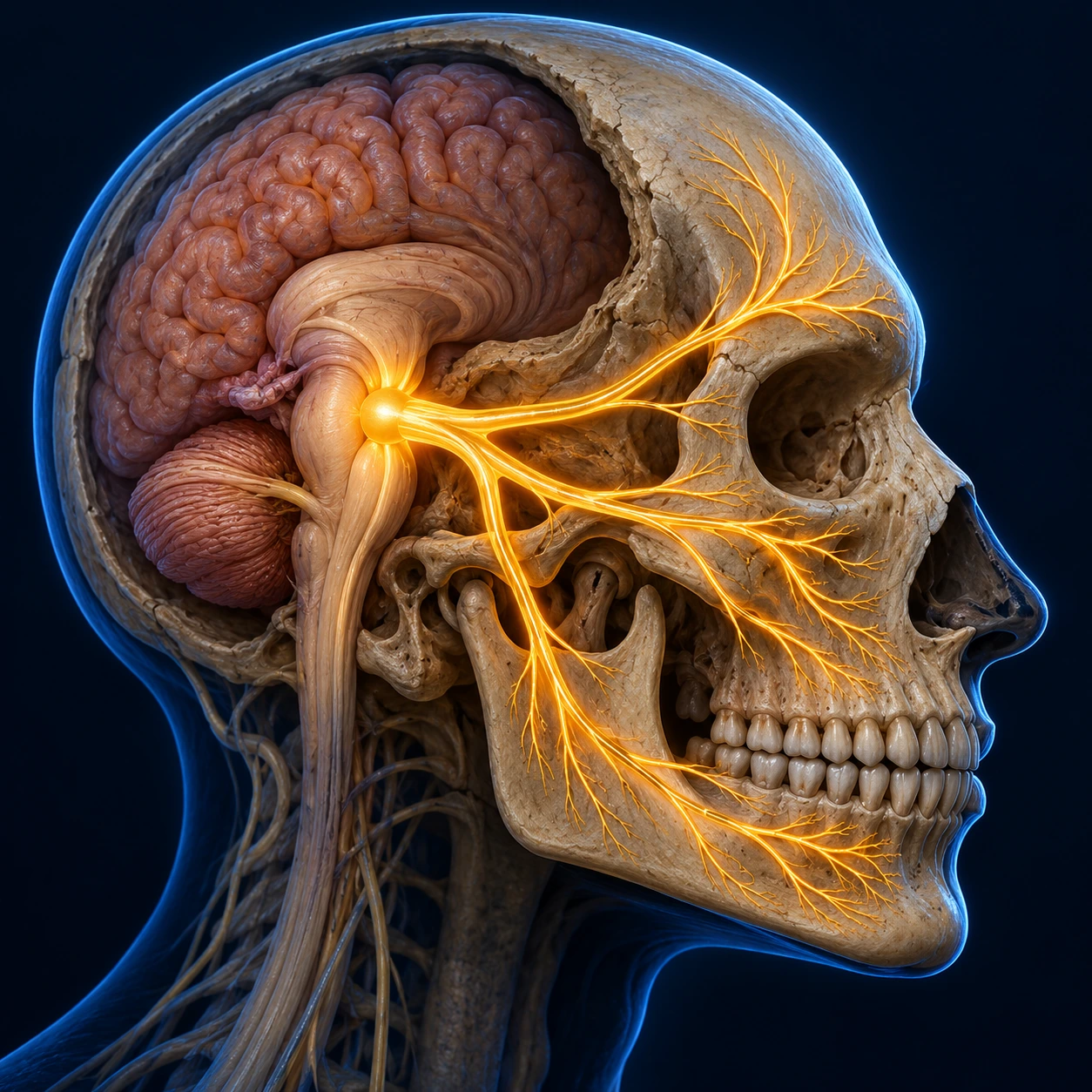
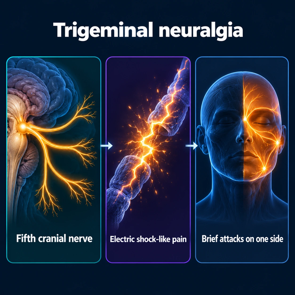
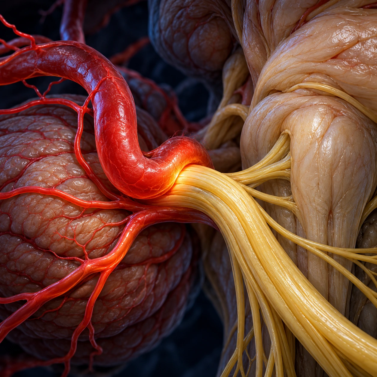
Table of Contents
1. Overview
Trigeminal neuralgia (TN) — sometimes called tic douloureux — is a chronic pain condition affecting the trigeminal nerve, the fifth cranial nerve (CN V), which carries sensation from the face to the brain. It produces some of the most intense pain that medicine recognizes: sudden, severe, electric-shock-like or stabbing jolts that shoot across one side of the face. The attacks last from a fraction of a second to about two minutes, but the pain in that brief window can be so overwhelming that people freeze mid-sentence, drop what they are holding, or grab their cheek and wait for it to pass.
For centuries TN carried the grim nickname “the suicide disease,” because in an earlier era — before effective drugs and modern surgery — some sufferers were driven to despair by relentless, untreatable pain. That label is outdated, and it deserves to be retired. Today, trigeminal neuralgia is a treatable condition. The great majority of patients get substantial relief from a single inexpensive medication, and for those who do not, there are surgical procedures — including one that can offer a lasting cure — with well-documented success rates. If you have just been diagnosed and you found that old nickname online, please know that the picture for someone diagnosed in this century is very different. The pain is real and it is severe, but it is also one of the most responsive facial pain syndromes we know.
What makes TN distinctive, and so often misdiagnosed, is its triggers. The pain is set off by light, ordinary touch to specific spots on the face — brushing your teeth, shaving, putting on makeup, a cold breeze on the cheek, chewing a meal, or simply talking. It is not the firm pressure of a bruise that hurts; it is the gentlest contact. Because the pain so often lands in the jaw and around the teeth, the very first stop for many patients is not a neurologist but a dentist — a detour that is worth understanding before it costs you teeth, which is discussed throughout this page.
2. Epidemiology
Trigeminal neuralgia is uncommon but not rare. Older figures often cited put the incidence at roughly 4 to 13 new cases per 100,000 people per year and lifetime prevalence near 0.3%. A 2026 pooled analysis of 17 studies and over 170 million people gave an incidence of 25.33 per 100,000 person-years (95% CI 11.87–54.02) and a lifetime prevalence of 108.43 per 100,000 people (95% CI 30.54–384.18), with wide variation between countries that the authors link partly to changing diagnostic criteria. It is more common in women than men (roughly 3:2) and rises sharply with age — most people are diagnosed after age 50, and the incidence keeps climbing into the seventies and eighties.
Age matters for more than statistics. The typical neurovascular form of TN clusters in older adults, where a blood vessel has gradually come to rest against the nerve. When TN appears in someone young — under 40 — or affects both sides of the face, clinicians take that seriously, because it raises the probability that the underlying cause is something other than simple age-related vessel contact, most importantly multiple sclerosis (see Etiology). The right side of the face is affected slightly more often than the left, and bilateral disease is uncommon (around 1–4% of cases) — when it occurs, it is again a flag to look harder for a secondary cause.
3. Pathophysiology
The trigeminal nerve has three branches: V1 (ophthalmic) serving the forehead and around the eye, V2 (maxillary) serving the cheek and upper jaw, and V3 (mandibular) serving the lower jaw. TN pain follows one or more of these territories — most often V2 and V3 together, which is exactly why the pain is so easily mistaken for a toothache.
In the most common form, the problem starts where the nerve root leaves the brainstem and is still wrapped in central (brain-type) myelin — a zone particularly vulnerable to injury. A pulsating artery (frequently the superior cerebellar artery) lies against the root and, beat after beat, year after year, wears away the insulating myelin sheath through chronic compression. With the insulation stripped, bare nerve fibers can short-circuit against each other (a phenomenon called ephaptic cross-talk), so a light, harmless touch signal jumps across to pain fibers and ignites a burst of electrical activity. Sander Love and colleagues documented this demyelination in the nerve roots of patients at surgery, anchoring the modern mechanistic picture. The result is the hallmark of TN: a trivial stimulus — a breeze, a bite of toast — producing a violent, paroxysmal jolt of pain, often followed by a brief refractory period during which the area cannot be triggered again.
This understanding explains why the most effective drugs are sodium-channel-blocking anticonvulsants: they calm hyperexcitable, misfiring nerve membranes, the same way they quiet the abnormal electrical discharges of a seizure. It also explains why physically relieving the pressure on the nerve — lifting the offending vessel off the root — can resolve the pain at its source.
4. Etiology and Risk Factors
Modern classification (ICHD-3 and the EAN guideline) divides TN into three groups, and the distinction guides both testing and treatment:
- Classical TN — caused by neurovascular compression with demonstrable morphological change (the nerve is distorted, indented, or atrophic where a vessel touches it). This is the single most common identifiable cause.
- Secondary TN — caused by an identifiable underlying disease: multiple sclerosis, a tumor in the path of the nerve (such as a vestibular schwannoma, meningioma, or epidermoid cyst), an arteriovenous malformation, or a brainstem lesion. Roughly 15% of TN is secondary.
- Idiopathic TN — no clear vascular contact and no other cause is found despite a thorough workup.
The multiple sclerosis connection is one of the most important things to know about this condition. TN can be the first symptom of multiple sclerosis (MS), sometimes years before any other sign appears. In multiple sclerosis, demyelinating plaques in the brainstem damage the trigeminal pathway from within, producing the same kind of misfiring as a compressing vessel. People with multiple sclerosis develop TN about 20 times more often than the general population. A pooled analysis of 19 studies and 30,348 people with multiple sclerosis found that about 3.4% (95% CI 1.5–5.9%) have TN, with very wide differences between studies. A younger patient, a patient with bilateral facial pain, or anyone with other neurological symptoms (numbness elsewhere, visual changes, weakness) raises the suspicion of a secondary cause. The 2019 European Academy of Neurology (EAN) guideline goes further: it states that magnetic resonance imaging should be part of the work-up in all TN patients, because no clinical characteristics can exclude secondary TN. Finding multiple sclerosis changes the whole plan.
The main risk factors, then, are increasing age, female sex, multiple sclerosis, and hypertension (which is associated with the vascular changes behind compression). TN is not caused by dental work, stress, or anything the patient did — an important point for people who blame themselves.
5. Clinical Presentation
The classic story is remarkably consistent, and recognizing it is most of the diagnosis. People describe attacks of sudden, one-sided, electric-shock or stabbing pain in the cheek, jaw, gums, lips, or around the eye. Each jolt is brief — seconds to about two minutes — but attacks can come in rapid volleys, sometimes dozens in a day. Between attacks many people are completely pain-free, although some have a lower-grade aching background pain on top of the jolts (a feature sometimes called “TN type 2” or concomitant continuous pain).
The defining feature is the trigger. Pain is provoked by light touch or movement of innocent everyday activities:
- Brushing teeth or touching the gums
- Chewing, eating, or even swallowing
- Talking, smiling, or moving the jaw
- Shaving, washing the face, or applying makeup
- A cold breeze, a draft, or wind on the cheek — many people cover that side of the face outdoors or refuse to sit near an air-conditioning vent
- Light touch to a specific “trigger zone” on the face
Because eating and talking set it off, people often lose weight, avoid meals, become socially withdrawn, and dread routine self-care. They may sit very still, terrified that the next sentence or sip will detonate another attack. That avoidance is rational, not psychological — but it takes a heavy emotional toll (see Complications).
The dental detour. Because the pain so often lands in the upper or lower teeth, an enormous number of patients see a dentist first and are convinced something is wrong with a tooth. The teeth and X-rays look essentially normal, but the pain is undeniable, so well-meaning treatment escalates: fillings, root canals, and eventually extractions — none of which help, because the problem was never in the tooth. It is heartbreakingly common for someone to lose one or several healthy teeth before TN is finally recognized. If facial or tooth pain is electric, lightning-fast, triggered by a breeze or light touch, and the dental exam keeps coming back clean, that is the pattern of trigeminal neuralgia, a nerve condition that a neurology assessment can identify before any irreversible dental procedure.
6. Diagnosis
TN is fundamentally a clinical diagnosis — it is made from the story and the pattern, not from a blood test. The international criteria (ICHD-3) require recurrent paroxysms of unilateral facial pain in the trigeminal distribution, lasting from a fraction of a second to two minutes, with shock-like, shooting, stabbing or sharp quality, precipitated by innocuous stimuli, and not better explained by another diagnosis. The neurological exam in classical TN is typically normal — importantly, there is no facial weakness or numbness in the simple case.
Imaging earns its place by answering two questions. A high-resolution magnetic resonance imaging (MRI) of the brain, ideally with thin sequences through the trigeminal nerve (and often MR angiography), is used to (1) find neurovascular compression — demonstrating a vessel touching and distorting the nerve root, which both confirms the classical form and maps the target for surgery — and (2) rule out secondary causes, above all multiple sclerosis plaques in the brainstem and tumors along the nerve. Practice guidelines (AAN–EFNS) specifically recommend that the evaluation look for these secondary causes, and note that the presence of sensory loss, bilateral symptoms, or abnormal trigeminal reflexes raises the likelihood of a structural cause that imaging looks for. The 2019 EAN guideline states that magnetic resonance imaging, using a combination of three high-resolution sequences, should be performed in all TN patients because no clinical characteristics can exclude secondary TN, and that trigeminal reflexes can be used if magnetic resonance imaging is not possible.
The differential diagnosis — the other things facial pain can be — is worth knowing because each is managed differently:
- Dental disease (cracked tooth, abscess, pulpitis): pain is usually constant or throbbing, hot/cold sensitive, and localizes to a tooth that the dentist can find.
- Temporomandibular joint (TMJ) disorder: aching pain around the jaw joint, worse with chewing, often with clicking and jaw-muscle tenderness — not lightning-like.
- Cluster headache and other trigeminal autonomic cephalalgias: severe one-sided pain around the eye, but lasting much longer (15–180 minutes) and accompanied by a red, tearing eye, a drooping lid, and a stuffy nostril.
- Migraine: throbbing, longer-lasting, with nausea and light sensitivity rather than touch-triggered electric jolts.
- Bell's palsy: this causes facial weakness — a drooping side of the face — not the paroxysmal pain of TN. It is a useful contrast: TN is a sensory/pain problem of CN V; Bell's palsy is a motor problem of CN VII (the facial nerve).
- Postherpetic neuralgia after shingles in a trigeminal branch: usually burning and constant, with a history of a rash.
7. Treatment
Here is the genuinely reassuring part: most people with TN respond to medication, and the first-line drug works well.
Medication — the first and best step for most
Carbamazepine is the established first-line treatment and is genuinely effective — it is one of the few situations in pain medicine where a drug response is so reliable that a good response actually supports the diagnosis. Across studies, roughly 70–90% of patients get meaningful relief when treatment is started; in a retrospective study of 200 outpatients (100 starting carbamazepine and 100 starting oxcarbazepine, with 22 later excluded as possible secondary cases), 98% on carbamazepine and 94% on oxcarbazepine responded at first. Oxcarbazepine, a close relative, is just as effective for many people and is often better tolerated, so clinicians frequently start with it.
These drugs are not without trade-offs, and being honest about them helps people stick with treatment:
- Side effects include drowsiness, dizziness, unsteadiness, double vision, and nausea — usually worst early and often eased by starting low and increasing slowly.
- Monitoring matters: carbamazepine can lower blood sodium (hyponatremia — even more so with oxcarbazepine) and, rarely, suppress blood counts or liver function, so periodic blood tests are standard. The carbamazepine label carries a boxed warning for aplastic anemia and agranulocytosis (loss of blood-making cells and of infection-fighting white cells): the risk is 5 to 8 times that of the general population, whose own risk is low, about 6 per million people a year for agranulocytosis and 2 per million a year for aplastic anemia.
- A serious skin-reaction risk (Stevens–Johnson syndrome) is strongly linked to a genetic marker, human leukocyte antigen-B*15:02, which is far more common in people of Han Chinese, Thai, and some other Southeast Asian ancestries — guidelines recommend testing for it before starting carbamazepine in those populations.
- Carbamazepine interacts with many medications. Its label lists drugs and foods that raise carbamazepine levels, including macrolide antibiotics (erythromycin, clarithromycin), azole antifungals, fluoxetine, verapamil, diltiazem and grapefruit juice. It also lowers the blood levels of many other drugs, including oral and other hormonal contraceptives, warfarin and lamotrigine, and its use with nefazodone is contraindicated.
When the first drug is not enough, or not tolerated, other agents are used alone or added on: baclofen, lamotrigine (a useful add-on, with evidence supporting it), and gabapentin or pregabalin. These have less robust evidence than carbamazepine but help many patients, especially those who cannot take the first-line drugs. It is commonly said that even drugs that work at first become less effective over years, and that doses needed to control attacks can creep up to levels that cause unacceptable grogginess. In the 200-outpatient study, late loss of effect was seen only in a very small minority (3 patients on carbamazepine and 2 on oxcarbazepine), and side effects were the main problem: 27% of carbamazepine responders and 18% of oxcarbazepine responders stopped or cut to an unsatisfactory dose. When that happens, it is not a failure — the 2019 EAN guideline recommends that patients be offered surgery if pain is not sufficiently controlled or medical treatment is poorly tolerated.
Botulinum toxin injection is also on the list: the 2019 EAN guideline states that lamotrigine, gabapentin, botulinum toxin type A, pregabalin, baclofen and phenytoin may be used alone or as add-ons. A 2024 review of 23 studies, 4 of them randomized, found that pain scores and attack frequency fell from baseline after the injections, and its authors state that more high-quality studies are needed.
Sudden severe flares. For acute worsening of pain, the 2019 EAN guideline states that an intravenous infusion of fosphenytoin or lidocaine can be used. These are given through a vein in a hospital or clinic, which matters when a flare stops a person eating and drinking.
Electroacupuncture. In a randomized factorial trial of 120 people, electroacupuncture, carbamazepine 300 mg a day and the two combined each lowered pain scores more than their sham or placebo controls, with the largest difference on the combination at 4 weeks (-3.7 points on a pain scale). By week 28 the difference was -0.8 for electroacupuncture, 0.2 for carbamazepine and -2.9 for the combination. Side effects were reported by 10.17% with electroacupuncture and 25.42% with carbamazepine.
When medication fails or stops being tolerated — procedures
Microvascular decompression (MVD) is the procedure that comes closest to a cure, because it fixes the actual cause. Pioneered by neurosurgeon Peter Jannetta, MVD is an operation under general anesthesia in which the surgeon opens a small window behind the ear, finds the blood vessel pressing on the nerve root, and lifts it away, placing a soft cushion (often polytetrafluoroethylene) between the vessel and the nerve so it can no longer pound on it. In the landmark long-term study by Barker, Jannetta and colleagues of more than 1,000 patients, about 70% remained pain-free without medication at 10 years, with immediate post-operative relief in roughly 80–90%. That study covered 1,185 patients: 30% had recurrences, 11% had a second operation, 2 patients died shortly after surgery (0.2%), 1 had a brain-stem stroke (0.1%), and 16 (1%) lost hearing on that side. Crucially, because nothing is deliberately injured, MVD usually preserves facial sensation — you keep feeling in your face. The trade-off is that it is brain surgery: risks include hearing loss, facial numbness, cerebrospinal fluid (CSF) leak, and, rarely, more serious complications, so it is generally offered to patients healthy enough for surgery, especially those with clear vascular compression on magnetic resonance imaging and good general health.
Ablative (lesioning) procedures work differently — instead of relieving pressure, they deliberately damage part of the pain pathway so it can no longer transmit the attacks. They are less invasive (and so suit older or frailer patients, or those who decline open surgery), but the honest trade-off is numbness: by design they trade pain for some loss of sensation, and the pain has a higher chance of returning over time.
- Stereotactic radiosurgery — a focused beam of radiation is aimed at the nerve root; no incision is needed. Relief is not immediate (it can take weeks to months), and in the foundational outcome series by Maesawa and colleagues most patients improved, though some later relapsed and a minority developed new facial numbness. Among those 220 patients, complete relief reached 70.3% at 1 year, complete or partial relief was 85.6% at 1 year and 55.8% at 5 years, and new or increased facial tingling or numbness affected 10.2% at 2 years.
- Percutaneous rhizotomy — a needle is passed through the cheek to the nerve and a lesion is made by balloon compression, glycerol injection, or radiofrequency heating. These give fast relief and are repeatable, at the cost of more facial numbness and, uncommonly, troublesome numb-but-painful sensations.
Choosing among these is a shared decision that weighs your age and health, what the magnetic resonance imaging shows, how much numbness you are willing to accept, and how durable you need the relief to be. Surgical outcome studies (such as Tyler-Kabara and colleagues) show that patients with the classic, typical pattern of TN tend to do better after surgery than those with atypical, constant pain (more than 5 years after decompression, excellent results in 73% with typical pain and 35% with atypical pain) — another reason careful diagnosis up front pays off.
The 2019 EAN guideline recommends microvascular decompression as first-line surgery in classical TN, gives no recommendation between the options in idiopathic TN, and states that neuroablative treatments are the preferred choice if magnetic resonance imaging shows no vessel contact. A 2026 network analysis of 11 mostly observational studies and 10,741 patients found that, against microvascular decompression, stereotactic radiosurgery had fewer complications (risk ratio 0.24) but less complete pain relief (risk ratio 0.52) and more recurrence (risk ratio 3.14). Its authors rate the certainty as low to very low and call the results hypothesis-generating.
8. Complications
The most serious complications of TN are not in the face but in the toll the disease takes on a life:
- Irreversible dental damage from procedures done in pursuit of a “bad tooth” that was never the problem — including extraction of healthy teeth.
- Weight loss and malnutrition from fear of chewing.
- Social withdrawal and isolation, because talking, smiling, and eating with others all become risky.
- Anxiety and depression, which are common and understandable. Living in dread of the next jolt — never knowing if a word, a sip, or a draft will trigger agony — is exhausting. Studies of TN consistently find high rates of mood symptoms and impaired quality of life.
It bears repeating plainly: the psychological toll of this condition is real and valid. The old “suicide disease” nickname grew out of genuine suffering in an era without treatment. Hopelessness is a treatable symptom of a treatable disease, and support exists: the 2019 EAN guideline recommends that patients are offered psychological and nursing support alongside medical and surgical care. Effective pain control very often lifts the mood that the pain was crushing.
9. Prognosis
The outlook for a person diagnosed today is good and steadily improving. Most people achieve good pain control with medication, at least initially. TN often follows a relapsing–remitting course, with attack-free periods of weeks, months, or even years between flares — a pattern that can be discouraging when a flare returns but also means relief is the norm, not the exception.
Over years, some people find medication less effective or harder to tolerate, and that is when surgery enters the picture — with microvascular decompression offering the best chance of long-term, drug-free freedom from pain (around 70% still pain-free at a decade). Even when one approach fades, there is almost always another lever to pull. The realistic message for newly diagnosed patients is not “you will suffer” but “this is treatable, and there is a sequence of options designed to keep you ahead of the pain.”
10. Prevention
There is no way to prevent trigeminal neuralgia itself — you cannot stop a vessel from drifting against a nerve over decades, and you cannot prevent the demyelination that drives it. So the realistic goals are about preventing attacks and preventing harm:
- Steering clear of known triggers during a flare. Many people learn to chew on the unaffected side, eat soft foods, use lukewarm water, protect the face from cold wind with a scarf, and shield the cheek from drafts and air-conditioning. These do not cure anything, but they reduce the number of attacks while medication takes hold.
- Medication taken consistently, not just reactively — TN drugs work best as steady prevention, keeping the nerve quiet, rather than as something taken only after pain hits.
- The dental detour. The single most preventable harm in TN is unnecessary, irreversible dental work. Prompt, accurate diagnosis — recognizing the electric, trigger-driven pattern and getting a neurology assessment before extractions or root canals — protects healthy teeth and shortens the road to real relief. If you are reading this before that first dentist appointment, that may be the most useful sentence on this page.
11. Recent Research and Advances
Several lines of work are reshaping how TN is diagnosed and treated. High-resolution magnetic resonance imaging protocols now visualize the trigeminal root and its vessel contact in fine detail, helping distinguish true compression with nerve distortion (which predicts surgical success) from incidental contact, and sharpening the classical / secondary / idiopathic distinction that the 2019 EAN guideline formalized. That guideline adds that vessel contact on imaging should not be used to confirm the diagnosis of TN; it may help decide if and when a person is referred for microvascular decompression.
Genetics and molecular pain biology are an active frontier. Rare variants in voltage-gated sodium-channel genes have been linked to some cases, which fits the channel-blocking logic of current drugs and points toward more selective, better-tolerated sodium-channel medicines in development — agents designed to quiet the misfiring nerve without the sedation and monitoring burden of carbamazepine.
Not every promising idea has held up. Erenumab, an antibody drug used for migraine, was reported to help in open-label studies, but in a double-blind trial of 80 people it did no better than placebo (35% responders against 45%, p = 0.36).
On the procedural side, refinements in stereotactic radiosurgery dosing and targeting, comparative long-term data across MVD, radiosurgery, and percutaneous techniques, and growing interest in neuromodulation are helping clinicians and patients match the right procedure to the right person. The 2020 Lancet Neurology review by Bendtsen and colleagues synthesizes much of this progress and reflects a field that has moved decisively away from the bleak picture behind the disease's old nickname.
Key Research Papers
Historical Background
Trigeminal neuralgia was clearly described in the 18th century by the English physician John Fothergill (1773), who reported the lancinating, touch-triggered facial pain that still bears the clinical picture we recognize today; the older term tic douloureux (“painful tic”) captured the wince attacks provoke. For nearly two centuries treatment was crude and often futile, which is how the “suicide disease” reputation took hold. The modern era began with neurosurgeon Peter Jannetta, who in the 1960s and 1970s developed and popularized microvascular decompression, demonstrating that a blood vessel compressing the nerve root was a common, fixable cause — transforming TN from an incurable affliction into an operable one. The subsequent arrival of stereotactic radiosurgery in the 1990s, alongside effective anticonvulsant drug therapy led by carbamazepine, completed the shift to today's full menu of medical and surgical options.
Key Research Papers
- Cruccu G, Di Stefano G, Truini A. Trigeminal Neuralgia. New England Journal of Medicine. 2020;383(8):754–762. — doi:10.1056/NEJMra1914484
- Bendtsen L, Zakrzewska JM, Heinskou TB, et al. Advances in diagnosis, classification, pathophysiology, and management of trigeminal neuralgia. The Lancet Neurology. 2020;19(9):784–796. — doi:10.1016/S1474-4422(20)30233-7
- Bendtsen L, Zakrzewska JM, Abbott J, et al. European Academy of Neurology guideline on trigeminal neuralgia. European Journal of Neurology. 2019;26(6):831–849. — doi:10.1111/ene.13950
- Cruccu G, Gronseth G, Alksne J, et al. AAN–EFNS guidelines on trigeminal neuralgia management. European Journal of Neurology. 2008;15(10):1013–1028. — doi:10.1111/j.1468-1331.2008.02185.x
- Gronseth G, Cruccu G, Alksne J, et al. Practice Parameter: the diagnostic evaluation and treatment of trigeminal neuralgia (an evidence-based review). Neurology. 2008;71(15):1183–1190. — doi:10.1212/01.wnl.0000326598.83183.04
- Barker FG, Jannetta PJ, Bissonette DJ, et al. The long-term outcome of microvascular decompression for trigeminal neuralgia. New England Journal of Medicine. 1996;334(17):1077–1083. — doi:10.1056/NEJM199604253341701
- Maesawa S, Salame C, Flickinger JC, et al. Clinical outcomes after stereotactic radiosurgery for idiopathic trigeminal neuralgia. Journal of Neurosurgery. 2001;94(1):14–20. — doi:10.3171/jns.2001.94.1.0014
- Tyler-Kabara EC, Kassam AB, Horowitz MH, et al. Predictors of outcome in surgically managed patients with typical and atypical trigeminal neuralgia. Journal of Neurosurgery. 2002;96(3):527–531. — doi:10.3171/jns.2002.96.3.0527
- Di Stefano G, La Cesa S, Truini A, Cruccu G. Natural history and outcome of 200 outpatients with classical trigeminal neuralgia treated with carbamazepine or oxcarbazepine. The Journal of Headache and Pain. 2014;15(1):34. — doi:10.1186/1129-2377-15-34
- Love S, Coakham HB. Trigeminal neuralgia: pathology and pathogenesis. Brain. 2001;124(12):2347–2360. — doi:10.1093/brain/124.12.2347
- Lambru G, Zakrzewska J, Matharu M. Trigeminal neuralgia: a practical guide. Practical Neurology. 2021;21(5):392–402. — doi:10.1136/practneurol-2020-002782
- Headache Classification Committee of the International Headache Society. The International Classification of Headache Disorders, 3rd edition (ICHD-3). Cephalalgia. 2018;38(1):1–211. — doi:10.1177/0333102417738202
- Jeong YD, Jo Y, Son Y, et al. Global Incidence and Prevalence of Trigeminal Neuralgia, 1945-2024: A Systematic Review and Meta-Regression Analysis. J Clin Neurol. 2026;22(1):102–112. — PubMed PMID: 41517817
- Houshi S, Tavallaei MJ, Barzegar M, et al. Prevalence of trigeminal neuralgia in multiple sclerosis: A systematic review and meta-analysis. Mult Scler Relat Disord. 2022;57:103472. — PubMed PMID: 34986455
- Hu X, Xia Y, Li J, et al. Efficacy and Safety of Botulinum Toxin Type A in the Treatment of Trigeminal Neuralgia: An Update on Systematic Review With Meta-analyses. Clin J Pain. 2024;40(6):383–392. — PubMed PMID: 38385501
- Li R, Sun J, Luo K, et al. Electroacupuncture and carbamazepine for patients with trigeminal neuralgia: a randomized, controlled, 2 × 2 factorial trial. J Neurol. 2024;271(8):5122–5136. — PubMed PMID: 38816482
- Fonseca PEO, Bonatti BF, de Oliveira HM, et al. From microvascular decompression to radiosurgery: a network meta-analysis of interventions for medication-refractory trigeminal neuralgia. Neurosurg Rev. 2026;49(1):261. — PubMed PMID: 41793527
- Schott Andersen AS, Maarbjerg S, Noory N, et al. Safety and efficacy of erenumab in patients with trigeminal neuralgia in Denmark: a double-blind, randomised, placebo-controlled, proof-of-concept study. Lancet Neurol. 2022;21(11):994–1003. — PubMed PMID: 36113495
PubMed Topic Searches
The links below open live PubMed searches so you can review the latest peer-reviewed literature on trigeminal neuralgia — its diagnosis, drug therapy, surgical options, and links to multiple sclerosis. New studies are indexed continually, so these searches stay current.
- PubMed: Trigeminal neuralgia — overview
- PubMed: Trigeminal neuralgia and carbamazepine
- PubMed: Trigeminal neuralgia and oxcarbazepine
- PubMed: Microvascular decompression for trigeminal neuralgia
- PubMed: Stereotactic radiosurgery for trigeminal neuralgia
- PubMed: Percutaneous rhizotomy for trigeminal neuralgia
- PubMed: Trigeminal neuralgia and multiple sclerosis
- PubMed: Neurovascular compression in trigeminal neuralgia
- PubMed: MRI in the diagnosis of trigeminal neuralgia
- PubMed: Trigeminal neuralgia, quality of life, and depression
- PubMed: Sodium-channel mechanisms in trigeminal neuralgia
- PubMed: Differential diagnosis of facial pain