Idiopathic Intracranial Hypertension
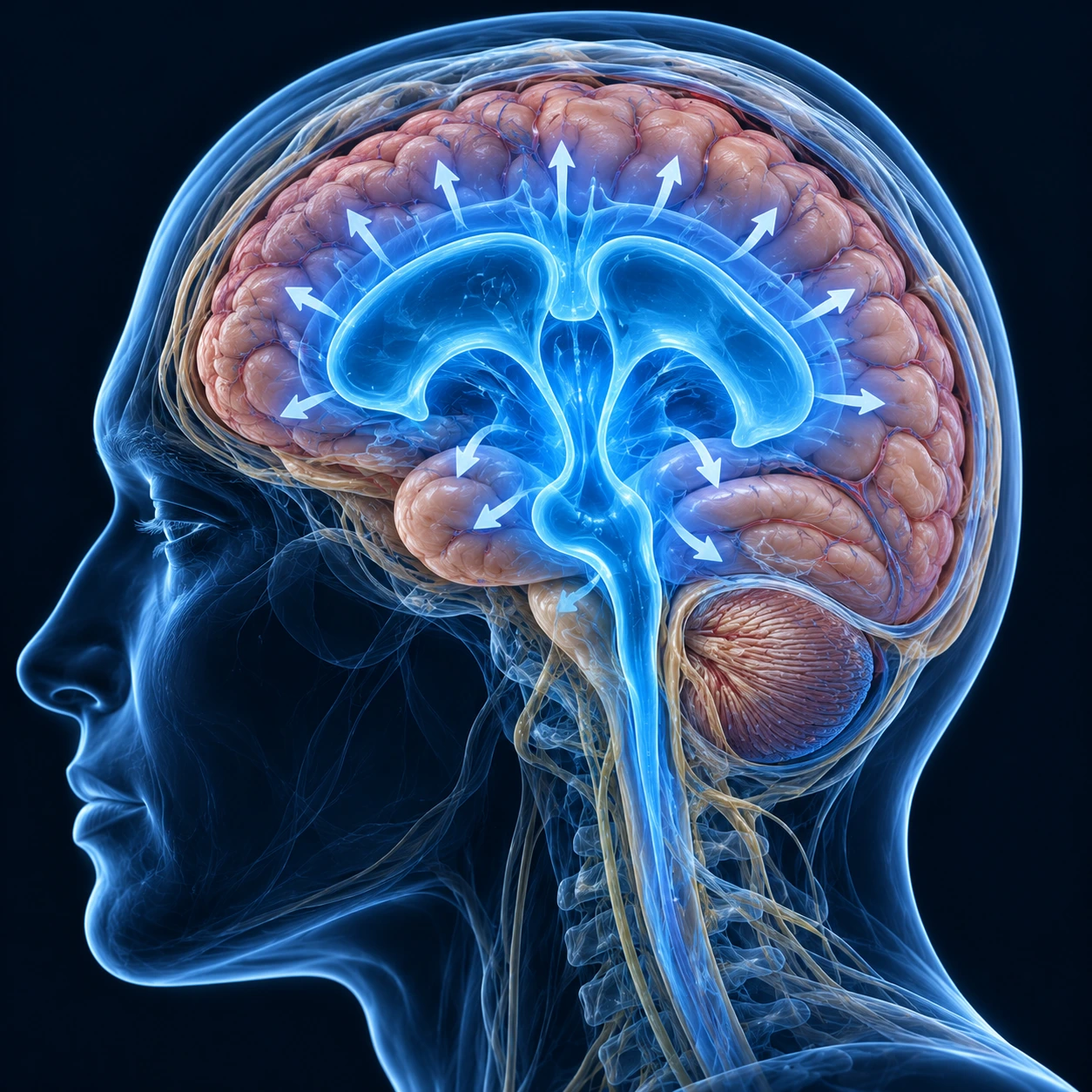
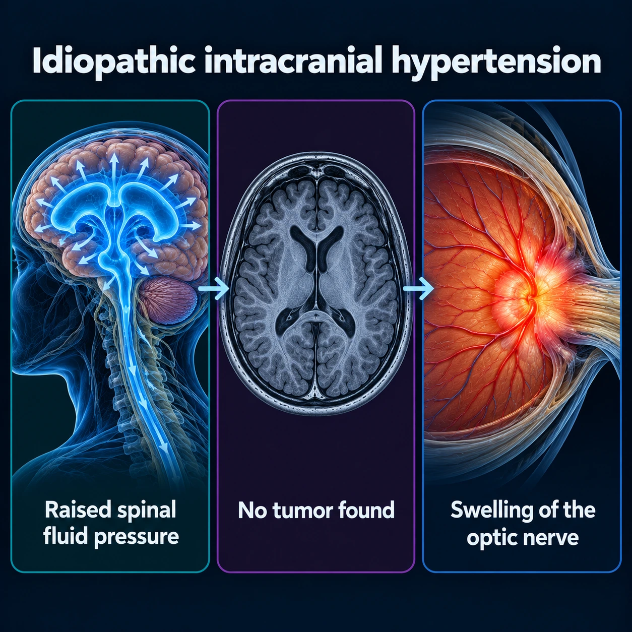
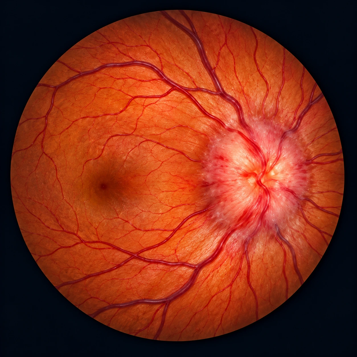
Table of Contents
Overview
Idiopathic intracranial hypertension (IIH), also called pseudotumor cerebri (PTC), is a condition of elevated cerebrospinal fluid (CSF) pressure exceeding 25 cmH₂O in adults (28 cmH₂O in children) in the absence of a structural lesion, hydrocephalus, or abnormal cerebrospinal fluid composition. The outdated term "benign intracranial hypertension" is no longer used because IIH is not benign — it can cause permanent vision loss from optic nerve damage. The condition mimics a brain tumor clinically (hence "pseudotumor cerebri") but neuroimaging reveals no mass lesion. IIH is classified as idiopathic only after secondary causes of elevated intracranial pressure have been excluded.
Demographics and Risk Factors
IIH predominantly affects women of reproductive age, with a female-to-male ratio of approximately 9:1. Obesity is present in 90% of cases, and the rising prevalence of IIH closely parallels the global obesity epidemic. The classic patient profile is an obese woman of childbearing age presenting with headache and visual disturbances.
Risk factors include:
- Obesity (body mass index (BMI) >30) — present in ~90% of idiopathic cases; weight gain preceding onset is common
- Female sex and reproductive age — hormonal factors implicated but not fully understood
- Recent significant weight gain (even without reaching obesity threshold)
- Medications reported in association: tetracyclines (doxycycline, minocycline), vitamin A/retinoids (isotretinoin for acne), oral contraceptives, corticosteroid withdrawal, recombinant growth hormone, excess levothyroxine, lithium. These are reported associations: the 2018 UK consensus guidelines find no clear evidence that such medications, the oral contraceptive included, are contraindicated in people with this condition. The consensus medication list names a levonorgestrel implant rather than the pill, and also names fluoroquinolones, tamoxifen and danazol.
- Men with IIH are less common but tend to have faster vision loss and more aggressive disease. The 2018 UK consensus guidelines call for more in-depth investigation for other causes in patients who are not female, not of childbearing age, or have a body mass index below 30.
In a UK study of 2,760 women with the condition, compared with women of the same age and body mass index, heart disease, heart failure and stroke together were about twice as common (adjusted hazard ratio 2.10). New cases more than tripled from 2005 to 2017, from 2.5 to 9.3 per 100,000 person-years.
Secondary Causes and Diagnosis of Exclusion
IIH is a diagnosis of exclusion. Secondary causes of elevated intracranial pressure are ruled out first.
Medications and toxins (reported associations): tetracyclines, vitamin A and retinoids (isotretinoin prescribed for acne), oral contraceptives, corticosteroid withdrawal, recombinant growth hormone, excess thyroid hormone replacement, lithium, anabolic steroids.
Venous outflow obstruction: cerebral venous sinus thrombosis (the 2018 UK consensus guidelines call CT or MR venography mandatory to exclude it, and treatment differs fundamentally), dural arteriovenous fistula.
Systemic conditions: hypothyroidism, hypoparathyroidism, iron deficiency anemia, systemic lupus erythematosus, chronic kidney disease, hypervitaminosis A.
Diagnostic workup for exclusion:
- Magnetic resonance imaging (MRI) brain with gadolinium — normal parenchyma, no mass, no hydrocephalus; may show empty sella turcica (an indirect sign of chronically elevated intracranial pressure remodeling the sella), bilateral optic nerve sheath distension, and flattening of the posterior globe
- MR venography — to exclude cerebral venous sinus thrombosis; transverse sinus stenosis is found in up to 90% of IIH, though whether this is cause or effect remains debated
- Lumbar puncture with opening pressure measurement (≥25 cmH₂O) and cerebrospinal fluid composition analysis — protein, glucose, and cell count are all normal in this condition
- Basic labs: complete blood count, comprehensive metabolic panel, thyroid-stimulating hormone, vitamin A level, and a thorough medication review
Modified Dandy Criteria
The Modified Dandy Criteria are used to confirm the diagnosis of IIH:
- Symptoms and/or signs of increased intracranial pressure: headache, papilledema, visual obscurations, diplopia from palsy of one or both sixth cranial nerves, pulsatile tinnitus
- Lumbar puncture opening pressure ≥25 cmH₂O with normal cerebrospinal fluid composition (protein, glucose, and cell count all normal)
- Normal neuroimaging: no intracranial mass, no hydrocephalus, no meningeal enhancement, no structural abnormality
- No other identifiable cause for elevated intracranial pressure
Note: IIH without papilledema is recognized as a subset where all criteria except papilledema are met. In a Utah series of 353 patients it made up 5.7%.
Clinical Features
Headache (present in ~94% of patients): Daily or near-daily headache, often worse in the morning, aggravated by Valsalva maneuvers (coughing, straining), and may worsen when supine. The character is throbbing, retro-orbital, or pressure-like and can mimic migraine or tension headache. Headache in the setting of papilledema is a recognized pointer to IIH.
Papilledema (bilateral optic disc swelling): The hallmark finding. Elevated cerebrospinal fluid pressure is transmitted along the optic nerve sheath, causing disc swelling visible on fundoscopy. If prolonged, papilledema leads to progressive optic nerve fiber loss and permanent visual field defects — the most feared complication of IIH.
Transient visual obscurations: Brief (seconds-long) episodes of graying, blackening, or blurring of vision provoked by changes in posture, Valsalva, or bending forward. These reflect transient optic nerve ischemia from pressure spikes.
Pulsatile tinnitus: A whooshing sound synchronous with the heartbeat, caused by turbulent venous flow through stenotic transverse sinuses under high pressure. This symptom is characteristic of IIH and is not common in most other headache disorders.
Diplopia from sixth cranial nerve palsy: Unilateral or bilateral sixth nerve palsy causing horizontal diplopia. This is a false localizing sign — the nerve is injured by elevated intracranial pressure regardless of where the pressure originates, not because there is a primary lesion of the nerve.
Visual field loss: Enlargement of the blind spot (enlarged physiological blind spot) is the earliest detectable change on perimetry. Progressive loss follows an inferior nasal pattern, then arcuate defect, and eventually tunnel vision. Visual field defects can become permanent if intracranial pressure is not controlled.
Cognitive symptoms: Some patients report brain fog, difficulty with concentration, and word-finding difficulties. These are underrecognized but can significantly impact quality of life.
Treatment
Weight loss (first-line for obese patients): The 2018 UK consensus guidelines call weight loss the only disease-modifying therapy in typical IIH. They state that the amount needed to put the disease into remission is not known, and that up to 15% of weight loss was required in one cohort. In the IIH Treatment Trial (IIHTT, 165 participants), everyone received a low-sodium weight-reduction diet, and the trial compared acetazolamide with placebo. Visual field (perimetric mean deviation) improved 1.43 dB with acetazolamide and 0.71 dB with placebo, a difference of 0.71 dB (95% CI 0 to 1.43 dB; P = .050), which its authors call modest, with clinical importance still to be determined. Weight fell 7.50 kg with acetazolamide and 3.45 kg with placebo.
Weight-loss surgery: A 2021 randomized trial of 66 women with a body mass index of 35 or more compared bariatric surgery with a community weight programme. Pressure in the head was 6.0 cm of cerebrospinal fluid lower at 12 months and 8.2 cm lower at 24 months after surgery, with weight 26.6 kg lower at 24 months and better quality of life.
Glucagon-like peptide-1 drugs: In a small 2023 placebo-controlled trial of 16 women, exenatide lowered pressure by 5.6 to 6.4 cm of cerebrospinal fluid. Its authors wrote that the data support moving to a larger phase 3 trial. A 2026 review of 8 studies and 13,243 patients linked this drug family to less optic nerve swelling and headache, but its authors call the evidence low-certainty because most of the studies were observational.
Acetazolamide: First-line pharmacological treatment. A carbonic anhydrase inhibitor that reduces cerebrospinal fluid production at the choroid plexus. Starting dose 500 mg twice daily, titrated to 2,000–4,000 mg/day as tolerated. The IIHTT (Wall et al., 2014) showed modest improvement in visual field, with improvement in papilledema grade and vision-related quality of life. Common side effects include paresthesias in hands and feet (nearly universal), dysgeusia (metallic taste of carbonated drinks), hypokalemia, kidney stone formation, and fatigue. Tolerance varies: in the trial 44% reached 4 g a day, and in another cohort 48% stopped because of side effects at a mean of 1.5 g. The label lists as contraindications hypersensitivity to acetazolamide (cross-sensitivity with sulfa drugs is possible), low blood sodium or potassium, marked kidney or liver disease, adrenal gland failure, hyperchloremic acidosis and cirrhosis.
Topiramate: An alternative or adjunct to acetazolamide. Also a carbonic anhydrase inhibitor with the added benefit of promoting weight loss. Useful when acetazolamide is not tolerated. The 2018 UK consensus guidelines record safety facts that matter because most patients are women of childbearing age: there is clear evidence of a higher rate of fetal abnormalities after topiramate use, and it can make the contraceptive pill and other hormonal contraceptives less effective. Its side effects include depression and slowed thinking.
Furosemide: The 2018 UK consensus guidelines state that the role of other diuretics such as furosemide is not certain, though some clinicians use them as alternative therapies.
Surgical options for vision-threatening IIH:
- Optic nerve sheath fenestration (ONSF): Surgical incisions made in the dura surrounding the optic nerve behind the globe, creating a pressure-release valve that protects the optic nerve from intracranial pressure transmission. Primarily prevents vision loss rather than treating headache. Indicated for rapid vision loss when medical treatment is insufficient or fails.
- Cerebrospinal fluid shunting (lumboperitoneal or ventriculoperitoneal shunt): Diverts cerebrospinal fluid to the peritoneal cavity to reduce intracranial pressure. Effective for both headache and vision protection but associated with high revision rates due to shunt malfunction and overshunting causing low-pressure headache. Used for severe refractory headache unresponsive to medication and vision loss.
- Transverse venous sinus stenting: An emerging endovascular procedure. Stenting stenotic transverse sinuses reduces the downstream venous pressure that contributes to elevated intracranial pressure. Promising results in select patients with demonstrable venous sinus stenosis and a measurable pressure gradient. A 2024 meta-analysis (36 studies, 1066 patients) reported improvement in tinnitus in 95%, optic nerve swelling in 89%, vision in 88% and headache in 79%, with treatment failure in 8.35% and complications in 5.35%, including subdural bleeding. The 2018 UK consensus guidelines add that narrowing next to the stent needs retreatment in about a third, and that the role of stenting is not yet established.
Repeat lumbar puncture: Provides temporary intracranial pressure reduction. Primarily used diagnostically or as a temporary bridge while awaiting surgical intervention. Not a long-term solution.
Vision Monitoring and Prognosis
Serial visual field testing with Humphrey automated perimetry (24-2 program) every 1–3 months during active disease is the standard follow-up. Optical coherence tomography (OCT) of the retinal nerve fiber layer (RNFL) provides objective measurement of optic nerve damage. Fundoscopy to grade papilledema is part of each visit.
With timely weight loss and medical treatment, most patients achieve intracranial pressure control and vision stabilization. Vision loss occurs in 10–25% of patients; severe vision loss in approximately 5%. Recurrence is common with weight regain. Chronic intractable headache persists in some patients even after intracranial pressure normalization. Pregnancy can trigger IIH exacerbation. The 2018 UK consensus guidelines state that it is difficult to make any safe recommendations on using acetazolamide during pregnancy and that its manufacturers do not recommend it, on the basis of birth defects seen in rodents.
Key Research Papers
- Wall M, et al. The idiopathic intracranial hypertension treatment trial: clinical profile at baseline. JAMA Neurol. 2014;71(6):693-701. — PubMed PMID: 24756302
- NORDIC Idiopathic Intracranial Hypertension Study Group Writing Committee, Wall M, McDermott MP, et al. Effect of acetazolamide on visual function in patients with idiopathic intracranial hypertension and mild visual loss: the idiopathic intracranial hypertension treatment trial. JAMA. 2014;311(16):1641-51. — PubMed PMID: 24756514
- Mollan SP, et al. Idiopathic intracranial hypertension: consensus guidelines on management. J Neurol Neurosurg Psychiatry. 2018;89(10):1088-1100. — PubMed PMID: 29903905
- Friedman DI, et al. Revised diagnostic criteria for the pseudotumor cerebri syndrome in adults and children. Neurology. 2013;81(13):1159-1165. — PubMed PMID: 23966248
- Markey KA, et al. Understanding idiopathic intracranial hypertension: mechanisms, management, and future directions. Lancet Neurol. 2016;15(1):78-91. — PubMed PMID: 26700907
- Celebisoy N, et al. Treatment of idiopathic intracranial hypertension: topiramate vs acetazolamide, an open-label study. Acta Neurol Scand. 2007;116(5):322-327. — PubMed PMID: 17922725
- Azzam AY, Mortezaei A, Morsy MM, et al. Venous sinus stenting for idiopathic intracranial hypertension: an updated meta-analysis. J Neurol Sci. 2024;459:122948. — PubMed PMID: 38457956
- Thurtell MJ, Wall M. Idiopathic intracranial hypertension (pseudotumor cerebri): recognition, treatment, and ongoing management. Curr Treat Options Neurol. 2013;15(1):1-12. — PubMed PMID: 23136035
- Rowe FJ, Sarkies NJ. The relationship between obesity and idiopathic intracranial hypertension. Int J Obes Relat Metab Disord. 1999;23(1):54-59. — PubMed PMID: 10094577
- Adderley NJ, Subramanian A, Nirantharakumar K, et al. Association between idiopathic intracranial hypertension and risk of cardiovascular diseases in women in the United Kingdom. JAMA Neurol. 2019;76(9):1088-1098. — PubMed PMID: 31282950
- Thambisetty M, et al. Fulminant idiopathic intracranial hypertension. Neurology. 2007;68(3):229-232. — PubMed PMID: 17224579
- McGeeney BE, Friedman DI. Pseudotumor cerebri pathophysiology. Headache. 2014;54(3):445-458. — PubMed PMID: 24433163
- Mollan SP, Mitchell JL, Ottridge RS, et al. Effectiveness of bariatric surgery vs community weight management intervention for the treatment of idiopathic intracranial hypertension: a randomized clinical trial. JAMA Neurol. 2021;78(6):678-686. — PubMed PMID: 33900360
- Mitchell JL, Lyons HS, Walker JK, et al. The effect of GLP-1RA exenatide on idiopathic intracranial hypertension: a randomized clinical trial. Brain. 2023;146(5):1821-1830. — PubMed PMID: 36907221
- Falcão L, Brenner LBO, Machado Magalhães PL, et al. Glucagon-like peptide-1 receptor agonists in idiopathic intracranial hypertension: a systematic review and meta-analysis. Headache. 2026;66(9):2117-2128. — PubMed PMID: 42619634
- Digre KB, Nakamoto BK, Warner JE, et al. A comparison of idiopathic intracranial hypertension with and without papilledema. Headache. 2009;49(2):185-93. — PubMed PMID: 19222592
Connections
- Neurology
- Migraine
- Normal Pressure Hydrocephalus
- Chiari Malformation
- Cerebral Venous Thrombosis
- Vitamin A (retinoid toxicity trigger)
- Epilepsy
- Multiple Sclerosis
- Headache — headache is the presenting symptom in ~94% of IIH patients, and separating it from migraine is the first diagnostic step.