Chiari Malformation
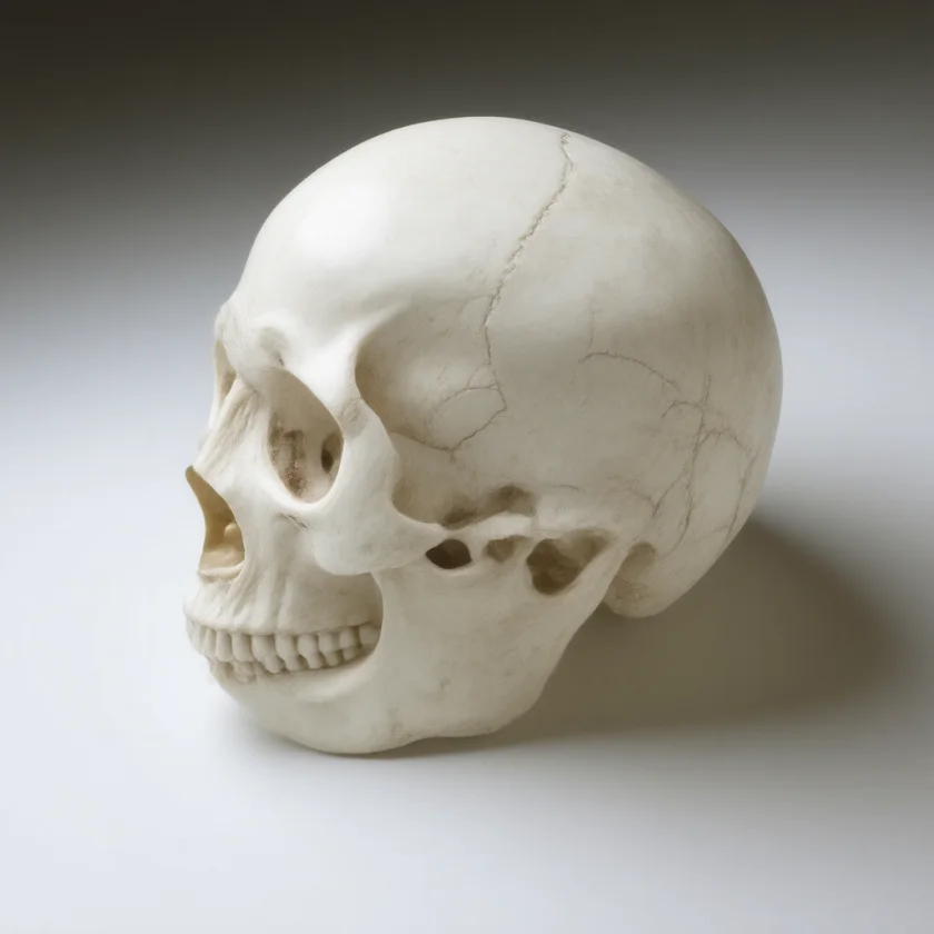

Overview and Classification
Chiari malformation (CM) is a structural defect characterized by herniation of the cerebellar tonsils (and in some types, the brainstem) through the foramen magnum into the cervical spinal canal. The foramen magnum is the opening at the base of the skull through which the brainstem passes into the spinal cord; when posterior fossa structures descend through it, they compress the brainstem, cerebellum, and cervical spinal cord while obstructing cerebrospinal fluid (CSF) flow.
Chiari malformations are classified into four types based on the structures involved and severity:
- Type I: Cerebellar tonsillar ectopia ≥5 mm below the foramen magnum; no brainstem herniation; the most common form, often presenting in adolescence or adulthood.
- Type II (Arnold-Chiari): Displacement of both the cerebellar vermis and brainstem through the foramen magnum; almost always associated with myelomeningocele (open spina bifida); diagnosed at birth.
- Type III: Rare; posterior fossa encephalocele containing herniated cerebellum and sometimes brainstem through the occipital bone; severe neurological consequences.
- Type IV: Extremely rare; absence of posterior fossa structures (cerebellar aplasia/hypoplasia); not technically herniation but classified with Chiari historically.
The eponym honors Hans Chiari, an Austrian pathologist who described the condition in 1891. Arnold-Chiari specifically refers to Type II, acknowledging Julius Arnold's early contributions.
Type I: The Adult Form
Chiari Type I is the most commonly diagnosed form. The cerebellar tonsils extend at least 5 mm below the McRae line (a reference line across the foramen magnum on magnetic resonance imaging, MRI). Many individuals with mild tonsillar ectopia (3–5 mm) remain asymptomatic throughout life; tonsillar descent greater than 5 mm is more likely to obstruct cerebrospinal fluid flow and produce symptoms.
Type I has no associated brain malformation and is often discovered incidentally on magnetic resonance imaging obtained for unrelated reasons (headache workup, trauma). It can manifest at any age but most commonly comes to clinical attention in the second through fourth decades. The exact cause is debated: a relatively small posterior fossa (insufficient space for the cerebellum) is the leading structural hypothesis, causing the tonsils to be “pushed” downward. Secondary Chiari can occur from intracranial hypotension (low cerebrospinal fluid pressure pulling the brain downward) — this distinction is clinically important because treatment differs.
In a cohort of 364 people with symptomatic Chiari I, 275 were women, the average age at onset was 24.9 years, 89 (24%) named an injury as the trigger and 43 (12%) reported a family history of Chiari I or syringomyelia. Measurements in 50 of these patients showed that the back of the skull (posterior cranial fossa) held a mean 13.4 ml less total volume than in matched controls, with normal brain volume, which supports the small-space explanation.
Type II: Arnold-Chiari and Myelomeningocele
Chiari Type II almost universally accompanies myelomeningocele, the most severe form of spina bifida in which the spinal cord is exposed through an opening in the lower back. The association is so strong that virtually all patients with myelomeningocele have radiographic evidence of Type II Chiari.
In Type II, both the cerebellar vermis and the brainstem (medulla) descend through the foramen magnum. This produces more severe brainstem dysfunction than Type I, including:
- Lower cranial nerve palsies (cranial nerves IX–XII): dysphagia, aspiration, hoarse cry, vocal cord paralysis in neonates.
- Respiratory dysfunction: apnea, stridor (life-threatening in severe cases).
- Hydrocephalus: nearly universal; caused by aqueductal stenosis and impaired cerebrospinal fluid flow; managed with ventriculoperitoneal shunt or endoscopic third ventriculostomy.
In utero surgical repair of myelomeningocele (Management of Myelomeningocele Study, MOMS trial, 2011) reduced the need for ventriculoperitoneal shunting (a tube draining brain fluid to the abdomen) and improved motor outcomes compared to postnatal repair: shunts were placed in 40% vs. 82% by 12 months. In utero repair does not cure the Chiari malformation but reduces its severity.
The surgery before birth also has costs: in the same trial it was associated with a higher risk of preterm delivery and of the uterus opening at delivery (uterine dehiscence). A follow-up of the full cohort of 183 found independent walking at 30 months in 44.8% vs. 23.9%, with no effect on cognitive development at that age. School-age brain scans published in 2023 (66 prenatal, 63 postnatal repairs) still showed lower rates of hindbrain herniation after prenatal repair.
Rare Types (III and IV)
Type III Chiari is characterized by a posterior fossa encephalocele — a sac protruding through a defect in the occipital bone (occiput) or upper cervical vertebrae containing cerebellar tissue and sometimes brainstem. It is extremely rare, associated with severe neurological impairment, and carries high mortality. Surgical repair is complex with limited neurological recovery.
Type IV (cerebellar aplasia or hypoplasia) is the rarest category. The cerebellum fails to develop, leaving the posterior fossa largely empty. It is not truly a herniation disorder and is often discussed separately in current classifications.
Clinical Features
Symptoms in Chiari Type I arise from compression at the foramen magnum and cerebrospinal fluid flow obstruction.
Cough Headache — The Classic Presentation
A brief but severe headache in the suboccipital region (base of skull, back of head) provoked by Valsalva maneuvers — coughing, sneezing, laughing, straining with bowel movements. The pain typically lasts seconds to a minute and radiates to the occiput, neck, or arms. Valsalva-triggered suboccipital headache is a recognized reason for brain magnetic resonance imaging with attention to the posterior fossa and foramen magnum.
Neck and Arm Symptoms
Neck pain, occipital neuralgia, paresthesias (tingling, numbness) radiating into the upper extremities, and upper extremity weakness. These can result from direct cervicomedullary compression or from syringomyelia (see below).
Lower Cranial Nerve Symptoms
Dysphagia (difficulty swallowing), dysarthria (slurred speech), hoarseness from vagal or hypoglossal nerve involvement; vocal cord paralysis in severe cases.
Ataxia and Coordination Difficulty
Cerebellar compression produces gait ataxia, incoordination of upper extremities, and balance problems.
Downbeat Nystagmus
The eyes drift upward and then beat downward. Downbeat nystagmus is the hallmark of foramen magnum pathology (Chiari, craniocervical instability, Arnold-Chiari) until proven otherwise. It results from disruption of cerebellar floccular pathways at the foramen magnum.
Central Sleep Apnea
Brainstem compression at the respiratory centers (pre-Bötzinger complex in medulla) can cause central sleep apnea — cessation of respiratory effort during sleep, distinct from obstructive sleep apnea. Chiari is one of the recognized causes of unexplained central sleep apnea.
Facial Pain and Sensory Disturbances
Trigeminal distribution numbness or pain from trigeminal nucleus compression.
Syringomyelia
Syringomyelia — a fluid-filled cavity (syrinx) within the spinal cord parenchyma — develops in 20–65% of patients with Chiari Type I. The figure depends on who is studied: 65% in a cohort of 364 people with symptoms, while a population-based group of 51 children had 12% at diagnosis and no new cavities during follow-up. The pathophysiology involves obstruction of pulsatile cerebrospinal fluid flow at the foramen magnum with each cardiac systole; this is believed to force cerebrospinal fluid into the spinal cord’s central canal and eventually the parenchyma, creating an expanding cavity.
The syrinx typically develops in the cervical cord and expands over months to years, damaging anterior horn cells (lower motor neurons) and crossing spinothalamic fibers (pain and temperature sensation).
Clinical presentation of syringomyelia follows a characteristic pattern:
- Cape distribution sensory loss: bilateral loss of pain and temperature sensation across the shoulders, upper chest, and upper back (like a cape or shawl draped over the shoulders); this is dissociated sensory loss — pain/temperature affected but vibration and proprioception preserved, because the anterior white commissure (where spinothalamic fibers cross) is damaged by the syrinx while the dorsal columns (vibration/proprioception) are spared.
- Upper extremity weakness and atrophy: from anterior horn cell damage.
- Scoliosis: especially in children; syrinx-associated scoliosis (not idiopathic) is identified with spinal magnetic resonance imaging, the test that shows an underlying Chiari/syrinx.
- Loss of deep tendon reflexes in arms (lower motor neuron pattern), hyperreflexia and spasticity in legs (upper motor neuron if cord compression).
Once Chiari decompression surgery restores cerebrospinal fluid flow at the foramen magnum, the syrinx typically collapses over weeks to months without direct surgical intervention on the syrinx itself.
Associated Conditions
Connective Tissue Disorders
Hypermobile Ehlers-Danlos syndrome (hEDS) and other connective tissue disorders predispose to craniocervical instability — excessive movement at the atlantoaxial or atlanto-occipital joints. Craniocervical instability can produce Chiari-like symptoms even without cerebellar tonsillar descent exceeding 5 mm, and some practitioners hold that it can produce true Chiari by pulling the brain downward. A 2017 review of the Ehlers-Danlos syndromes reports Chiari I as a comorbid condition that may be complicated by craniocervical instability, and calls for stronger evidence. This is an important and sometimes under-recognized overlap.
Postural Orthostatic Tachycardia Syndrome
Many patients with hEDS have both postural orthostatic tachycardia syndrome and craniocervical instability; some have true Chiari. The hEDS–postural orthostatic tachycardia syndrome–Chiari overlap is being increasingly recognized.
Tethered Spinal Cord
In Type II Chiari, tethered cord (where the spinal cord is anchored and cannot rise normally with growth) often coexists. Surgical release improves some neurological function.
Scoliosis
Syringomyelia-associated scoliosis in children with Chiari I is a different condition from idiopathic adolescent scoliosis, and treatment differs; examination for Chiari/syrinx is how the two are told apart.
Diagnosis
Magnetic resonance imaging is the gold-standard diagnostic modality:
- Magnetic resonance imaging brain (sagittal T1): measures tonsillar position relative to McRae line; ≥5 mm below = diagnostic threshold for Type I, but clinical context matters (3–5 mm with symptoms is significant).
- Magnetic resonance imaging complete spine: to detect syrinx (the entire spinal cord is imaged), tethered cord.
- Phase-contrast magnetic resonance imaging (cerebrospinal fluid flow study): dynamic imaging of cerebrospinal fluid pulsatility at the foramen magnum. In a retrospective study of 44 children, abnormal flow behind the brainstem alone was not linked to symptom recurrence after surgery (p = 0.10), and recurrence did not differ with the degree of tonsil descent (p = 0.55); abnormal flow both in front of and behind the brainstem was linked to fewer recurrences, and the authors concluded this imaging may be a useful tool for choosing surgical candidates.
- Flexion-extension magnetic resonance imaging or upright magnetic resonance imaging: to evaluate craniocervical instability, especially in connective tissue disorder patients where instability may be dynamic (not visible on standard supine magnetic resonance imaging).
Neurophysiological studies: somatosensory evoked potentials, brainstem auditory evoked potentials — can document subclinical brainstem or cord dysfunction.
Sleep study (polysomnography): to characterize sleep apnea (central vs. obstructive) and quantify respiratory events.
Treatment
Asymptomatic Type I (Incidental Finding)
The usual practice described is no surgery, with serial magnetic resonance imaging every 3–5 years (earlier if new symptoms develop) and teaching about warning symptoms.
A 2017 systematic review found that most people with no symptoms stayed that way (93.3%), even with syringomyelia. Among people with symptoms who did not have surgery, 27%–47% improved after 15 months; headaches and nausea often improved, while balance problems (ataxia) and sensory disturbance tended not to. A 2025 World Federation of Neurosurgical Societies Spine Committee consensus reports conservative management as appropriate for people with no or mild symptoms.
Symptomatic Type I / Type I with Syringomyelia
The primary surgical treatment is posterior fossa decompression (PFD):
- Suboccipital craniectomy: removal of a portion of the occipital bone to enlarge the foramen magnum.
- C1 laminectomy: removal of the posterior arch of the first cervical vertebra.
- Duraplasty: opening the dura and patching it with autologous or synthetic graft to expand the dural sac volume, restoring cerebrospinal fluid flow.
Outcomes of posterior fossa decompression: Cough headache resolves in 80–90% of patients. Syrinx collapses or stabilizes in ~70% within months. Neurological deficits (weakness, sensory loss) improve variably — duration of deficits before surgery is the strongest predictor of recovery. Long-term data show durable outcomes in most patients.
A 2025 meta-analysis of 20 studies and 3,063 patients found that decompression with duraplasty reduced syrinx width by a mean of 2.46 mm and syrinx length by 1.68 vertebral segments.
In a consortium of 692 children with Chiari I and a syrinx, decompression with duraplasty gave more headache relief (89.6% vs. 80.8%) and fewer repeat operations (8.3% vs. 17.9%) than bone removal alone, but more complications in the first 6 months (24.3% vs. 13.7%). This applies to children and was not a randomized comparison.
A randomized trial published in 2026 enrolled 162 people aged 21 or younger with Chiari I and a syrinx at 38 centers, with each center assigned to one operation. Complications within 6 months occurred in 14% with duraplasty and 6% without (not a significant difference, P = 0.11). At 24 months, clinical improvement was 58% vs. 46%, the syrinx had shrunk by a mean of 3.08 mm vs. 1.22 mm, and repeat decompression was 3% vs. 14%; quality-of-life changes were similar. The authors state that larger trials are needed.
Type II (Arnold-Chiari)
Management is multifaceted:
- In utero myelomeningocele repair (MOMS trial): reduces but does not eliminate need for ventriculoperitoneal shunting; improves motor outcomes.
- Postnatal myelomeningocele closure if not done prenatally.
- Ventriculoperitoneal shunt for hydrocephalus (nearly universal).
- Posterior fossa decompression if brainstem compression is symptomatic (apnea, dysphagia, vocal cord paralysis).
Craniocervical Stabilization
For patients with documented craniocervical instability (especially in hEDS), cervical fusion (occipital-cervical or atlantoaxial) may be necessary to provide stability and stop dynamic compression.
Post-Operative Monitoring
Magnetic resonance imaging at 3–6 months for syrinx regression, then annually for 2 years, then as needed.
Key Research Papers
- Milhorat TH, et al. Chiari I malformation redefined: clinical and radiographic findings for 364 symptomatic patients. Neurosurgery. 1999;44(5):1005–1017. — PubMed PMID: 10232534
- Aitken LA, et al. Chiari type I malformation in a pediatric population. Pediatr Neurol. 2009;40(6):449–454. — PubMed PMID: 19433279
- Pindrik J, Johnston JM Jr. Clinical presentation of Chiari I malformation and syringomyelia in children. Neurosurg Clin N Am. 2015;26(4):509–514. — PubMed PMID: 26408059
- Oakes WJ. Chiari malformations, hydromyelia, syringomyelia. In: Wilkins RH, Rengachary SS, eds. Neurosurgery. McGraw-Hill; 1996. — Search PubMed
- Alperin N, et al. Magnetic resonance imaging-based measurements of cerebrospinal fluid and blood flow as indicators of intracranial compliance in patients with Chiari malformation. J Neurosurg. 2005;103(1):46–52. — PubMed PMID: 16121972
- Adzick NS, et al. A randomized trial of prenatal versus postnatal repair of myelomeningocele. N Engl J Med. 2011;364(11):993–1004. — PubMed PMID: 21306277
- Henderson FC Sr, et al. Neurological and spinal manifestations of the Ehlers-Danlos syndromes. Am J Med Genet C Semin Med Genet. 2017;175(1):195–211. — PubMed PMID: 28220607
- Meadows J, et al. Asymptomatic Chiari Type I malformations identified on magnetic resonance imaging. J Neurosurg. 2000;92(6):920–926. — PubMed PMID: 10839250
- McGirt MJ, et al. Correlation of hindbrain CSF flow and outcome after surgical decompression for Chiari I malformation. Childs Nerv Syst. 2008;24(7):833–840. — PubMed PMID: 18205006
- Langridge B, et al. Chiari malformation type 1: a systematic review of natural history and conservative management. World Neurosurg. 2017;104:213–219. — PubMed PMID: 28435116
- Costa F, et al. Chiari malformation: diagnosis, classifications, natural history, and conservative management. World Federation of Neurosurgical Societies Spine Committee recommendations. Spine (Phila Pa 1976). 2025;50(11):767–778. — PubMed PMID: 39925305
- Limbrick DD Jr, Shannon CN, Bayman EO, et al. Decompression with or without Duraplasty for Chiari I and Syringomyelia. N Engl J Med. 2026;394(20):2015–2025. — PubMed PMID: 42202320
- Akbari SHA, et al. Complications and outcomes of posterior fossa decompression with duraplasty versus without duraplasty for pediatric patients with Chiari malformation type I and syringomyelia: a study from the Park-Reeves Syringomyelia Research Consortium. J Neurosurg Pediatr. 2022;30(1):39–51. — PubMed PMID: 35426814
- Prasad AS, et al. Efficacy of neurosurgical intervention in syrinx resolution in patients presenting with Chiari malformation type I and syringomyelia: a systematic review and radiological meta-analysis. Neurosurg Rev. 2025;48(1):724. — PubMed PMID: 41111088
- Farmer DL, et al. The Management of Myelomeningocele Study: full cohort 30-month pediatric outcomes. Am J Obstet Gynecol. 2018;218(2):256.e1–256.e13. — PubMed PMID: 29246577
- George E, et al. Long-term imaging follow-up from the Management of Myelomeningocele Study. AJNR Am J Neuroradiol. 2023;44(7):861–866. — PubMed PMID: 37385677