CADASIL — Cerebral Autosomal Dominant Arteriopathy with Subcortical Infarcts and Leukoencephalopathy
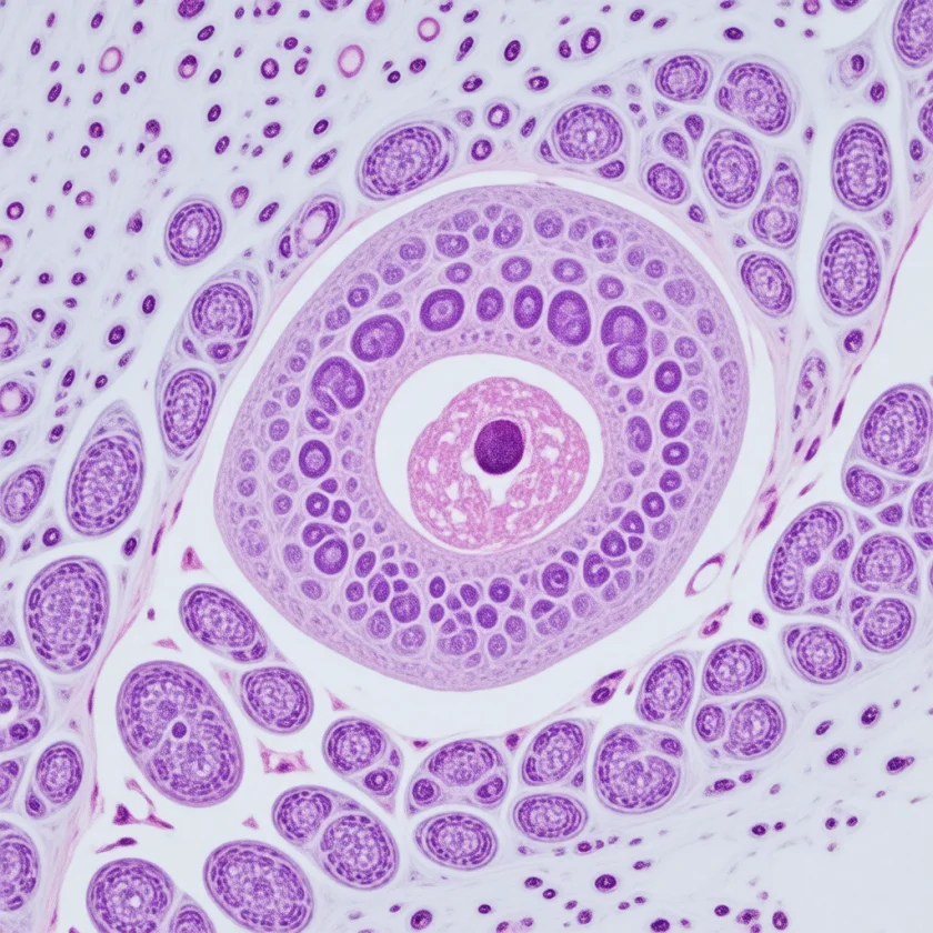
CADASIL is the most common inherited cause of stroke and vascular dementia in adults. It is caused by mutations in the NOTCH3 gene that damage small blood vessels deep in the brain, producing a characteristic pattern of white matter injury, recurrent strokes, and progressive decline — all beginning decades before old age. Yet because it is rare and its early symptoms (migraine with aura, mood disturbances) overlap with everyday conditions, the average diagnosis takes many years from first symptoms. If you or a family member has unexplained early strokes, a family history of dementia or stroke before age 60, and migraines with aura, this page explains the biology, what doctors look for, what the magnetic resonance imaging (MRI) will show, and what can be done to reduce harm and protect other family members.
Table of Contents
- CADASIL and Its Cause
- The NOTCH3 Mutation and Vascular Pathology
- Clinical Features and Disease Progression
- Magnetic Resonance Imaging Findings — The Diagnostic Fingerprint
- How CADASIL Is Diagnosed
- Management and Treatment
- Inheritance, Genetic Counseling, and Family Testing
- Prognosis and Long-Term Outlook
- Key Research Papers
- Connections
- Featured Videos
CADASIL and Its Cause
CADASIL stands for Cerebral Autosomal Dominant Arteriopathy with Subcortical Infarcts and Leukoencephalopathy. The name contains the entire disease summary: it is a disorder of small brain arteries (arteriopathy) caused by a dominant gene mutation (one defective copy is enough to cause disease) that produces small, deep brain infarcts (subcortical infarcts) and widespread damage to the brain's white matter (leukoencephalopathy). The condition was first fully characterized in 1993 by Tournier-Lasserve and colleagues, who mapped it to chromosome 19, and the causative gene, NOTCH3, was identified in 1996.
CADASIL affects small and medium penetrating arteries throughout the body, but the brain is by far the most vulnerable organ because it depends so critically on a continuous, precisely regulated blood supply. The arteries most affected are those that supply the deep white matter — the vast network of insulated nerve-fiber tracts that connect different regions of the cerebral cortex with each other and with subcortical structures. When these small arteries fail, the white matter they supply undergoes a slow ischemic injury over years and decades.
The estimated prevalence is approximately 2–5 per 100,000, but many cases go undiagnosed, and some studies suggest the true prevalence may be several times higher. The disease is found in all ethnic groups worldwide. Because it is autosomal dominant, each child of an affected parent has a 50% chance of inheriting the mutation. Not every carrier develops the full disease: a 2016 study of 60,706 people's gene data found disease-type NOTCH3 changes in 3.4 of every 1,000 people — about 100 times more than the known number of CADASIL cases would predict — which suggests many such changes cause a much milder illness that may go unrecognized.
The NOTCH3 Mutation and Vascular Pathology
The NOTCH3 gene encodes a large cell-surface receptor protein involved in regulating vascular smooth muscle cell differentiation and survival. The NOTCH3 receptor has a series of EGF-like (epidermal growth factor-like) repeating domains in its extracellular portion. CADASIL mutations — of which more than 300 distinct mutations have been identified — all share one key feature: they add or remove a cysteine residue in one of these EGF-like domains (specifically, in EGF-like domains 1 through 34).
The addition or subtraction of a cysteine disrupts the precise disulfide bond pairing that holds each EGF-like domain in its correct three-dimensional shape. The resulting misfolded NOTCH3 extracellular domain (ECD) accumulates on the surface of vascular smooth muscle cells and pericytes instead of being properly processed. Over time, this NOTCH3 ECD deposits in the walls of small arteries throughout the body — it can even be detected in blood vessels of the skin, muscle, and peripheral nerves, which is why a skin biopsy can sometimes diagnose CADASIL.
The downstream consequences in the brain's small arteries include:
- Vascular smooth muscle cell loss — the smooth muscle cells that control vessel diameter and blood flow regulation progressively degenerate, leaving the vessel wall unable to autoregulate blood supply properly.
- Thickening and fibrosis of vessel walls — the walls of affected arteries become thickened, fibrotic, and tortuous. The lumen (the channel through which blood flows) narrows.
- Granular osmiophilic material (GOM) — electron microscopy of affected arteries reveals dense deposits of abnormal material (called GOM deposits) adjacent to smooth muscle cells. This finding is considered pathognomonic of CADASIL and can be identified in skin biopsies.
- Loss of cerebrovascular reactivity — the brain normally dilates blood vessels in response to increased neural activity and constricts them when activity is lower. In CADASIL, this neurovascular coupling is impaired, making the brain vulnerable to even transient blood pressure drops and to physiological stressors like dehydration, fever, or low oxygen.
The net result is a progressive reduction in blood flow to the brain's white matter, producing areas of ischemia, small complete infarcts, and eventual widespread white matter signal change on magnetic resonance imaging — all while the brain's larger arteries and the cortex itself are largely spared.
Clinical Features and Disease Progression
CADASIL follows a characteristic clinical trajectory across the lifespan, though the timing and severity vary considerably even within the same family carrying the same mutation. The disease unfolds in overlapping phases.
A 1998 German study of 102 people with biopsy-proven CADASIL from 29 families gives a picture in plain numbers: 71% had repeated strokes or mini-strokes (TIAs), starting at an average age of 46.1; 38% had migraine, with aura in 87% of them; 28% had dementia; and 55% of those over 60 could not walk without help — yet 14% of the over-60s had no disability at all. The median survival was 64 years for men and 69 years for women, and the course varied markedly even within the same family.
Migraine with Aura (Age 20s–30s — Often the First Symptom)
Migraine with aura is the earliest and most common initial manifestation, affecting approximately 35% of CADASIL patients and typically beginning in the 20s or 30s. The auras can be visual (zig-zag fortification spectra, scotomata), sensory, aphasic, or motor. Some patients experience unusually severe, prolonged, or treatment-resistant migraines. Importantly, migraine with aura is also common in the general population — so its presence alone does not suggest CADASIL. However, in someone with a family history of early stroke or dementia, early-onset migraine with aura is one of the reasons clinicians consider a CADASIL evaluation.
Transient Ischemic Attacks and Subcortical Strokes (Age 40s–50s)
The hallmark of CADASIL is recurrent ischemic events — TIAs and subcortical lacunar infarcts — in the absence of traditional cardiovascular risk factors. The average age of first stroke or transient ischemic attack (TIA) is around 45 years. Because the affected vessels are small penetrating arteries rather than large arteries, the strokes produce classical lacunar syndromes:
- Pure motor hemiparesis — weakness of one side without sensory loss or language disturbance, from a small infarct in the internal capsule or pons.
- Pure sensory stroke — unilateral sensory loss, from a thalamic or internal capsule lacune.
- Dysarthria-clumsy hand syndrome — slurred speech plus clumsiness of one hand, from a small pontine or internal capsule infarct.
- Ataxic hemiparesis — limb weakness combined with ipsilateral cerebellar-like clumsiness.
These events accumulate over years, and while each individual infarct may produce only mild or transient symptoms, their cumulative effect produces steadily worsening neurological function.
Cognitive Impairment and Executive Dysfunction (Age 50s–60s)
Cognitive decline in CADASIL is predominantly a subcortical pattern, reflecting white matter disconnection rather than cortical neuron death. The most prominent early deficits are in executive function — the ability to plan, organize, multitask, and shift attention — and in processing speed. Memory is affected but tends to be less severely impaired than in Alzheimer's disease, and the pattern is one of retrieval difficulty (information encoded but hard to access) rather than complete failure to store new memories.
Neuropsychiatric symptoms are common and include depression (present in up to 50% of patients), apathy (distinct from depression — a loss of motivation and initiation without sadness), and in later stages, emotional lability and pseudobulbar affect (involuntary laughing or crying disproportionate to emotional state).
Dementia and Pseudobulbar Palsy (Advanced Disease)
In the advanced stage, typically reaching in the 60s though highly variable, the cumulative white matter injury and lacunar strokes produce frank dementia. Pseudobulbar palsy — difficulty swallowing (dysphagia), dysarthria, drooling, and exaggerated jaw jerk reflex, caused by bilateral injury to the corticobulbar tracts — appears in many patients with advanced disease. Gait disturbance becomes prominent as subcortical motor pathways are disrupted; patients develop a slow, shuffling gait with small steps (marche à petit pas) that resembles Parkinson's disease but does not respond to dopaminergic therapy. Urinary incontinence is common. Dependency and eventual immobility follow.
Magnetic Resonance Imaging Findings — The Diagnostic Fingerprint
Brain magnetic resonance imaging in CADASIL has a distinctive and highly characteristic appearance that raises strong suspicion for the diagnosis even before genetic testing. The abnormalities are best seen on FLAIR (fluid-attenuated inversion recovery) and T2-weighted sequences.
White Matter Hyperintensities
Extensive, confluent areas of bright signal on FLAIR/T2 sequences in the deep white matter are the most obvious finding. Unlike age-related white matter changes (which are patchy and periventricular in early stages), CADASIL white matter disease is more extensive and involves specific regions that are highly predictive:
Anterior Temporal Pole — The Key Diagnostic Clue
Involvement of the anterior temporal poles (the tips of the temporal lobes) is the single most diagnostically useful magnetic resonance imaging finding in CADASIL. White matter signal change in this region is present in the vast majority of CADASIL patients and is unusual in other causes of white matter disease — including hypertensive leukoencephalopathy, multiple sclerosis, and Binswanger disease. In a 2001 study it was found in 19 patients with CADASIL and in none with ordinary age- and blood-pressure-related white matter disease, and its presence in a patient with suspected white matter disease is a common reason for CADASIL genetic testing.
External Capsule Involvement
Signal change in the external capsule (a white matter structure lateral to the basal ganglia, separate from the internal capsule) is also characteristic of CADASIL. Combined anterior temporal pole and external capsule involvement has high sensitivity and specificity for CADASIL among all hereditary small vessel diseases.
Lacunar Infarcts
Multiple small, punched-out areas of completed infarction (dark on T1, bright on T2/FLAIR, with DWI restriction in acute phase) are seen in the basal ganglia, thalamus, internal capsule, brainstem, and centrum semiovale. Their number correlates with clinical disability and cognitive impairment.
Microbleeds
Cerebral microbleeds — tiny foci of old hemorrhage best seen on susceptibility-weighted imaging (SWI) or gradient echo sequences — are found in approximately 25–38% of CADASIL patients (31% of symptomatic carriers in a 2001 Dutch study) and increase in number over time. Their presence indicates severe small vessel disease and is a marker of higher risk for symptomatic intracerebral hemorrhage. This has important implications for antiplatelet and anticoagulant decisions.
How CADASIL Is Diagnosed
Diagnosis rests on a combination of clinical history, family history, magnetic resonance imaging findings, and genetic confirmation. The diagnostic pathway typically proceeds as follows:
Clinical Suspicion
Clinicians consider CADASIL in a patient with: recurrent subcortical ischemic strokes or TIAs before age 60 without traditional risk factors (no hypertension, no atrial fibrillation, no carotid disease, no diabetes, non-smoker); magnetic resonance imaging showing white matter disease disproportionate to age or risk factors — especially with anterior temporal pole or external capsule involvement; a family history of similar strokes, dementia, or premature neurological decline; or migraine with aura in conjunction with any of the above.
Genetic Testing — NOTCH3 Sequencing
NOTCH3 gene sequencing is the gold-standard diagnostic test. Most diagnostic laboratories sequence the entire coding region or specifically screen exons 2–24, which encode the EGF-like domains where pathogenic cysteine mutations cluster. A cysteine-altering missense mutation in an EGF-like domain is considered diagnostic. Pathogenic variants can also be found in exons outside this range, so comprehensive sequencing is preferred. Each first-degree relative of a confirmed case has a 50% chance of carrying the variant; because some variants cause mild or no disease, a positive test does not by itself predict how severe the illness will be (see Inheritance, below).
Skin Biopsy
Before genetic testing became widely available, skin biopsy with electron microscopy looking for granular osmiophilic material (GOM) deposits adjacent to vascular smooth muscle cells was the primary diagnostic method. It remains a useful confirmatory test — particularly in atypical cases or where genetic testing is unavailable — but its sensitivity depends heavily on biopsy technique and laboratory experience. An NOTCH3 immunostain on the biopsy can detect accumulated NOTCH3 extracellular domain even when GOM is not conclusive.
Neuropsychological Testing
Formal neuropsychological evaluation characterizes the pattern and severity of cognitive impairment, documents baseline function, tracks progression, and guides decisions about driving, work capacity, and daily function. A subcortical cognitive profile (executive dysfunction + processing speed >> memory) is expected and is documented by formal testing.
Differential Diagnosis
Other conditions that can mimic CADASIL include: multiple sclerosis (periventricular white matter lesions; younger women; optic neuritis; relapsing-remitting course; anti-MOG or anti-AQP4 antibodies); hypertensive leukoencephalopathy (confluent white matter change, but posterior predominance and typically spares anterior temporal poles); cerebral amyloid angiopathy (cortical/subcortical microbleeds + lobar hemorrhages; older patients; amyloid-PET positive); MELAS (mitochondrial encephalomyopathy, lactic acidosis, stroke-like episodes; young patients; elevated lactate; ragged red fibers; stroke-like episodes in cortex, not subcortical; maternal inheritance); Fabry disease (lysosomal storage disorder; alpha-galactosidase deficiency; early strokes + posterior circulation; acroparesthesias + angiokeratomas; elevated lyso-Gb3).
Management and Treatment
There is currently no disease-modifying treatment that slows or reverses the vascular degeneration caused by NOTCH3 mutations. Management focuses on stroke prevention, symptom treatment, risk factor modification, and preserving quality of life. This is an area of active research, with approaches aimed at the NOTCH3 protein itself so far tested in laboratory animals.
Antiplatelet Therapy for Stroke Prevention
Aspirin (75–100 mg daily) is the most widely used antiplatelet agent in CADASIL for prevention of recurrent ischemic events, by analogy with its use in other ischemic stroke syndromes. However, the evidence base specific to CADASIL is limited — it has not been tested in randomized controlled trials in this specific disease. Given the high rate of microbleeds in CADASIL (25–38%), which signals risk for intracerebral hemorrhage, the risk-benefit balance of antiplatelet therapy is weighed individually. Patients with numerous microbleeds may be at higher hemorrhagic risk, and decisions about aspirin take into account the microbleed burden on magnetic resonance imaging. Dual antiplatelet therapy and anticoagulation are generally avoided unless there is a coexisting cardiac indication, because they amplify hemorrhagic risk without established additional benefit in CADASIL-specific ischemia.
Oral Contraceptives and Smoking in CADASIL
Combined oral contraceptive pills (estrogen + progestogen) substantially increase stroke risk in women who already carry a genetic predisposition to small vessel disease, and they are contraindicated in CADASIL. Progestogen-only methods and non-hormonal contraception are safer. Smoking accelerates vascular damage. These are among the highest-yield modifiable exposures in CADASIL management.
Blood Pressure Control
Although classic CADASIL does not require hypertension to progress (the arteriopathy is genetically driven), hypertension worsens small vessel disease and accelerates white matter injury. The usual blood pressure aim in CADASIL is the normal range (<130/80 mmHg for most patients). Even patients without overt hypertension benefit from regular monitoring and aggressive lifestyle management.
The type of blood pressure medicine may matter. In the 2023 TREAT-SVDs trial, which compared three medicines for 4 weeks each, the brain blood vessels' ability to widen on demand (cerebrovascular reactivity) in people with CADASIL improved with amlodipine or losartan compared with atenolol; in people with ordinary small vessel disease there was no difference. Only 17 people with CADASIL were in the main analysis, and whether this changes strokes or memory is not yet known.
Migraine Management
Migraine treatment in CADASIL requires caution. Triptans and ergotamine derivatives cause vasoconstriction and are generally avoided or used with caution in CADASIL because vasoconstriction in already-vulnerable small brain arteries could theoretically trigger an ischemic event (though robust clinical evidence for harm from triptans specifically in CADASIL is limited). Non-vasoconstricting preventive therapies — such as topiramate, amitriptyline, and propranolol — are preferred. Acute attacks can be treated with nonsteroidal anti-inflammatory drugs (NSAIDs) and antiemetics.
Cognitive and Psychiatric Symptom Management
Depression and apathy are common and treatable. selective serotonin reuptake inhibitors (SSRIs) are first-line for depression. Apathy — the specific syndrome of reduced motivation and initiation — does not reliably respond to selective serotonin reuptake inhibitors (which may worsen it) and may benefit from stimulant-class approaches (methylphenidate) or dopaminergic agents, though evidence in CADASIL specifically is scarce. Cognitive rehabilitation, occupational therapy, and memory strategies can improve function and quality of life even when structural disease is progressive.
Lifestyle and Vascular Risk Factor Management
Regular aerobic exercise improves cerebrovascular health and has been shown in observational data to slow white matter progression in vascular cognitive impairment generally. Maintaining healthy weight, avoiding sleep apnea (which causes repeated nocturnal hypoxia and exacerbates cerebrovascular stress), and ensuring adequate hydration (dehydration impairs cerebral perfusion in CADASIL's dysfunctional small arteries) are practical, accessible interventions.
Experimental and Emerging Therapies
Notch3 pathway modulation is the subject of active preclinical research. One approach involves reducing mutant NOTCH3 ECD accumulation. Genetic therapies targeting NOTCH3 expression are in development. Antibodies against the NOTCH3 extracellular domain have so far been tested in mice: in a 2018 study in CADASIL mice, an antibody protected the brain arteries' blood-flow responses, although it did not reduce the deposits or stop white matter lesions.
The first completed randomized trial of a supplement in CADASIL, published in 2026, gave a tocotrienol-rich vitamin E complex or placebo to 51 patients for 2 years. It did not slow the disease: treatment failure (a stroke, worsening disability or cognitive decline) occurred in 68.0% on vitamin E and 61.5% on placebo, and the analysis met its pre-set futility criteria; the supplement was well tolerated. Trials such as this one and TREAT-SVDs have been run at specialist academic centres in Europe.
Inheritance, Genetic Counseling, and Family Testing
CADASIL is transmitted as an autosomal dominant condition — which means a single copy of the mutant NOTCH3 gene is sufficient to cause disease, and an affected person has a 50% chance of passing the mutation to each child, regardless of the child's sex. Not every carrier develops the classic disease, however (reduced penetrance). Disease-type NOTCH3 changes are found in about 3.4 of every 1,000 people in large gene databases, far more than the number of people diagnosed with CADASIL, and people whose change lies outside EGF-like domains 1–6 can have few symptoms well into their 70s. In a 2022 Dutch study, seven relatives from CADASIL families, aged 50 to 72, carried a variant in domains 7–34 and had a normal brain MRI (the oldest, aged 72, had early changes).
Where the change sits in the gene matters most. In 664 patients, changes in the first six EGF-like domains brought the first stroke about 12 years earlier, shorter survival and more white matter damage than changes in domains 7–34; the authors call variant position “the most important determinant of CADASIL disease severity.” A 2023 study sorted all the domains into high, medium and low risk, adding domains 8, 11 and 26 to the high-risk group; in the general population, people with a high-risk change had about 11 times the odds of stroke (odds ratio 10.81) of people with a low-risk change.
Genetic counseling is an essential part of CADASIL management. Key elements include:
- Testing of first-degree relatives — parents, siblings, and children of a confirmed CADASIL patient each have 50% probability of carrying the mutation. Testing can be offered proactively, as asymptomatic carriers can take preventive measures (contraception, smoking cessation, blood pressure control) decades before symptoms develop.
- Pre-symptomatic testing — relatives who want to know their status before symptoms appear are usually offered pre-test and post-test counseling from a certified genetic counselor, because a positive result carries significant psychological, financial, and insurance implications.
- Prenatal and preimplantation genetic testing — CADASIL can be detected in a fetus via amniocentesis or chorionic villus sampling. Preimplantation genetic testing (PGT) allows selection of unaffected embryos before implantation during IVF. These options are open to affected individuals and couples who wish to conceive.
- Variant of uncertain significance (VUS) — not all detected NOTCH3 sequence variants are definitively pathogenic. Variants that change a cysteine or introduce a new cysteine in the EGF-like domains are almost always pathogenic. Other variants require careful interpretation using population databases, functional data, and co-segregation analysis in the family.
Prognosis and Long-Term Outlook
CADASIL is a progressive condition with no curative treatment at present. The overall course spans decades, but the trajectory varies considerably. Key prognostic observations from natural history studies include:
- The mean age at death has been reported as approximately 65–70 years in affected individuals, compared with a general-population life expectancy in the same era — though this figure is influenced by era of study and ascertainment bias, and many patients live into their 70s.
- Cognitive decline is inevitable in most patients but progresses at variable rates. Some individuals maintain relatively preserved cognition until late in life, while others are significantly impaired in their 50s.
- The number and volume of white matter hyperintensities on magnetic resonance imaging, and the number and location of lacunar infarcts, correlate with both current cognitive function and future decline rate.
- Functional independence — the ability to manage daily activities without assistance — is typically maintained into the late 50s or 60s but eventually declines as cumulative motor, cognitive, and neuropsychiatric deficits accumulate.
- The position of the NOTCH3 change is the main known predictor of severity: changes in EGF-like domains 1–6 (and, per a 2023 study, domains 8, 11 and 26) are linked with earlier stroke and shorter survival than changes elsewhere. Even so, significant variability exists within families carrying identical mutations.
From the patient's perspective: the most important message is that early diagnosis enables meaningful preventive action. Avoiding oral contraceptives and smoking, controlling blood pressure, maintaining cardiovascular fitness, and monitoring for modifiable risk factors can each reduce the rate of vascular events and white matter progression. A diagnosis is not just bad news — it is information that lets a person and their family make choices that protect brain health for as long as possible.
Key Research Papers
- Tournier-Lasserve E, Joutel A, Melki J, et al. Cerebral autosomal dominant arteriopathy with subcortical infarcts and leukoencephalopathy maps to chromosome 19q12. Nature Genetics. 1993;3(3):256–259. — PubMed PMID: 8485581
- Joutel A, Corpechot C, Ducros A, et al. Notch3 mutations in CADASIL, a hereditary adult-onset condition causing stroke and dementia. Nature. 1996;383(6602):707–710. — PubMed PMID: 8878478
- Dichgans M, Mayer M, Uttner I, et al. The phenotypic spectrum of CADASIL: clinical findings in 102 cases. Annals of Neurology. 1998;44(5):731–739. — PubMed PMID: 9818928
- Joutel A, Andreux F, Gaulis S, et al. The ectodomain of the Notch3 receptor accumulates within the cerebrovasculature of CADASIL patients. Journal of Clinical Investigation. 2000;105(5):597–605. — PubMed PMID: 10712431
- O'Sullivan M, Jarosz JM, Martin RJ, et al. MRI hyperintensities of the temporal lobe and external capsule in patients with CADASIL. Neurology. 2001;56(5):628–634. — PubMed PMID: 11245715
- Lesnik Oberstein SA, van den Boom R, van Buchem MA, et al. Cerebral microbleeds in CADASIL. Neurology. 2001;57(6):1066–1070. — PubMed PMID: 11571335
- Chabriat H, Joutel A, Dichgans M, Tournier-Lasserve E, Bousser MG. Cadasil. The Lancet Neurology. 2009;8(7):643–653. — PubMed PMID: 19539236
- Rutten JW, Dauwerse HG, Gravesteijn G, et al. Archetypal NOTCH3 mutations frequent in public exome: implications for CADASIL. Annals of Clinical and Translational Neurology. 2016;3(11):844–853. — PubMed PMID: 27844030
- Ghezali L, Capone C, Baron-Menguy C, et al. Notch3(ECD) immunotherapy improves cerebrovascular responses in CADASIL mice. Annals of Neurology. 2018;84(2):246–259. — PubMed PMID: 30014602
- Rutten JW, Van Eijsden BJ, Duering M, et al. The effect of NOTCH3 pathogenic variant position on CADASIL disease severity: NOTCH3 EGFr 1-6 pathogenic variant are associated with a more severe phenotype and lower survival compared with EGFr 7-34 pathogenic variant. Genetics in Medicine. 2019;21(3):676–682. — PubMed PMID: 30032161
- Hack RJ, Gravesteijn G, Cerfontaine MN, et al. Cerebral Autosomal Dominant Arteriopathy With Subcortical Infarcts and Leukoencephalopathy Family Members With a Pathogenic NOTCH3 Variant Can Have a Normal Brain Magnetic Resonance Imaging and Skin Biopsy Beyond Age 50 Years. Stroke. 2022;53(6):1964–1974. — PubMed PMID: 35300531
- Hack RJ, Gravesteijn G, Cerfontaine MN, et al. Three-tiered EGFr domain risk stratification for individualized NOTCH3-small vessel disease prediction. Brain. 2023;146(7):2913–2927. — PubMed PMID: 36535904
- Kopczak A, Stringer MS, van den Brink H, et al. Effect of blood pressure-lowering agents on microvascular function in people with small vessel diseases (TREAT-SVDs): a multicentre, open-label, randomised, crossover trial. The Lancet Neurology. 2023;22(11):991–1004. — PubMed PMID: 37863608
- Chabriat H, Biard L, Guey S, et al. Tocotrienol-rich vitamin E complex in CADASIL (T3CAD): a randomised, double-blind, placebo-controlled trial. eClinicalMedicine. 2026;94:103853. — PubMed PMID: 42005919
Search PubMed for more research: