HELLP Syndrome
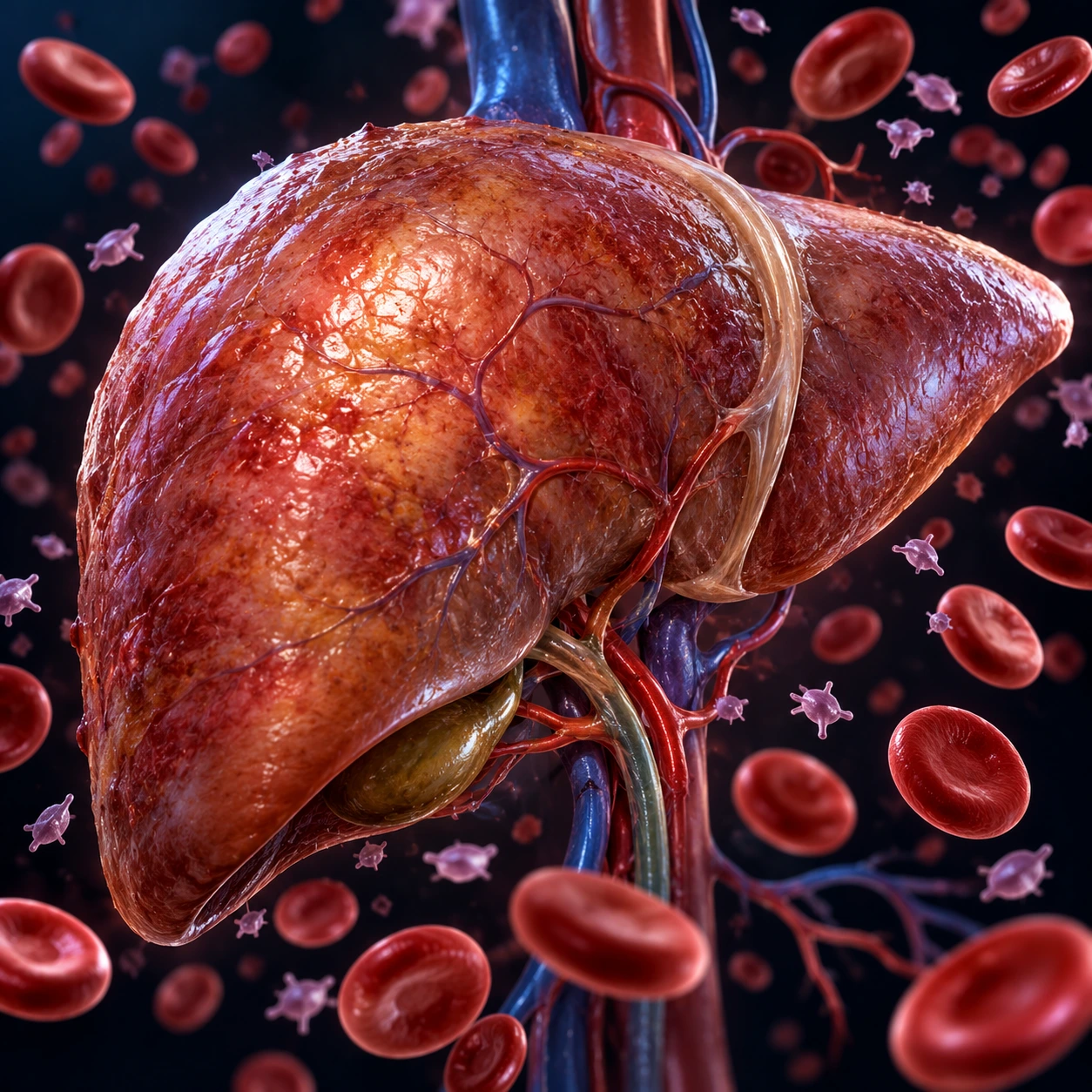
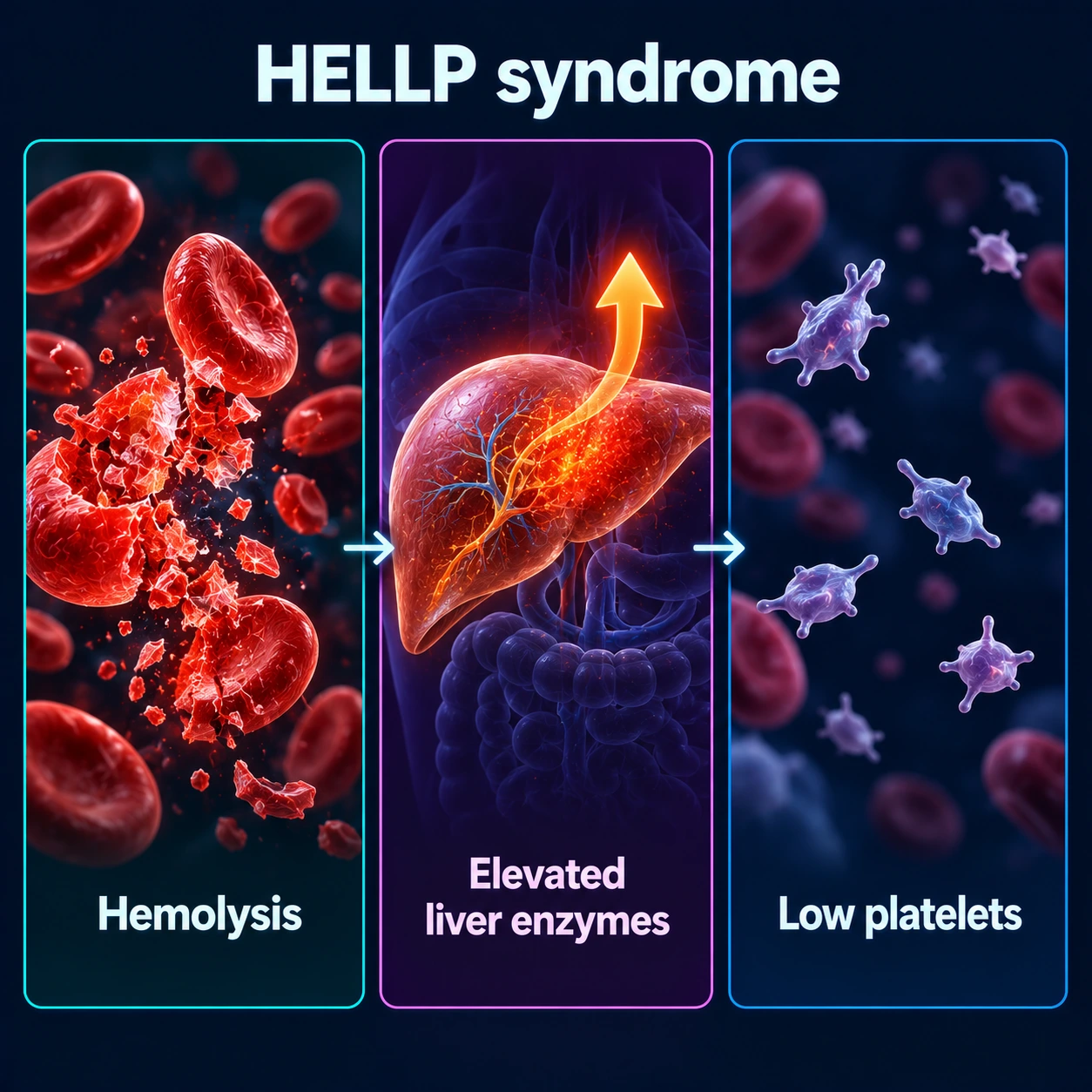
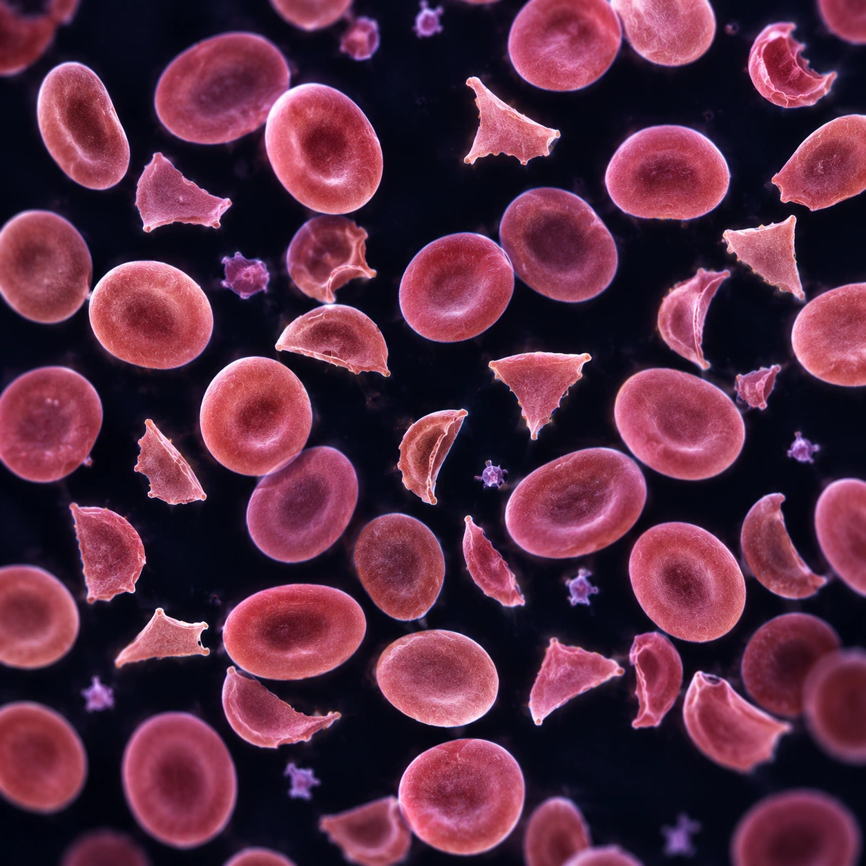
Table of Contents
- Overview
- Acronym and Classification Systems
- Epidemiology and Risk Factors
- Pathophysiology
- Clinical Presentation
- Diagnosis and Laboratory Criteria
- Differential Diagnosis
- Management — Antepartum
- Management — Postpartum and Complications
- Fetal and Neonatal Outcomes
- Prognosis and Recurrence
- Key Research Papers
- Featured Videos
Overview
HELLP syndrome is a severe, life-threatening complication of pregnancy defined by the triad of Hemolysis, Elevated Liver enzymes, and Low Platelets. It most commonly arises in the third trimester of pregnancy, though it can appear postpartum, and is widely recognized as a variant or severe complication of preeclampsia. Approximately 10–20% of women with severe preeclampsia will develop HELLP syndrome, yet in up to 15% of HELLP cases the condition occurs entirely without the classic hallmarks of preeclampsia — namely hypertension and proteinuria — making timely recognition exceptionally challenging.
A 2009 review of the published literature (Haram et al.) put the incidence at 0.5–0.9% of all pregnancies and 10–20% of cases of severe preeclampsia; about 70% of cases develop before delivery, most between the 27th and 37th week, and the rest within 48 hours of delivery. The syndrome was first formally described and named by Dr. Louis Weinstein in 1982, who recognized the constellation of laboratory findings as a distinct and dangerous entity rather than a subset of standard preeclampsia. Prior to Weinstein's landmark paper, many women with HELLP were misdiagnosed as having gallbladder disease, gastritis, peptic ulcer disease, viral hepatitis, or idiopathic thrombocytopenic purpura — misdiagnoses that delayed potentially life-saving intervention.
Risk is highest in multiparous women and those of older maternal age. White and Hispanic women appear to be at somewhat higher risk than Black women, though HELLP can affect any pregnant patient. Because its presenting symptoms are so nonspecific — right upper quadrant pain, nausea, malaise — the diagnosis rests on blood tests ordered when a third-trimester patient seems unwell, and it is missed when those tests are not done. Speed matters: HELLP can deteriorate to hepatic rupture, disseminated intravascular coagulation (DIC), eclampsia, acute kidney injury, and maternal death within hours if not identified and managed promptly.
Acronym and Classification Systems
The acronym HELLP encapsulates the three defining laboratory abnormalities:
- H — Hemolysis: microangiopathic hemolytic anemia with schistocytes (fragmented red blood cells) visible on peripheral blood smear, elevated lactate dehydrogenase (LDH), low haptoglobin, and elevated indirect bilirubin.
- EL — Elevated Liver Enzymes: elevated aspartate aminotransferase (AST) and alanine aminotransferase (ALT), often with lactate dehydrogenase also elevated from hepatic injury (distinct from the hemolysis-driven lactate dehydrogenase elevation).
- LP — Low Platelets: thrombocytopenia below 100,000/µL, arising from platelet consumption at sites of endothelial injury.
Two formal classification systems exist, each with distinct clinical utility:
Tennessee Classification (Sibai)
Developed by Dr. Baha Sibai, the Tennessee system is the most widely cited in clinical practice and research. It distinguishes between:
- Complete (Full) HELLP: all three criteria — hemolysis, elevated liver enzymes, and low platelets — are present simultaneously. This is the classic and most dangerous form.
- Partial HELLP: only one or two of the three criteria are met (sometimes termed ELLP, HEL, or LP syndrome depending on which elements are present). Partial HELLP carries significant morbidity and frequently evolves into full HELLP if untreated, yet is often overlooked precisely because it does not meet the full definition. Studies have shown that partial HELLP may actually have a worse perinatal outcome in some cohorts because the diagnosis is delayed.
Mississippi Triple-Class System (Martin)
Developed by Dr. James Martin, this system classifies HELLP according to platelet nadir, providing more granular prognostic stratification:
- Class 1 (Most Severe): platelet count falls below 50,000/µL. This class is associated with the highest rates of maternal morbidity, DIC, transfusion requirement, and perinatal complications. Urgent delivery is mandated regardless of gestational age.
- Class 2 (Moderate): platelet nadir between 50,000 and 100,000/µL. Moderate severity; close monitoring and typically delivery within 24–48 hours.
- Class 3 (Mildest / Partial): platelet nadir between 100,000 and 150,000/µL. May represent an early or evolving form; can progress to Class 1 or 2 without intervention. The Mississippi system is particularly useful for research and for communicating disease severity between care teams in tertiary referral centers.
Understanding both classification systems is important because different institutions and published studies reference one or the other, and management thresholds — especially around platelet transfusion, delivery timing at borderline gestational ages, and corticosteroid use — are often tied to class designation.
Epidemiology and Risk Factors
HELLP syndrome affects approximately 0.5–0.9% of all pregnancies worldwide (Haram et al., 2009). In women with severe preeclampsia, the incidence rises sharply to 10–20%. In the United States, HELLP complicates an estimated 48,000 pregnancies annually. Globally, it is a major contributor to maternal mortality, particularly in low- and middle-income countries where access to intensive care and blood product support is limited.
The syndrome predominantly affects women in the third trimester, with the peak incidence between 27 and 37 weeks of gestation. However, approximately 10% of cases present before 27 weeks, and about 30% of cases manifest in the postpartum period — typically within 48 hours of delivery but occasionally beyond that window. Postpartum HELLP is the most treacherous subset because healthcare providers and patients are often no longer vigilant for obstetric complications once delivery has occurred.
Established risk factors include:
- Prior preeclampsia or HELLP: the strongest single predictor; recurrence risk for HELLP in subsequent pregnancies is 2–27%.
- Multiparity: counterintuitively, HELLP (unlike classic preeclampsia, which is more common in nulliparas) has a higher incidence in multiparous women.
- Older maternal age: risk increases with advancing age.
- White or Hispanic race/ethnicity: higher incidence compared to Black women, though Black women who develop HELLP may have more severe outcomes due to socioeconomic and access disparities.
- Antiphospholipid syndrome: prothrombotic state amplifies placental pathology and HELLP risk.
- Thrombophilias: factor V Leiden, prothrombin gene mutation, protein C/S deficiency — contribute to impaired placental circulation.
- Chronic hypertension: pre-existing hypertension approximately doubles the risk of developing superimposed preeclampsia and HELLP.
- Multiple gestation: twins and higher-order multiples carry elevated preeclampsia and HELLP risk due to greater placental mass and associated hemodynamic demands.
Pathophysiology
HELLP syndrome arises from the same fundamental endothelial dysfunction cascade that underlies preeclampsia, but with particular tropism for the hepatic vasculature. The sequence of events is now understood to begin long before clinical symptoms appear:
Step 1 — Abnormal placentation: In normal pregnancy, cytotrophoblast cells invade the maternal spiral arteries during the first and early second trimester, remodeling them into wide, low-resistance vessels capable of sustaining high placental blood flow. In HELLP and preeclampsia, this invasion is shallow and incomplete. The spiral arteries remain narrow, high-resistance vessels, resulting in chronic placental hypoperfusion and ischemia throughout pregnancy.
Step 2 — Release of anti-angiogenic factors: The ischemic placenta responds by releasing excessive quantities of two potent anti-angiogenic proteins into the maternal circulation: sFlt-1 (soluble FMS-like tyrosine kinase 1) and sEng (soluble endoglin). sFlt-1 acts as a decoy receptor that sequesters free VEGF (vascular endothelial growth factor) and PlGF (placental growth factor), stripping the maternal endothelium of survival signals it depends on. sEng similarly antagonizes TGF-beta signaling. Together, these circulating factors cause widespread maternal endothelial dysfunction.
Step 3 — Endothelial injury and platelet activation: Damaged maternal endothelium becomes thrombogenic — it loses its normal anti-platelet and anti-coagulant properties and begins to activate platelets and the coagulation cascade. Platelets adhere to injured vessel walls, aggregate, and are consumed at a rate that exceeds production, producing thrombocytopenia. Fibrin strands form in the microvasculature, and red blood cells traveling through these fibrin meshes are sheared apart, creating the characteristic schistocytes (fragmented red blood cells visible on peripheral smear) that define microangiopathic hemolytic anemia.
Step 4 — Hepatic pathology: The liver's sinusoidal vasculature is exquisitely sensitive to endothelial injury in HELLP. Fibrin deposits accumulate in the periportal and perisinusoidal spaces, obstructing hepatic blood flow and causing hepatocyte necrosis. This leads to elevated aspartate aminotransferase and alanine aminotransferase. As the liver swells, the hepatic capsule (Glisson's capsule) is stretched, producing the characteristic severe right upper quadrant pain that is the most common presenting symptom. In 0.9–2% of HELLP cases, hepatic swelling progresses to subcapsular hematoma — a hemorrhagic collection between the liver and its capsule. Rupture of this hematoma, occurring in approximately 1% of HELLP cases, is catastrophic and carries a maternal mortality rate of 35–60%.
Step 5 — Platelet nadir and recovery timeline: A critical but often misunderstood feature of HELLP is that platelet counts continue to fall for 24–48 hours after delivery before the process begins to reverse. This occurs because the anti-angiogenic factors and activated complement pathways remain in the maternal circulation even after placental removal, continuing to drive endothelial injury temporarily. Recovery — rising platelets, falling liver enzymes and lactate dehydrogenase — typically begins 48–72 hours postpartum and is complete within 96–120 hours in uncomplicated cases.
Clinical Presentation
HELLP syndrome is notorious for its nonspecific presentation. The symptoms so closely mimic common benign conditions — gallbladder disease, gastritis, peptic ulcer disease, viral hepatitis, or a stomach flu — that misdiagnosis occurs in a substantial proportion of cases, sometimes for days. In a pregnant or recently postpartum woman these complaints are the ones that blood tests are used to sort out, because only the laboratory results separate HELLP from the harmless look-alikes.
Core symptoms and their frequencies:
- Malaise and fatigue (90%): typically the earliest symptom, often dismissed as normal late-pregnancy discomfort; can precede the laboratory abnormalities by days.
- Right upper quadrant (RUQ) or epigastric pain (75–90%): the most diagnostically important symptom; caused by hepatic swelling and capsular stretch, or by an evolving subcapsular hematoma. Pain in the upper right abdomen or over the stomach in a third-trimester or recently postpartum patient is a sign hospitals treat as urgent, because HELLP cannot be ruled out without blood tests.
- Nausea and vomiting (36–84%): often accompanies epigastric pain and leads to misdiagnosis as gastroenteritis or severe morning sickness (hyperemesis gravidarum).
- Headache (33–61%): caused by hypertension and cerebral edema; may presage eclamptic seizure.
- Visual disturbances (10–20%): blurred vision, photopsia, scotomata — from hypertensive retinopathy and cerebral edema.
- Edema: present in the majority, though fluid retention is so common in late pregnancy that it rarely raises diagnostic concern on its own.
Physical examination findings: RUQ or epigastric tenderness on palpation (the most reliable exam finding); hypertension in approximately 85% of patients (systolic ≥140 or diastolic ≥90 mmHg); absent hypertension in up to 15% — a crucial point because providers who look only for high blood pressure will miss a minority of cases entirely. Jaundice, when present, signals significant hemolysis and hepatic injury and indicates severe disease. Edema is common. Neurological examination is usually normal unless eclampsia has supervened.
Proteinuria: present in approximately 86% of cases, but absent in 14% — another reminder that the classic preeclampsia triad (hypertension + proteinuria + edema) is not a requirement for HELLP diagnosis.
Timing — when HELLP presents:
- Antepartum (70%): most commonly between 27 and 37 weeks, though cases at 23–24 weeks are documented. The earlier the gestational age at onset, the worse the perinatal prognosis.
- Postpartum (30%): typically within 48 hours of delivery, but occasionally up to 7 days postpartum. This subset is the most dangerous because the obstetric team and patient have often mentally transitioned out of "high-risk" mode. Postpartum HELLP can be the first and only presentation — the antepartum course was entirely unremarkable. Upper right abdominal pain, nausea and feeling unwell within a week of delivery are signs emergency departments treat as urgent, with a complete blood count (CBC) and liver blood tests (liver function tests, LFTs) done straight away.
Diagnosis and Laboratory Criteria
The diagnosis of HELLP syndrome rests entirely on laboratory criteria. There is no single pathognomonic clinical finding; the diagnosis is confirmed when the characteristic constellation of laboratory abnormalities is present in a pregnant or peripartum patient.
Tennessee Criteria (Most Widely Used)
1. Hemolysis — at least two of the following:
- Peripheral blood smear demonstrating schistocytes (fragmented, helmet-shaped red blood cells) — the smear is the essential test, and the diagnosis is not made without it
- Lactate dehydrogenase greater than 600 U/L
- Haptoglobin below 25 mg/dL (haptoglobin is consumed when it binds free hemoglobin released by lysed red cells)
- Indirect (unconjugated) bilirubin greater than 1.2 mg/dL
2. Elevated Liver Enzymes:
- Aspartate aminotransferase greater than 70 U/L (some institutions use the threshold of twice the upper limit of normal)
- Alanine aminotransferase elevated; may exceed 2,000 U/L in fulminant cases — extreme alanine aminotransferase elevation signals massive hepatocyte necrosis
- Lactate dehydrogenase greater than 600 U/L (overlaps with hemolysis marker; elevated lactate dehydrogenase in this context reflects both hepatic necrosis and red cell hemolysis)
3. Low Platelets:
- Platelet count below 100,000/µL (Mississippi Class 2) at minimum for full HELLP diagnosis
- Mississippi Class 1: nadir below 50,000/µL — highest risk of bleeding, DIC, and transfusion
- Critical timing point: the platelet nadir occurs 24–48 hours after delivery; a platelet count that is borderline at the time of delivery may plummet in the immediate postpartum period
Additional Laboratory Tests
A full laboratory work-up for HELLP covers:
- Coagulation panel (prothrombin time (PT), activated partial thromboplastin time (aPTT), fibrinogen, D-dimer): DIC develops in 4–38% of HELLP cases; prothrombin time and activated partial thromboplastin time may be normal early but fibrinogen consumption and D-dimer elevation signal evolving coagulopathy
- Serum creatinine and blood urea nitrogen (BUN): assess for associated acute kidney injury (AKI), which complicates up to 10% of severe HELLP
- Serum glucose: expected to be normal in HELLP — this is a critical differentiating feature from acute fatty liver of pregnancy (AFLP), where hypoglycemia is characteristic
- Uric acid: elevated in preeclampsia and HELLP; not diagnostic but useful as a severity marker
- Urine protein: 24-hour urine protein or spot urine protein-to-creatinine ratio to quantify proteinuria
- Blood type and crossmatch: done early, because transfusion is often needed
- ADAMTS13 activity: if TTP (thrombotic thrombocytopenic purpura) cannot be clinically excluded — ADAMTS13 activity below 10% suggests TTP rather than HELLP, with critical implications for treatment
A blood test for two placental proteins has been studied as a way of telling who is about to get much worse. Across 18 United States centres, 1,014 pregnant women admitted between 23 and 35 weeks with a hypertensive disorder of pregnancy had the ratio of soluble fms-like tyrosine kinase 1 (sFlt-1) to placental growth factor (PlGF) measured in their blood. In the validation group, a ratio of 40 or higher had a 65% positive and a 96% negative predictive value for going on to develop preeclampsia with severe features, and performed better than the standard clinical tests (area under the curve 0.92 against under 0.75). Serious maternal complications occurred in 16.1% of women with a ratio of 40 or more against 2.8% of those below it (Thadhani et al., 2022).
Monitoring Frequency
In active HELLP syndrome, laboratory values are checked every 6–12 hours. The disease can evolve from partial to full HELLP, and from Class 3 to Class 1, within a single day. Repeated complete blood counts and liver function tests show the trajectory of the disease and set the timing of interventions such as platelet transfusion and delivery.
Imaging
Routine liver imaging is not required for diagnosis of uncomplicated HELLP. However, hepatic ultrasound or computed tomography (CT) abdomen is indicated when subcapsular hematoma is suspected — particularly in any HELLP patient with sudden worsening of RUQ pain, hemodynamic instability, or unexplained drop in hemoglobin. Computed tomography with contrast provides the most accurate characterization of hematoma size, extension, and presence of active hemorrhage or rupture.
Differential Diagnosis
The combination of hemolytic anemia, thrombocytopenia, and hepatic injury in a pregnant or peripartum patient raises a differential that includes several conditions requiring fundamentally different treatments. Misidentification can be fatal.
1. Thrombotic Thrombocytopenic Purpura (TTP): Like HELLP, TTP produces microangiopathic hemolytic anemia (schistocytes on smear) and thrombocytopenia. The key distinguishing feature is ADAMTS13 activity below 10% in TTP (normal in HELLP). TTP is also characterized by more prominent neurological symptoms (altered consciousness, stroke, focal deficits), fever, and severe renal impairment in a pentad pattern, though not all five features need be present. TTP is not pregnancy-specific but is more common in pregnancy. Critically, TTP does NOT respond to delivery — it requires urgent plasma exchange (plasmapheresis). Delivering a preterm fetus for a condition that requires plasma exchange, not obstetric intervention, represents a serious management error.
2. Hemolytic Uremic Syndrome (HUS): HUS shares microangiopathic hemolytic anemia and thrombocytopenia with HELLP but is dominated by acute kidney injury rather than hepatic involvement. Typical HUS follows Shiga-toxin-producing E. coli (usually O157:H7) infection with diarrheal prodrome. Atypical HUS (aHUS) results from complement pathway dysregulation and may recur in subsequent pregnancies. ADAMTS13 is normal in HUS. Treatment of aHUS is eculizumab (complement inhibitor), not delivery.
3. Acute Fatty Liver of Pregnancy (AFLP): AFLP is the most dangerous mimicker of HELLP because both are obstetric liver emergencies occurring in the third trimester, and both are treated with delivery. Key differentiating features:
- Hypoglycemia: characteristic of AFLP (absent or mild in HELLP); reflects hepatic failure impairing gluconeogenesis
- Hyperammonemia and hepatic encephalopathy: more prominent in AFLP
- Aspartate aminotransferase:alanine aminotransferase ratio often greater than 1 in AFLP (nonspecific but suggestive)
- Coagulopathy: PT is markedly prolonged in AFLP (hepatic synthetic failure); in HELLP, coagulopathy occurs only when DIC develops
- Liver biopsy: AFLP shows microvesicular steatosis (fat droplets filling hepatocytes); HELLP shows periportal fibrin deposition and hepatocyte necrosis — biopsy is rarely needed clinically but is diagnostically definitive
- LCHAD deficiency: the fetus carrying long-chain 3-hydroxyacyl-CoA dehydrogenase (LCHAD) deficiency generates metabolites toxic to the maternal liver — a causal link between fetal genotype and AFLP in the mother
A screening study put numbers on that fetal link and showed it belongs to acute fatty liver of pregnancy rather than to HELLP. Among 108 samples referred for molecular screening, mutations causing long-chain 3-hydroxyacyl-CoA dehydrogenase deficiency in the baby were found in 5 of the 27 families (19%) where the mother had acute fatty liver of pregnancy, the common maternal change being the E474Q mutation. Of the 81 mothers with HELLP syndrome, only one carried a single copy of E474Q and no affected newborn was found (Yang et al., 2002).
4. Immune Thrombocytopenic Purpura (ITP): ITP causes isolated thrombocytopenia without hemolysis and without liver enzyme elevation. Antiplatelet antibodies (anti-GPIIb/IIIa) are present in most cases. ITP is chronic and does not cause the acute deterioration seen in HELLP. Treatment is steroids, intravenous immunoglobulin (IVIG), or rituximab — not delivery.
5. Antiphospholipid Syndrome (APS): Can cause thrombocytopenia, pregnancy loss, and endothelial activation, and may precipitate preeclampsia and HELLP. APS and HELLP can coexist; positive antiphospholipid antibody titers in the setting of HELLP are the usual reason for a hematology referral.
6. Acute cholecystitis, choledocholithiasis, Budd-Chiari syndrome: These are the conditions ultrasound rules out in a pregnant patient with upper right abdominal pain. Ultrasound will demonstrate gallstones, biliary dilation, or hepatic vein thrombosis in these conditions, while HELLP will show a normal biliary tree (or subcapsular hematoma in advanced cases).
Management — Antepartum
Delivery is the only definitive cure for HELLP syndrome. All management before delivery is supportive — stabilizing the mother, protecting the fetus where gestational age allows, and preparing for safe delivery. The gestational age at presentation is the central variable that drives decision-making.
Delivery Threshold by Gestational Age
Greater than or equal to 34 weeks gestation: immediate delivery. At or beyond 34 weeks, fetal lung maturity is sufficient and the risks of continued intrauterine exposure to a deteriorating maternal environment outweigh any benefit of further expectant management. Induction of labor is appropriate if the cervix is favorable and the maternal condition is stable. Cesarean section is indicated for standard obstetric indications (malpresentation, failed induction, fetal distress) — HELLP alone is not an indication for cesarean.
Less than 34 weeks gestation: short-term stabilization followed by delivery within 24–48 hours. The primary reason to delay delivery briefly at less than 34 weeks is to administer corticosteroids for fetal lung maturity. Betamethasone 12 mg intramuscular every 24 hours for two doses accelerates pulmonary surfactant synthesis in the fetal lung, substantially reducing the risk of respiratory distress syndrome (RDS) in the premature neonate. This 24–48 hour window is the maximum acceptable delay — if the mother's condition deteriorates at any point during this period, delivery goes ahead immediately regardless of whether the full corticosteroid course has been completed.
Before 27–28 weeks: the decision is individualized and involves a high-stakes conversation between the obstetric team, neonatology, and the patient. Fetal viability, parental wishes, and the severity of maternal disease all factor in. Extreme prematurity carries high neonatal morbidity and mortality, but maternal deterioration in severe HELLP Class 1 at any gestational age demands delivery.
Maternal Stabilization (Concurrent with Delivery Planning)
1. Magnesium sulfate for seizure prophylaxis: Given to all patients with HELLP or severe preeclampsia. Loading dose: 4–6 g intravenously over 15–20 minutes. Maintenance: 1–2 g/hour by continuous infusion. Magnesium prevents eclamptic seizures by mechanisms including N-methyl-D-aspartate (NMDA) receptor antagonism and cerebral vasodilation. Toxicity monitoring is essential: loss of deep tendon reflexes occurs at serum magnesium 7–10 mEq/L, respiratory depression at greater than 12 mEq/L. Calcium gluconate 1 g intravenous (IV) is the antidote and is kept at the bedside throughout. Magnesium leaves the body through the kidneys, so the infusion is reduced or stopped when urine output falls below 25–30 mL/hour.
2. Antihypertensive therapy: Blood pressure at or above 160 mmHg systolic or 110 mmHg diastolic is a hypertensive emergency in pregnancy — this threshold is associated with maternal stroke, the leading cause of maternal death in severe preeclampsia and HELLP. First-line agents include:
- Labetalol intravenous: 20 mg bolus, may repeat 40 mg, then 80 mg at 10-minute intervals to a maximum of 300 mg total. As a beta blocker it can trigger bronchospasm in asthma and slow the heart further when the pulse is already low, so other agents are used in those situations.
- Nifedipine immediate-release oral: 10–20 mg every 20–30 minutes; onset within 20 minutes. Widely used in many centers as the first choice for acute blood pressure (BP) reduction in obstetric patients.
- Hydralazine intravenous: 5–10 mg every 20 minutes; more unpredictable response than labetalol or nifedipine; reflex tachycardia is common.
Target blood pressure is less than 160/110 mmHg; the 2009 Haram review states the aim of keeping blood pressure below 155/105 mmHg. Pressures below about 150/100 are not driven down hard, because too much antihypertensive treatment reduces blood flow through the placenta and worsens the baby's condition. Angiotensin-converting enzyme inhibitors (ACE inhibitors) and angiotensin receptor blockers (ARBs) are strictly contraindicated in pregnancy due to fetotoxicity (renal dysgenesis, oligohydramnios, neonatal renal failure).
3. Platelet transfusion: Indications are not standardized across centers but reasonable consensus thresholds are:
- Platelet count below 20,000/µL: transfuse to prevent spontaneous bleeding
- Platelet count below 50,000/µL before operative delivery (cesarean section): transfuse to reduce surgical hemorrhage risk
- Platelet count before neuraxial anesthesia (spinal or epidural): the 2021 interdisciplinary consensus statement of the Society for Obstetric Anesthesia and Perinatology concluded that the risk of a spinal epidural hematoma with a platelet count of 70,000 × 106/L or above is likely to be very low in obstetric patients whose low platelets are due to hypertensive disorders of pregnancy, gestational thrombocytopenia or immune thrombocytopenia, in the absence of other risk factors. Below that count the statement sets no fixed cut-off: the decision weighs the individual's bleeding history, other illnesses, the airway examination and the risks of general anesthesia, which is why some patients are given a general anesthetic instead
Each apheresis unit of platelets typically raises the count by 30,000–50,000/µL transiently. Because platelets are rapidly consumed in active HELLP, the benefit is short-lived; transfusion is timed to immediately precede the procedure rather than given hours before.
Corticosteroids for HELLP (Dexamethasone)
High-dose dexamethasone has been used to accelerate biochemical recovery in HELLP and may facilitate brief prolongation of pregnancy at very preterm gestational ages to complete the fetal lung maturity course. The most commonly used regimen is dexamethasone 10 mg intravenous every 12 hours antepartum. The trial evidence does not support it. In a double-blind placebo-controlled trial of 132 women (Fonseca et al., 2005), dexamethasone shortened hospital stay only from 8.2 to 6.5 days, which was not statistically significant (P = .37), and there was no significant difference in how quickly platelet counts recovered (hazard ratio 1.2, 95% CI 0.8–1.8), nor in lactate dehydrogenase (hazard ratio 0.9) or aspartate aminotransferase (hazard ratio 0.6, 95% CI 0.4–1.1), nor in the rate of complications; the authors concluded their results do not support using dexamethasone for HELLP. A 2023 meta-analysis of 7 randomized trials covering 485 treated women likewise found no significant benefit over placebo for maternal illness (relative risk 1.36, 95% CI 0.45–4.10), eclampsia (1.16, 0.76–1.77), acute kidney failure (0.71, 0.41–1.22), fluid on the lungs (0.34, 0.10–1.15) or low urine output (1.08, 0.75–1.54), and none for the newborns (Sun et al., 2023). The 2009 Haram review describes standard corticosteroid treatment as of uncertain clinical value for the mother's HELLP syndrome. Dexamethasone for fetal lung maturity (betamethasone preferred) is different from dexamethasone for HELLP treatment — these are separate indications with different agents and goals.
Management — Postpartum and Complications
Delivery resolves the underlying cause of HELLP by removing the dysfunctional placenta, but the disease does not resolve immediately. The postpartum period carries its own dangers and requires sustained vigilance.
Postpartum Monitoring and Supportive Care
Platelet counts and liver function tests are monitored every 6–12 hours after delivery because the platelet nadir — the lowest point of the platelet count during the illness — characteristically occurs 24–48 hours after delivery. A patient whose platelets were 60,000/µL at delivery may have a nadir of 25,000/µL on postpartum day 1. Recovery begins thereafter, with platelets rising and liver enzymes falling, and most women achieve full biochemical recovery within 96–120 hours postpartum.
Magnesium sulfate is continued for 24–48 hours postpartum for ongoing seizure prophylaxis. During this period, careful fluid management is essential — postpartum women are at risk for pulmonary edema from fluid mobilization and third-space fluid shifts, and magnesium further impairs myocardial contractility. Strict intake and output monitoring with a Foley catheter is standard in the immediate postpartum management of HELLP.
Antihypertensive therapy: Hypertension frequently persists or worsens in the first several postpartum days before gradually resolving over weeks. Oral antihypertensives (nifedipine XL, labetalol, or methyldopa for breastfeeding women) are continued until blood pressure normalizes — typically within 6–12 weeks postpartum, though some women require longer-term therapy.
Subcapsular Hepatic Hematoma
Subcapsular hematoma of the liver is a rare but potentially lethal complication occurring in 0.9–2% of HELLP cases. Sudden severe worsening of upper right abdominal pain, a steep fall in hemoglobin with no obvious source of bleeding, or a drop in blood pressure and rising pulse are the signs that point to it, and hospitals treat them as urgent. Confirmatory imaging is hepatic ultrasound at the bedside for unstable patients, or computed tomography abdomen with contrast for stable patients (provides superior anatomic detail).
Management depends on whether rupture has occurred:
- Unruptured subcapsular hematoma: conservative management with strict bed rest, serial imaging, blood pressure control, avoidance of maneuvers that increase intra-abdominal pressure (Valsalva, vigorous palpation), and close hemodynamic monitoring. Delivery (if not yet delivered) is essential. Most unruptured hematomas resolve spontaneously over weeks.
- Ruptured hematoma (hepatic rupture): a surgical emergency with maternal mortality of 35–60%. Management approach:
- Activate massive transfusion protocol immediately (packed red blood cells, fresh frozen plasma, cryoprecipitate, and platelets in a 1:1:1 ratio)
- Interventional radiology — hepatic artery embolization: preferred first-line intervention in centers with 24/7 IR capability; can control hemorrhage without the risks of open surgery in a coagulopathic patient
- Surgical intervention: exploratory laparotomy, hepatorrhaphy (surgical repair of liver laceration), perihepatic packing with planned re-look laparotomy in 24–48 hours; right lobe of the liver is predominantly involved
- Liver transplantation: required in the rare cases of fulminant hepatic failure following hepatic rupture with irreversible hepatic necrosis; outcomes are poor but cases of successful transplantation are documented
Plasma Exchange for Refractory HELLP
In the minority of patients whose HELLP syndrome does not resolve within 72–96 hours postpartum — particularly those with Class 1 disease or where TTP cannot be confidently excluded — plasma exchange (plasmapheresis) is an important therapeutic option. Plasma exchange removes circulating anti-angiogenic factors, microangiopathic mediators, and complement activation products, while simultaneously replacing ADAMTS13 and other protective plasma proteins. Multiple case series and small trials support its use in refractory postpartum HELLP, and it is the definitive treatment if retrospective ADAMTS13 testing reveals activity below 10% consistent with TTP.
Disseminated Intravascular Coagulation (DIC)
DIC develops in 4–38% of HELLP cases and represents a shift from the localized coagulopathy of platelet consumption to a systemic, uncontrolled activation of both coagulation and fibrinolysis. Signs include prolonged PT and aPTT, low fibrinogen (below 200 mg/dL is concerning; below 100 mg/dL requires urgent replacement), elevated D-dimer, and clinical bleeding from venipuncture sites, surgical wounds, or mucosal surfaces. Treatment is delivery plus replacement of consumed coagulation factors with fresh frozen plasma, cryoprecipitate (for fibrinogen), and platelet transfusion.
Fetal and Neonatal Outcomes
Fetal and neonatal outcomes in HELLP syndrome are primarily determined by two variables: gestational age at delivery and placental function. Because HELLP demands early delivery — often preterm — the neonatal consequences are substantially shaped by the hazards of prematurity.
Perinatal mortality ranges from 6% to 37% across published series, reflecting wide variation in disease severity and gestational age distribution across study populations. The highest mortality occurs in deliveries before 28 weeks of gestation, where prematurity-related complications dominate. In high-resource settings with modern neonatal intensive care, perinatal survival at 28 weeks or greater has improved dramatically over the past two decades.
Intrauterine growth restriction (IUGR) is present in a significant proportion of fetuses exposed to HELLP, because the same placental insufficiency that drives maternal disease also reduces oxygen and nutrient delivery to the fetus. IUGR fetuses are at heightened risk for fetal distress during labor, intrapartum cesarean section, and neonatal metabolic complications.
Neonatal thrombocytopenia occurs in 15–38% of neonates born to mothers with HELLP syndrome. The mechanism involves transplacental transfer of anti-endothelial antibodies and activated complement, as well as direct effects of the maternal microangiopathic process on the fetal platelet-producing megakaryocytes. Newborn platelet counts are checked at birth and followed over the first several days of life; they usually recover on their own, and babies with very low counts (below 20,000/µL) may be given a platelet transfusion.
Respiratory distress syndrome (RDS) affects premature neonates delivered before lung maturity is achieved. Antenatal corticosteroids (betamethasone) substantially reduce RDS incidence and severity but do not eliminate it, particularly at very early gestational ages. Surfactant therapy administered by neonatologists at birth has transformed outcomes for premature infants with RDS.
Long-term neurodevelopmental outcomes for survivors of HELLP-associated prematurity are generally favorable when birth occurs at or after 32 weeks with appropriate NICU care. Deliveries before 28 weeks carry meaningful risks of intraventricular hemorrhage, periventricular leukomalacia, and long-term neurocognitive impairment. Babies born before 34 weeks in the setting of HELLP are usually enrolled in early developmental follow-up so that delays are picked up and treated early.
Prognosis and Recurrence
Maternal mortality from HELLP syndrome ranges from 1–3% in high-income countries with modern critical care capabilities to as high as 25% in resource-limited settings where timely access to platelet transfusion, intensive care, and surgical management of hepatic rupture is unavailable. The causes of maternal death include intracranial hemorrhage (from hypertensive emergency), hepatic rupture with exsanguination, respiratory failure from pulmonary edema, and multi-organ failure in the setting of severe DIC. Eclampsia (seizures) complicates 7–9% of HELLP cases and independently worsens prognosis.
Major maternal complications, measured in a single large series: the figures below come from a prospective cohort of 442 pregnancies with HELLP syndrome managed at one centre from 1977 to 1992 (Sibai et al., 1993). Of those pregnancies, 309 (70%) developed before delivery and 133 (30%) after it; women whose HELLP began after delivery had significantly more fluid on the lungs and more kidney failure.
- Disseminated intravascular coagulation: 21% (other series report 4–38%)
- Placental abruption (premature separation of placenta from uterine wall): 16%
- Acute kidney failure: 7.7%
- Pulmonary edema (fluid on the lungs): 6%
- Transfusion with blood or blood products: 55%
- Laparotomy for major bleeding inside the abdomen: 2%
- Subcapsular hepatic hematoma: 0.9% (0.9–2% across series)
- Retinal detachment: 0.9%
- Maternal death: 1.1% (five patients)
- Eclampsia: 7–9% across published series
- Hepatic rupture: approximately 1% across published series
Abruption was strongly linked to disseminated intravascular coagulation (p < 0.0001), kidney failure (p < 0.001) and fluid on the lungs (p < 0.01) in that series, and fluid on the lungs was closely associated with kidney failure (p < 0.0001).
Recurrence risk in subsequent pregnancies: The risk of HELLP recurrence in a subsequent pregnancy is 2–27%, with wide variation attributable to underlying maternal thrombophilia, antiphospholipid syndrome, and other risk factors. Even when full HELLP does not recur, women who have had HELLP are at substantially higher risk for preeclampsia, gestational hypertension, and IUGR in future pregnancies. A later pregnancy after HELLP is therefore handled as high-risk: early prenatal care, repeated blood pressure checks, and low-dose aspirin started at 12–16 weeks, which reduces but does not eliminate the risk of preeclampsia returning.
Long-term cardiovascular risk: HELLP syndrome, like preeclampsia, is now understood to be not merely an obstetric complication but also a marker of lifetime cardiovascular vulnerability. Women who have had HELLP carry a significantly elevated lifetime risk of hypertension, coronary artery disease, stroke, and end-stage renal disease compared to age-matched women with uncomplicated pregnancies. The placental dysfunction that drives HELLP may reveal pre-existing subclinical endothelial dysfunction that will manifest as cardiovascular disease decades later. Because of that raised risk, a history of HELLP is treated as information that belongs in ordinary primary care: blood pressure, cholesterol and fasting blood sugar are the measurements that pick up the problems early, and they are cheap tests. High blood pressure and the cluster called metabolic syndrome are the conditions found most often in these women.
Key Research Papers
- Weinstein L. Syndrome of hemolysis, elevated liver enzymes, and low platelet count: a severe consequence of hypertension in pregnancy. Am J Obstet Gynecol. 1982;142(2):159–167. — PubMed PMID: 7055180
- Sibai BM, Ramadan MK, Usta I, Salama M, Mercer BM, Friedman SA. Maternal morbidity and mortality in 442 pregnancies with hemolysis, elevated liver enzymes, and low platelets (HELLP syndrome). Am J Obstet Gynecol. 1993;169(4):1000–1006. — PubMed PMID: 8238109
- Sibai BM. Diagnosis, controversies, and management of the syndrome of hemolysis, elevated liver enzymes, and low platelet count. Obstet Gynecol. 2004;103(5):981–991. — PubMed PMID: 15121574
- Martin JN Jr, Rinehart BK, May WL, Magann EF, Terrone DA, Blake PG. The spectrum of severe preeclampsia: comparative analysis by HELLP (hemolysis, elevated liver enzyme levels, and low platelet count) syndrome classification. Am J Obstet Gynecol. 1999;180(6):1373–1384. — PubMed PMID: 10368474
- Haram K, Svendsen E, Abildgaard U. The HELLP syndrome: clinical issues and management. A review. BMC Pregnancy Childbirth. 2009;9:8. — PubMed PMID: 19245695
- Audibert F, Friedman SA, Frangieh AY, Sibai BM. Clinical utility of strict diagnostic criteria for the HELLP (hemolysis, elevated liver enzymes, and low platelets) syndrome. Am J Obstet Gynecol. 1996;175(2):460–464. — PubMed PMID: 8765269
- Sibai BM, Ramadan MK. Acute renal failure in pregnancies complicated by hemolysis, elevated liver enzymes, and low platelets. Am J Obstet Gynecol. 1993;168(6 Pt 1):1682–1687; discussion 1687–1690. — PubMed PMID: 8317509
- Vigil-De Gracia P. Maternal deaths due to eclampsia and HELLP syndrome. Int J Gynaecol Obstet. 2009;104(2):90–94. — PubMed PMID: 19027902
- Barton JR, Sibai BM. Diagnosis and management of hemolysis, elevated liver enzymes, and low platelets syndrome. Clin Perinatol. 2004;31(4):807–833, vii. — PubMed PMID: 15519429
- Bauer ME, Arendt K, Beilin Y, et al. The Society for Obstetric Anesthesia and Perinatology interdisciplinary consensus statement on neuraxial procedures in obstetric patients with thrombocytopenia. Anesth Analg. 2021;132(6):1531–1544. — PubMed PMID: 33861047
- Thadhani R, Lemoine E, Rana S, et al. Circulating angiogenic factor levels in hypertensive disorders of pregnancy. NEJM Evid. 2022;1(12):EVIDoa2200161. — PubMed PMID: 38319832
- Yang Z, Yamada J, Zhao Y, Strauss AW, Ibdah JA. Prospective screening for pediatric mitochondrial trifunctional protein defects in pregnancies complicated by liver disease. JAMA. 2002;288(17):2163–2166. — PubMed PMID: 12413376
- Magann EF, Bass D, Chauhan SP, Sullivan DL, Martin RW, Martin JN Jr. Antepartum corticosteroids: disease stabilization in patients with the syndrome of hemolysis, elevated liver enzymes, and low platelets (HELLP). Am J Obstet Gynecol. 1994;171(4):1148–1153. — PubMed PMID: 7943088
- O'Brien JM, Shumate SA, Satchwell SL, Milligan DA, Barton JR. Maternal benefit of corticosteroid therapy in patients with HELLP (hemolysis, elevated liver enzymes, and low platelet count) syndrome: impact on the rate of regional anesthesia. Am J Obstet Gynecol. 2002;186(3):475–479. — PubMed PMID: 11904610
- Fonseca JE, Méndez F, Cataño C, Arias F. Dexamethasone treatment does not improve the outcome of women with HELLP syndrome: a double-blind, placebo-controlled, randomized clinical trial. Am J Obstet Gynecol. 2005;193(5):1591–1598. — PubMed PMID: 16260197
- Sun WJ, Hu J, Zhang Q, Shan JM. Administration of corticosteroid therapy for HELLP syndrome in pregnant women: evidences from seven randomized controlled trials. Hypertens Pregnancy. 2023;42(1):2276726. — PubMed PMID: 37937841
Connections
- Acute Fatty Liver of Pregnancy
- Nephrology & Hepatology
- Liver Disease
- Acute Kidney Injury
- Peripartum Cardiomyopathy
- Thrombocytopenia
- Budd-Chiari Syndrome
- Preeclampsia
- Autoimmune Hepatitis
- Hypertension — pre-existing chronic hypertension roughly doubles the risk of HELLP, and survivors carry an elevated lifetime risk of it afterward.