Gitelman Syndrome
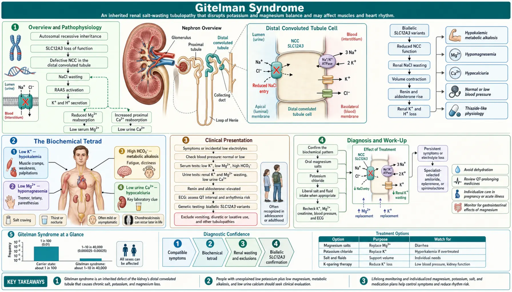
Most common inherited renal tubular disorder (prevalence ~1:40,000); autosomal recessive; SLC12A3 mutation causing defective thiazide-sensitive NaCl cotransporter (NCC) in distal convoluted tubule (DCT).
Table of Contents
- Overview and Pathophysiology
- The Biochemical Tetrad
- Clinical Presentation
- Diagnosis and Work-Up
- Differentiating Gitelman from Bartter Syndrome
- Cardiovascular Risks and QT Prolongation
- Treatment and Management
- Prognosis and Quality of Life
- Long-Term Outcomes in 587 Patients
- Key Research Papers
- Connections
Overview and Pathophysiology
Gitelman syndrome is the most common inherited salt-losing tubulopathy, caused by loss-of-function mutations in SLC12A3, which encodes the thiazide-sensitive NaCl cotransporter (NCC) in the distal convoluted tubule (DCT). The DCT reabsorbs approximately 5–7% of filtered NaCl. NCC inactivation leads to a cascade of downstream electrolyte disturbances:
- Salt wasting → volume depletion → activation of the renin–angiotensin–aldosterone system (RAAS) → secondary aldosteronism → increased activity of the epithelial sodium channel (ENaC) → K+ and H+ excretion → hypokalemia + metabolic alkalosis
- Reduced luminal Na+ → increased Na/Ca exchanger activity on basolateral membrane → increased intracellular Ca2+ pumped out → net hypocalciuria
- Impaired Mg2+ reabsorption via TRPM6 (Mg2+ channel in DCT, NCC-dependent) → hypomagnesemia
The resulting phenotype closely mimics chronic thiazide diuretic use — this pharmacological parallel is central to understanding the mechanism and guides both diagnosis and treatment.
Inheritance is autosomal recessive. Heterozygous carriers are usually asymptomatic but may have mild electrolyte changes. More than 400 pathogenic variants in SLC12A3 have been identified; compound heterozygosity is common.
The Biochemical Tetrad
Gitelman syndrome is defined by four characteristic laboratory findings that together constitute the diagnostic biochemical profile:
- Hypokalemia: typically 2.5–3.2 mEq/L, with elevated urine potassium (inappropriate kaliuresis despite low serum K+, driven by aldosterone). The urinary K/Cr ratio is elevated, confirming renal potassium wasting rather than extrarenal losses.
- Metabolic alkalosis: elevated HCO3- (usually 28–36 mEq/L), arising from aldosterone-driven H+ secretion and from contraction alkalosis secondary to volume depletion.
- Hypomagnesemia: serum Mg2+ <0.7 mmol/L; caused by impaired TRPM6-mediated Mg2+ reabsorption in the DCT. Present in approximately 70% of patients. Clinically important because hypomagnesemia causes neuromuscular irritability and potentiates cardiac arrhythmias independent of potassium.
- Hypocalciuria: urine calcium-to-creatinine ratio usually below 0.2 mmol/mmol, and 24-hour urine calcium rarely above 0.5 mg/kg/day (Knoers and Levtchenko, 2008). This is the KEY DISTINGUISHING FEATURE from Bartter syndrome, which produces hypercalciuria. The mechanism: NCC inactivation increases basolateral Na/Ca exchange, enhancing transcellular Ca2+ reabsorption.
All four findings, combined with normal blood pressure, elevated plasma renin activity, and elevated aldosterone, constitute the complete diagnostic biochemical profile of Gitelman syndrome.
Clinical Presentation
Onset typically occurs in late childhood, adolescence, or adulthood — unlike Bartter syndrome, which often presents in infancy or early childhood. A substantial proportion of patients are diagnosed incidentally when routine laboratory panels reveal unexplained hypokalemia.
Symptomatic patients commonly report:
- Fatigue and general weakness — hypokalemia reduces skeletal muscle adenosine triphosphate (ATP) production and impairs membrane repolarization
- Muscle cramps and tetany — hypomagnesemia increases neuromuscular irritability; alkalosis reduces ionized calcium, compounding this effect
- Salt craving — a behavioral response to chronic salt wasting; patients often describe an instinctive preference for salty foods
- Palpitations — hypokalemia prolongs cardiac action potential duration and QT interval; hypomagnesemia further destabilizes cardiac rhythm
- Paresthesias — tingling or numbness in extremities, attributable to hypomagnesemia and alkalosis-induced reduction in ionized calcium
- Nocturia and mild polyuria — present but less severe than in Bartter syndrome; reflects impaired urinary concentrating ability
- Normal blood pressure and normal growth — important distinguishing features; Bartter patients often exhibit failure to thrive and growth retardation
- Chondrocalcinosis — calcium pyrophosphate crystal deposition in cartilage, particularly in patients with long-standing hypomagnesemia; may cause acute pseudogout attacks
Gitelman syndrome does not cause polyhydramnios (excess amniotic fluid) or nephrocalcinosis, both of which are associated with antenatal Bartter syndrome.
Diagnosis and Work-Up
No single test diagnoses Gitelman syndrome — confirmation requires integration of clinical presentation, biochemical pattern, and genetic testing. A systematic approach:
- Step 1 — Confirm the biochemical tetrad: Serum K+, Mg2+, bicarbonate, and spot urine calcium-to-creatinine ratio (or 24-hour urine calcium). Hypocalciuria on a random urine specimen (Ca/Cr usually <0.2 mmol/mmol) is strongly suggestive.
- Step 2 — Confirm secondary hyperaldosteronism: Plasma renin activity (elevated) and serum aldosterone (elevated) in the absence of hypertension distinguishes Gitelman/Bartter from primary hyperaldosteronism (which suppresses renin).
- Step 3 — Exclude diuretic use: Urine thiazide and loop diuretic screen by mass spectrometry. Surreptitious diuretic use is the most important mimicker.
- Step 4 — Genetic testing: SLC12A3 sequencing identifies pathogenic variants in approximately 85–90% of clinically diagnosed cases. Next-generation sequencing panels covering SLC12A3 plus other tubulopathy genes are preferred. Some patients are compound heterozygotes with one allele carrying a deep intronic or splice-site variant.
- Step 5 — If SLC12A3 negative: Consider CLCNKB sequencing (Bartter Type III / mixed phenotype). Some CLCNKB mutations produce a Gitelman-like phenotype including hypocalciuria.
Historical note: The thiazide response test (thiazide causes less natriuresis in Gitelman patients than in healthy controls because the target NCC is already non-functional) was used prior to widespread genetic testing. It has been largely replaced by genetic analysis but remains a conceptual teaching tool.
Differentiating Gitelman from Bartter Syndrome
Gitelman and Bartter syndromes share hypokalemia, metabolic alkalosis, normal blood pressure, and elevated renin/aldosterone. The key distinguishing features are:
| Feature | Gitelman Syndrome | Bartter Syndrome (Types I/II/IV) | Bartter Type III |
|---|---|---|---|
| Urine calcium | Hypocalciuria (hallmark) | Hypercalciuria | Variable |
| Serum magnesium | Low (~70% of patients) | Usually normal | Usually normal |
| Age of onset | Adolescence/adulthood | Neonatal/early childhood | Childhood/adolescence |
| Clinical severity | Mild; often incidental | Severe; growth failure | Moderate |
| Nephrocalcinosis | Absent | Present (antenatal types) | Less common |
| Polyhydramnios | Absent | Present (Types I/II/IV) | Absent |
| Causative gene | SLC12A3 (DCT NCC) | SLC12A1, KCNJ1, BSND (thick ascending limb, TAL) | CLCNKB (TAL/DCT) |
Both conditions share elevated urine chloride (confirming renal Cl wasting rather than vomiting-related alkalosis, which causes low urine Cl). The hypocalciuria in Gitelman is arguably the single most reliable bedside biochemical discriminator.
Cardiovascular Risks and QT Prolongation
Although Gitelman syndrome is generally a mild condition, its electrolyte disturbances carry real cardiac risk that deserves attention in clinical management:
Hypokalemia and QT prolongation: K+ channels (particularly IKr, encoded by KCNH2) are responsible for phase 3 cardiac repolarization. When serum K+ falls, IKr current decreases, delaying repolarization and prolonging the QTc interval. A QTc >450 ms (males) or >460 ms (females) indicates elevated risk.
How often the QT is prolonged: Knoers and Levtchenko (2008) report that in about 50% of people with Gitelman syndrome the QT interval on the electrocardiogram is slightly to moderately prolonged, but in the great majority this is not linked to clinically relevant heart rhythm problems. Sudden cardiac arrest has been reported in only a few patients, and the authors describe heart screening in such cases to look for other triggers or underlying conditions.
Hypomagnesemia: Mg2+ stabilizes cardiac ion channels. Hypomagnesemia independently prolongs QT and, more dangerously, predisposes to early afterdepolarizations — the trigger for torsades de pointes (TdP), a potentially fatal polymorphic ventricular tachycardia. The combination of low potassium and low magnesium is synergistically arrhythmogenic.
Drug interactions: Many commonly prescribed medications prolong QT (antipsychotics including haloperidol and quetiapine; macrolide antibiotics; fluoroquinolones; methadone; ondansetron). These drugs carry substantially increased arrhythmia risk in patients with uncorrected Gitelman electrolyte disturbances. For this reason, medication lists are checked for QT-prolonging drugs, and electrolytes are corrected before such drugs are started.
Clinical monitoring: electrocardiogram (ECG) is recommended at baseline and during acute illness (when dehydration and vomiting can acutely worsen low potassium and low magnesium). Routine electrocardiogram monitoring at annual follow-up is reasonable in patients with persistent hypomagnesemia.
Treatment priority: In usual care, magnesium is replaced before or together with potassium. A 2007 review by Huang and Kuo explains why: magnesium deficiency makes low potassium worse and harder to correct with potassium alone. In the kidney cells that release potassium into the urine, magnesium inside the cell normally acts as a brake on the potassium channel called ROMK (renal outer medullary potassium channel). When magnesium inside the cell falls, the brake comes off, the channel lets more potassium out into the urine, and part of any potassium supplement is lost. The review adds that magnesium deficiency alone does not always cause low potassium; more sodium reaching the end of the kidney tubule or high aldosterone levels may be needed for the extra loss, and aldosterone is raised in Gitelman syndrome (see the tetrad above).
Treatment and Management
Treatment targets: serum K+ >3.5 mEq/L and serum Mg2+ >0.7 mmol/L. Complete normalization is often unachievable given ongoing renal wasting, but symptom control is the realistic goal.
1. Magnesium Supplementation (First Priority)
- Magnesium is corrected before or together with potassium — low magnesium inside kidney cells releases magnesium’s brake on the ROMK potassium channel, so supplemental K+ is lost in the urine more quickly
- Preparations: Knoers and Levtchenko (2008) report that magnesium oxide and magnesium sulfate are absorbed significantly less well than magnesium chloride, magnesium lactate and magnesium aspartate, and the authors favour magnesium chloride because it also replaces lost chloride; magnesium citrate and magnesium glycinate are also used, and people describe glycinate as gentler on the bowel
- Typical dose: 300–600 mg elemental magnesium per day in divided doses (splitting doses reduces osmotic diarrhea)
- Approximately 20% of patients fail to achieve target Mg2+ despite supplementation due to ongoing renal wasting
- In a 2025 European survey of 587 patients, 94% took potassium supplements but only 50% took magnesium
2. Potassium Supplementation
- Oral potassium chloride (KCl) is preferred: 40–100 mEq/day, adjusted to response
- Sustained-release or microencapsulated formulations (potassium chloride extended-release tablets) are better tolerated and reduce gastrointestinal (GI) irritation
- Intravenous (IV) potassium chloride for severe hypokalemia (<2.5 mEq/L), symptomatic episodes, or inability to tolerate oral therapy
3. Dietary Modifications
- High-sodium diet: counteracts ongoing salt wasting and reduces RAAS activation
- High-potassium foods: bananas, avocado, sweet potato, spinach, white beans
- Magnesium-rich foods: nuts (almonds, cashews), seeds (pumpkin, chia), legumes, dark chocolate, whole grains
4. Aldosterone Antagonists
- Spironolactone or eplerenone: block aldosterone at the collecting duct → reduce epithelial sodium channel (ENaC)-mediated K+ and H+ secretion
- Useful adjunct when K+ supplementation alone is insufficient
- High potassium (hyperkalemia) is a possible side effect (uncommon at Gitelman baseline levels but possible with high doses)
5. Amiloride
- K+-sparing diuretic that directly blocks ENaC; also has modest Mg2+-retaining effect via TRPM6 upregulation
- Useful alternative or addition to aldosterone antagonists, particularly when Mg2+ repletion is the primary challenge
6. Indomethacin
- Nonsteroidal anti-inflammatory drugs (NSAIDs) reduce prostaglandin-mediated renin secretion, thereby reducing aldosterone-driven losses
- Less commonly required in Gitelman than in Bartter syndrome; reserved for patients with persistent symptoms despite Mg/K supplementation
- Long-term nonsteroidal anti-inflammatory drug (NSAID) use carries kidney and gastrointestinal risks
The only randomised comparison: In a 2015 crossover trial by Blanchard and colleagues, 30 patients took slow-release indomethacin 75 mg, eplerenone 150 mg and amiloride 20 mg once daily, 6 weeks each, on top of unchanged potassium and magnesium supplements. Blood potassium started at 2.8 mmol/L and rose by 0.38 mmol/L with indomethacin, 0.19 mmol/L with amiloride and 0.15 mmol/L with eplerenone; 15 patients reached normal potassium (6 on indomethacin, 6 on amiloride, 3 on eplerenone). Indomethacin worked best but lowered kidney filtration (eGFR). Eight patients stopped early: 6 because of stomach and gut intolerance of indomethacin and 2 because of low blood pressure on eplerenone. Amiloride and eplerenone increased sodium loss.
Prognosis and Quality of Life
The overall prognosis of Gitelman syndrome is generally favorable. Most patients lead full, productive lives with appropriate electrolyte supplementation.
Acute decompensation risk: gastrointestinal illnesses involving vomiting or diarrhea can precipitate acute crises — additional fluid and electrolyte losses on top of the chronic baseline deficit can drive K+ below 2.0 mEq/L and Mg2+ below 0.4 mmol/L, with risk of muscle paralysis, tetany, and arrhythmia. Muscle weakness or paralysis, tetany (muscle spasms) and an irregular or racing heartbeat during such an illness are signs that hospitals treat as urgent.
Long-term renal function: Generally preserved. Gitelman syndrome does not cause nephrocalcinosis, and structural renal damage is not a characteristic feature. Renal function is checked periodically to detect unrelated comorbid renal disease.
Chondrocalcinosis: Chronic hypomagnesemia may contribute to calcium pyrophosphate (CPP) crystal deposition in cartilage, causing joint pain and episodic pseudogout. This can significantly impact quality of life and may require rheumatologic co-management.
Cardiac outcomes: Low risk with maintained electrolytes. In people whose potassium or magnesium stays low, QT-prolonging drugs add to the rhythm risk described above, and closer heart monitoring is usual.
Pregnancy: Requires close electrolyte monitoring throughout gestation. Electrolyte disturbances may worsen due to volume expansion and increased renal demands. Neonatal outcomes are generally good; affected fetuses do not exhibit the polyhydramnios or prematurity seen in antenatal Bartter syndrome.
Annual monitoring: Knoers and Levtchenko (2008) describe yearly outpatient follow-up, generally by kidney specialists. Tests commonly listed:
- Serum electrolytes (K+, Mg2+, HCO3-)
- 24-hour urine calcium and potassium
- Serum creatinine and estimated glomerular filtration rate (eGFR)
- Electrocardiogram (especially if Mg2+ remains low or QT-prolonging drugs are prescribed)
- Blood pressure
Long-Term Outcomes in 587 Patients
A 2025 survey (Wieërs and colleagues) collected data from doctors in Europe on 587 people with Gitelman syndrome in 13 countries (25% of them children) and compared patients in the Netherlands with the general population:
- Genetics: 93% had genetic testing, and 94% of those had variants in SLC12A3.
- Growth: children were shorter and lighter than average, and lower body weight continued into adulthood.
- Blood tests: besides the expected electrolyte changes, blood phosphate was lower; people with higher blood magnesium tended to have higher blood potassium.
- Symptoms: doctors most often reported muscle cramps, salt craving and muscle weakness. Patients scored worse than the general population for fatigue, physical and mental (cognitive) function, and ranked salt craving and heavy thirst with heavy urination (polydipsia-polyuria) as their most severe symptoms. Symptoms were heavier in adult women and in people with lower blood magnesium.
- Treatment: 94% took potassium and 50% magnesium; potassium-sparing medicines (used by 33%) raised blood potassium only slightly (3.2 vs. 3.1 mmol/L).
- Adults over time: 15% had chondrocalcinosis (calcium crystals in joint cartilage) and 28% had albumin or protein in the urine, but chronic kidney disease and high blood pressure were less common than in the general population, and diabetes was about as common.
Key Research Papers
- — Simon DB et al. Gitelman's variant of Bartter's syndrome, inherited hypokalaemic alkalosis, is caused by mutations in the thiazide-sensitive Na-Cl cotransporter. Nat Genet. 1996. — PubMed PMID: 8528245
- — Lemmink HH et al. Novel mutations in the thiazide-sensitive NaCl cotransporter gene in patients with Gitelman syndrome with predominant localization to the C-terminal domain. Kidney Int. 1998. — PubMed PMID: 9734597
- — Knoers NV, Levtchenko EN. Gitelman syndrome. Orphanet J Rare Dis. 2008. — PubMed PMID: 18667063
- — Jiang L et al. Clinical severity of Gitelman syndrome determined by serum magnesium. Am J Nephrol. 2014. — PubMed PMID: 24776766
- — Walsh PR et al. Clinical and diagnostic features of Bartter and Gitelman syndromes. Clin Kidney J. 2018. — PubMed PMID: 29942493
- — Cruz DN et al. Gitelman's syndrome revisited: an evaluation of symptoms and health-related quality of life. Kidney Int. 2001. — PubMed PMID: 11168953
- — Vargas-Poussou R et al. Spectrum of mutations in Gitelman syndrome. J Am Soc Nephrol. 2011. — PubMed PMID: 21415153
- — Blanchard A et al. Gitelman syndrome: consensus and guidance from a Kidney Disease: Improving Global Outcomes (KDIGO) Controversies Conference. Kidney Int. 2017. — PubMed PMID: 28003083
- — Schlingmann KP et al. Salt wasting and deafness resulting from mutations in two chloride channels. N Engl J Med. 2004. — PubMed PMID: 15044642
- — Riveira-Munoz E et al. Transcriptional and functional analyses of SLC12A3 mutations. J Am Soc Nephrol. 2007. — PubMed PMID: 17329572
- — Huang CL, Kuo E. Mechanism of hypokalemia in magnesium deficiency. J Am Soc Nephrol. 2007. — PubMed PMID: 17804670
- — Blanchard A et al. Indomethacin, amiloride, or eplerenone for treating hypokalemia in Gitelman syndrome. J Am Soc Nephrol. 2015. — PubMed PMID: 25012174
- — Wieërs MLAJ et al. Clinical Characteristics, Symptoms, and Long-Term Outcomes in Gitelman Syndrome. Kidney Int Rep. 2025. — PubMed PMID: 41278357
Connections
- Nephrology & Hepatology
- The Sodium–Potassium Pump — interactive animation
- Bartter Syndrome — similar salt-wasting tubulopathy affecting the thick ascending limb
- Nephrogenic Diabetes Insipidus — renal tubular dysfunction impairing water reabsorption
- Alport Syndrome — hereditary nephropathy affecting the glomerular basement membrane
- Magnesium — key supplementation target in Gitelman hypomagnesemia
- Potassium — hypokalemia management and repletion strategies
- Lab Tests — electrolyte panels, urine calcium, renin and aldosterone assays