Bartter Syndrome
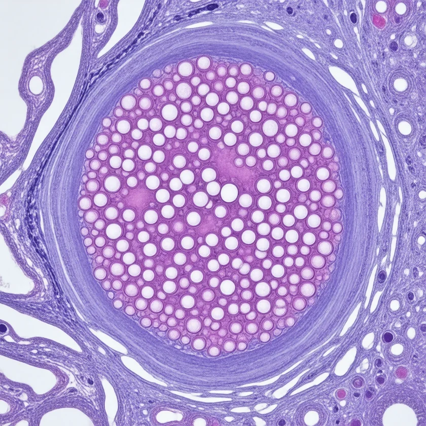
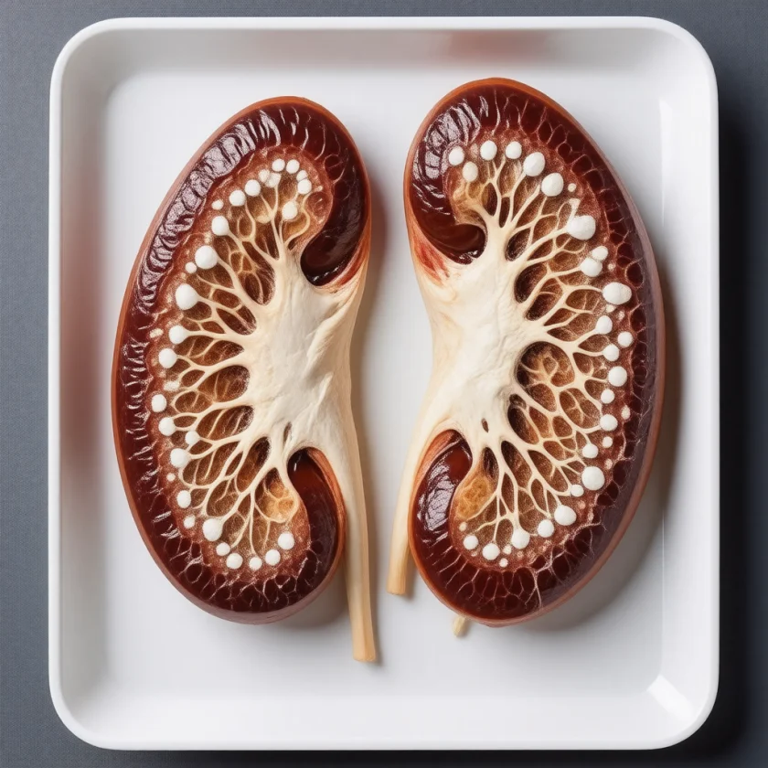

Bartter syndrome is a group of inherited renal tubular disorders caused by mutations, mostly autosomal recessive with X-linked and autosomal dominant forms, that disrupt ion transport in the thick ascending limb (TAL) of the loop of Henle, leading to salt wasting, hypokalemia, metabolic alkalosis, and hyperreninemia with low to normal blood pressure.
Interactive Visualization The Sodium–Potassium Pump — why you have a voltage Run the pump that gives every cell its charge — three sodium out, two potassium in, on Mg-ATP — then drop the magnesium and see exactly why low potassium refuses to correct. Launch →Table of Contents
Overview and Pathophysiology
Bartter syndrome is a group of mostly autosomal recessive renal tubular disorders, with X-linked and autosomal dominant forms, caused by defective ion transport in the thick ascending limb (TAL) of the loop of Henle. The TAL normally reabsorbs approximately 25% of filtered NaCl via the Na-K-2Cl cotransporter (NKCC2). Defects in NKCC2 or its associated channels lead to a cascade of downstream consequences: salt wasting causes volume depletion, which activates the renin-angiotensin-aldosterone system (RAAS), producing secondary hyperaldosteronism. The resulting aldosterone excess drives potassium excretion in the collecting duct, producing hypokalemia and metabolic alkalosis.
The phenotype closely mimics chronic furosemide use, because furosemide exerts its diuretic effect by blocking NKCC2 — the same transporter defective in Bartter syndrome. Critically, increased NaCl delivery to the macula densa stimulates prostaglandin E2 (PGE2) overproduction, which amplifies renin release and contributes to the severe polyuria seen especially in antenatal forms. Despite markedly elevated renin and aldosterone, blood pressure remains normal or low because the primary defect prevents volume retention.
The 2021 European expert consensus describes Bartter syndrome as a rare inherited salt-losing disorder of the kidney tubules with secondary hyperaldosteronism, low blood potassium and low blood chloride with metabolic alkalosis (hypokalemic and hypochloremic metabolic alkalosis), and low to normal blood pressure.
Genetic Types (I through V)
Five numbered genetic types of Bartter syndrome are described, each caused by a mutation in a different component of the TAL transport machinery; a further X-linked form, found in 2016, is listed after them:
- Type I (Antenatal, Severe): Mutation in SLC12A1 encoding the NKCC2 cotransporter itself. Loss of the primary NaCl uptake mechanism produces the most severe salt-wasting phenotype, typically presenting prenatally with massive polyhydramnios.
- Type II (Antenatal): Mutation in KCNJ1 encoding ROMK, the apical potassium channel that recycles K+ back into the tubular lumen to sustain NKCC2 function. Without ROMK, luminal K+ is rapidly depleted and NKCC2 stalls. Severity is similar to Type I. Paradoxically, neonates may initially present with transient hyperkalemia before hypokalemia develops.
- Type III (Classic Bartter, Most Common): Mutation in CLCNKB encoding the basolateral chloride channel ClC-Kb, which exports Cl- from the tubular cell into the interstitium. Type III shows the most variable phenotype, ranging from a mild Gitelman-like presentation to severe antenatal disease depending on the specific mutation.
- Type IV (Bartter with Sensorineural Deafness): Mutation in BSND encoding Barttin, an essential beta-subunit required for membrane trafficking and function of both ClC-Ka and ClC-Kb chloride channels. Because ClC-K channels are also expressed in the stria vascularis of the cochlea, loss of Barttin causes bilateral sensorineural hearing loss in addition to severe renal salt wasting. A digenic form (combined loss-of-function of both CLCNKA and CLCNKB) produces an identical phenotype.
- Type V: Gain-of-function mutation in CASR encoding the calcium-sensing receptor (CaSR). A constitutively active CaSR suppresses NKCC2 and ROMK activity, mimicking Bartter physiology. This type is associated with autosomal dominant hypocalcemia and hypercalciuria alongside the Bartter biochemistry.
- Transient Antenatal Form (MAGED2, X-Linked): A 2016 study found mutations in MAGED2, on the X chromosome, in 13 infants with a transient form of antenatal Bartter syndrome and in two families with unexplained polyhydramnios (excess amniotic fluid). The first signs were more severe than in the other antenatal types, with earlier polyhydramnios and earlier labor. Four patients died around birth and 11 survived, and all symptoms disappeared on their own during follow-up in the infants who survived.
Antenatal Bartter Syndrome (Types I and II)
Antenatal Bartter syndrome (Types I and II) presents prenatally or in the neonatal period and represents the most severe end of the clinical spectrum. Massive fetal polyuria — caused by failure of NaCl reabsorption in the TAL — leads to polyhydramnios (excessive accumulation of amniotic fluid), frequently the first clinical clue detected on routine obstetric ultrasound. Premature birth is common, often occurring before 34 weeks gestation.
Postnatally, affected infants display life-threatening salt wasting with severe dehydration. Serum sodium may be initially elevated (hypernatremia from free water deficit) before transitioning to hyponatremia as the pattern evolves. Profound hypercalciuria is characteristic of Types I and II, leading to nephrocalcinosis — calcium deposits in the renal medulla visible on ultrasound — which can progress to chronic kidney disease if untreated. Failure to thrive and poor weight gain are universal. Elevated prostaglandin E2 levels contribute to episodic fever.
Antenatal diagnosis is possible through amniocentesis (elevated amniotic fluid chloride concentration) or fetal urine sampling, and genetic testing of chorionic villus or amniocyte DNA confirms the mutation. Early initiation of indomethacin after delivery — to suppress prostaglandin synthesis — is the cornerstone of treatment and dramatically improves outcomes.
Classic Bartter Syndrome (Type III)
Type III Bartter syndrome, caused by CLCNKB mutations, is the most common form and typically presents in early childhood, though milder cases may not be recognized until adulthood. The cardinal clinical features are:
- Hypokalemia — often marked (K+ 2.0–3.0 mEq/L), causing muscle weakness, cramps, fatigue, and constipation
- Metabolic alkalosis — elevated bicarbonate (HCO3 often 30–40 mEq/L), sometimes producing tetany from alkalosis-induced reduction in ionized calcium
- Low to normal blood pressure — a key clinical clue, one Gitelman syndrome shares; despite massively elevated renin and aldosterone, blood pressure is low to normal because the primary defect prevents volume expansion
- Polyuria and polydipsia — from impaired urinary concentrating ability
- Salt craving and growth retardation in children
Type III typically does not cause the severe nephrocalcinosis seen in antenatal forms, though some patients develop mild hypercalciuria. The phenotypic variability of Type III is explained by the range of CLCNKB mutations — some cause near-complete loss of ClC-Kb function (severe, antenatal-like), while others preserve partial function (mild, Gitelman-like). This overlap complicates clinical classification and underscores the importance of genetic testing.
A study of 115 patients with CLCNKB mutations sorted them into three clinical pictures: 29.5% antenatal or neonatal (polyhydramnios or diagnosis in the first month of life), 44.5% classic disease diagnosed in childhood, and 26.0% a Gitelman-like picture found by chance in childhood or adulthood. Severe mutations, and missense mutations leaving little channel function, went with a younger age at diagnosis.
Laboratory Findings and Diagnosis
The biochemical signature of Bartter syndrome is distinctive:
- Serum potassium: Low — typically 2.0–3.0 mEq/L
- Serum bicarbonate: Elevated — metabolic alkalosis, HCO3 commonly 30–40 mEq/L
- Serum sodium: Normal to mildly low
- Serum magnesium: Normal to mildly low (mild hypomagnesemia, unlike the marked hypomagnesemia of Gitelman syndrome)
- Plasma renin activity: Markedly elevated
- Plasma aldosterone: Elevated (secondary hyperaldosteronism)
- Blood pressure: Low to normal — this blood pressure in the presence of elevated RAAS activity is a strong clue, although Gitelman syndrome shows the same picture
Urine studies are critical for differential diagnosis:
- Urine chloride: Elevated (>20 mEq/L) — distinguishes Bartter from surreptitious vomiting or bulimia, where urine Cl is low
- Urine calcium: Hypercalciuria in Types I, II, and IV; variable in Type III; low in Gitelman syndrome
- Urinary prostaglandin E2: Elevated, reflecting macula densa overproduction
In a single-center study of 45 children with Bartter or Gitelman syndrome, those with CLCNKB mutations had the lowest serum potassium and magnesium and the highest serum bicarbonate. Fractional excretion of chloride (a measure of how much filtered chloride the kidney lets go into the urine) was above 0.5% in every patient before supplements, and the authors describe it as a useful screening test for kidney salt loss in children with hypokalemic alkalosis.
Renal ultrasound is used to check for nephrocalcinosis, particularly in antenatal-onset cases. Definitive diagnosis is established by next-generation sequencing (NGS) panel targeting SLC12A1, KCNJ1, CLCNKB, BSND, CLCNKA, and CASR.
Differential Diagnosis
Several conditions share features with Bartter syndrome; these are the features that separate them:
- Gitelman Syndrome: Also presents with hypokalemia + metabolic alkalosis + normal blood pressure, but is distinguished by hypocalciuria (vs. Bartter hypercalciuria), significant hypomagnesemia, typically milder symptoms, and later onset (often adolescence or adulthood). Caused by SLC12A3 mutation affecting the NaCl cotransporter (NCC) in the distal convoluted tubule (DCT). The overlap with Type III Bartter can be difficult to resolve without genetic testing.
- Loop Diuretic Abuse (Furosemide/Bumetanide): Produces biochemically identical findings to Bartter syndrome. A urine drug screen, clinical history, and genetic testing separate the two. It is described in people with access to diuretics (eating disorder patients, healthcare workers, athletes).
- Chronic Vomiting / Bulimia Nervosa: Causes hypokalemia and metabolic alkalosis, but urine chloride is low (<20 mEq/L) because the kidneys conserve Cl- in volume depletion from vomiting. Bartter syndrome has high urine Cl.
- Primary Hyperaldosteronism (Conn Syndrome): Shares hypokalemia and metabolic alkalosis but presents with hypertension and suppressed (not elevated) plasma renin activity. Bartter has low to normal blood pressure and markedly elevated renin.
- Liddle Syndrome: Hypokalemia + alkalosis + hypertension from constitutively active ENaC channels; renin and aldosterone are both suppressed. Easily distinguished from Bartter by the presence of hypertension.
- Secondary Hyperaldosteronism from Other Causes: Heart failure, nephrotic syndrome, and cirrhosis can elevate aldosterone, but these are associated with edema, low urine sodium, and the clinical context of the primary disease.
Treatment and Management
Treatment is lifelong and requires a multi-drug regimen targeting both electrolyte replacement and the upstream prostaglandin-driven renin excess:
- Potassium supplementation: Large oral doses are required — often 5–10 mEq/kg/day in children — because the primary tubular defect continuously wastes potassium. Most patients cannot be fully corrected. Intravenous replacement is needed during crises. Potassium chloride is preferred (also corrects the alkalosis). In a study of 54 Korean patients, 94% took potassium chloride and 68% potassium-sparing diuretics, with a mean potassium chloride dose of 5.0 mEq/kg/day under age 18 and 2.1 mEq/kg/day over 18.
- Indomethacin (1–3 mg/kg/day): A non-selective nonsteroidal anti-inflammatory drug (NSAID) that inhibits prostaglandin synthesis, dramatically reducing polyuria, salt wasting, and hypokalemia. Cornerstone therapy for antenatal and childhood Bartter syndromes. It is commonly taken with food, and monitoring covers kidney function (glomerular filtration rate (GFR), creatinine), complete blood count (CBC), and gastrointestinal (GI) symptoms. Can cause premature closure of ductus arteriosus if used prenatally.
- Aldosterone antagonists: Spironolactone (2–5 mg/kg/day) or eplerenone block aldosterone's action at the collecting duct principal cells, reducing K+ wasting. Eplerenone is preferred in adolescent males to avoid spironolactone's antiandrogenic side effects (gynecomastia).
- Amiloride: A potassium-sparing diuretic that directly blocks ENaC in the collecting duct, independent of aldosterone, providing additive K+-sparing benefit.
- Cyclooxygenase-2 (COX-2) selective inhibitors (celecoxib): Alternative to indomethacin with reduced gastrointestinal toxicity; useful in patients who develop gastric ulceration or bleeding on indomethacin.
- Angiotensin-converting enzyme inhibitors (ACE inhibitors): Reduce angiotensin II-driven aldosterone production, providing additional electrolyte stabilization. Blood pressure is monitored during their use.
- High-sodium diet and oral rehydration: To compensate for ongoing salt wasting. Sodium supplements in severe cases.
Monitoring described for this condition includes regular electrolyte panels (every 3–6 months when stable), renal function tests, blood pressure, growth parameters in children, and annual renal ultrasound (nephrocalcinosis surveillance), with audiologic evaluation at diagnosis and periodically for Type IV patients.
What is not known: the 2021 European expert consensus states that, as with many rare diseases, there is a paucity of clinical studies that could guide diagnosis and treatment. The authors of the Korean study describe a large potassium need, along with potassium-sparing drugs, throughout life, while noting that patients tend to improve with age.
Prognosis and Long-Term Outcomes
With consistent treatment, many patients with Bartter syndrome reach near-normal growth and quality of life, but long-term studies give a mixed picture. A single-center study of 45 children with Bartter or Gitelman syndrome reported a satisfactory prognosis during childhood, with growth impaired but within the normal range (median height standard deviation score -1.2). In 54 Korean patients, at a median 8 years of follow-up, 41% had short stature (height below the 3rd percentile) and 11% had chronic kidney disease stage G3 to G5. Cognitive development is unaffected. Life expectancy approaches normal with good adherence to therapy. Several long-term risks are followed over time:
- Progressive nephrocalcinosis: Particularly in Types I and II. In the single-center study, nephrocalcinosis at presentation was found in most children with SLC12A1 and KCNJ1 mutations, in one child with CLCNKB mutations, and in neither of the 2 children with BSND mutations; in the Korean study it was common and improved with age in some patients. Calcium deposits in the renal medulla can reduce glomerular filtration rate over decades, eventually leading to chronic kidney disease (CKD). Early indomethacin reduces hypercalciuria and slows nephrocalcinosis progression. Some patients eventually require renal replacement therapy.
- Kidney decline, including in Type III: Among 77 people with Type III followed for a median of 8 years, chronic renal failure was found in 19 (25%), one needing hemodialysis and four a kidney transplant, and it occurred in all clinical pictures. In the single-center study, median kidney filtration at last follow-up was 71 mL/min/1.73 m2 with SLC12A1 mutations and 62 mL/min/1.73 m2 with KCNJ1 mutations, and pathological albuminuria (protein in the urine) was found in 31 of the 45 children; the authors point to the need to monitor glomerular as well as tubular function.
- Indomethacin nephrotoxicity: Long-term nonsteroidal anti-inflammatory drug use carries the risk of interstitial nephritis and hemodynamic nephrotoxicity. Periodic monitoring of glomerular filtration rate, urinalysis, and blood pressure is the described practice. The indomethacin label calls for the lowest effective dosage for the shortest duration consistent with treatment goals.
- Electrolyte crises: Intercurrent illness (vomiting, diarrhea, fever) can precipitate severe hypokalemia with cardiac arrhythmia risk. These are episodes hospitals treat as urgent, giving intravenous replacement during acute illness.
- Growth outcomes: Children treated early with indomethacin typically achieve catch-up growth. Those diagnosed late or undertreated may have permanent short stature.
- Sensorineural deafness in Type IV: Permanent, bilateral, and progressive. Care described includes early audiology referral and hearing aids or cochlear implant evaluation as needed.
- Pregnancy: Care is shared between kidney and pregnancy specialists; volume changes of pregnancy worsen electrolyte instability. The indomethacin label says to limit NSAID use between about 20 and 30 weeks of pregnancy because of the risk of low amniotic fluid and fetal kidney problems, and to avoid it from about 30 weeks on because of those risks and premature closing of the fetal ductus arteriosus.
Genetic counseling is available to families. In the autosomal recessive forms, both parents of an affected child are obligate carriers of the mutation and each sibling has a 25% risk of being affected.
Key Research Papers
- Simon DB, Karet FE, Hamdan JM, et al. Bartter's syndrome, hypokalaemic alkalosis with hypercalciuria, is caused by mutations in the Na-K-2Cl cotransporter NKCC2. Nat Genet. 1996;13(2):183-188. — PubMed PMID: 8640224
- Simon DB, Karet FE, Rodriguez-Soriano J, et al. Genetic heterogeneity of Bartter's syndrome revealed by mutations in the K+ channel, ROMK. Nat Genet. 1996;14(2):152-156. — PubMed PMID: 8841184
- Simon DB, Bindra RS, Mansfield TA, et al. Mutations in the chloride channel gene, CLCNKB, cause Bartter's syndrome type III. Nat Genet. 1997;17(2):171-178. — PubMed PMID: 9326936
- Birkenhäger R, Otto E, Schürmann MJ, et al. Mutation of BSND causes Bartter syndrome with sensorineural deafness and kidney failure. Nat Genet. 2001;29(3):310-314. — PubMed PMID: 11687798
- Hebert SC. Bartter syndrome. Curr Opin Nephrol Hypertens. 2003;12(5):527-532. — PubMed PMID: 12920401
- Fremont OT, Chan JC. Understanding Bartter syndrome and Gitelman syndrome. World J Pediatr. 2012;8(1):25-30. — PubMed PMID: 22282380
- Kleta R, Bockenhauer D. Bartter syndromes and other salt-losing tubulopathies. Nephron Physiol. 2006;104(2):p73-p80. — PubMed PMID: 16785747
- Seys E, Andrini O, Keck M, et al. Clinical and Genetic Spectrum of Bartter Syndrome Type 3. J Am Soc Nephrol. 2017;28(8):2540-2552. — PubMed PMID: 28381550
- Walsh PR, Tse Y, Ashton E, et al. Clinical and diagnostic features of Bartter and Gitelman syndromes. Clin Kidney J. 2018;11(3):302-309. — PubMed PMID: 29942493
- Proesmans W. Threading through the mizmaze of Bartter syndrome. Pediatr Nephrol. 2006;21(7):896-902. — PubMed PMID: 16773399
- Konrad M, Nijenhuis T, Ariceta G, et al. Diagnosis and management of Bartter syndrome: executive summary of the consensus and recommendations from the European Rare Kidney Disease Reference Network Working Group for Tubular Disorders. Kidney Int. 2021;99(2):324-335. — PubMed PMID: 33509356
- Laghmani K, Beck BB, Yang SS, et al. Polyhydramnios, Transient Antenatal Bartter's Syndrome, and MAGED2 Mutations. N Engl J Med. 2016;374(19):1853-1863. — PubMed PMID: 27120771
- Choi N, Kim SH, Bae EH, et al. Long-term outcome of Bartter syndrome in 54 patients: A multicenter study in Korea. Front Med (Lausanne). 2023;10:1099840. — PubMed PMID: 36993809
Connections
- Nephrology & Hepatology
- The Sodium–Potassium Pump — interactive animation
- Gitelman Syndrome — hypokalemia + alkalosis + hypocalciuria; closest differential
- Nephrogenic Diabetes Insipidus — renal tubular dysfunction causing polyuria
- Alport Syndrome — hereditary nephropathy with progressive kidney disease
- Potassium — primary electrolyte deficiency in Bartter syndrome
- Magnesium — electrolyte management and repletion
- Lab Tests — electrolyte panels and renal function testing