Immunoglobulin A (IgA) Deficiency (Selective Immunoglobulin A (IgA) Deficiency)
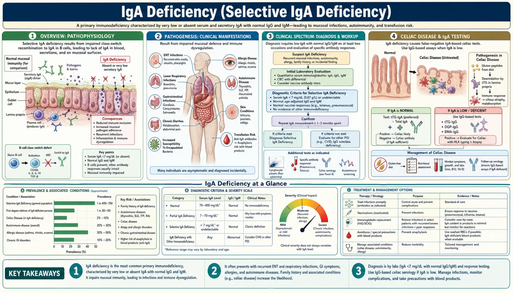
Overview
Selective immunoglobulin A (IgA) deficiency (SIgAD) is the most common primary immunodeficiency worldwide, affecting approximately 1 in 300–500 individuals of Caucasian descent and 1 in 16,000 Japanese individuals — a striking ethnic variation that points to underlying genetic architecture. A 2010 review gives the range as about 1 in 14,840 to 1 in 18,500 in Japan and 1 in 2,600 to 1 in 5,300 in China, compared with about 1 in 223 to 1 in 1,000 in United States community studies (Yel, 2010). It is defined as a serum immunoglobulin A level below 7 mg/dL with normal serum immunoglobulin G (IgG) and immunoglobulin M (IgM) in patients aged 4 years or older, after excluding secondary causes.
Immunoglobulin A is the principal secretory immunoglobulin of the body's mucosal surfaces. Secretory immunoglobulin A (sIgA) is the dominant antibody in saliva, tears, colostrum, breast milk, intestinal secretions, and the respiratory mucosa — the very surfaces that act as the body's first-line barrier against pathogens. Despite being the most abundant antibody class produced each day (approximately 3–5 grams secreted daily across all mucosal surfaces combined), its absence is remarkably well tolerated in many individuals because serum immunoglobulin G and immunoglobulin M can partially compensate.
Partial immunoglobulin A deficiency is defined as serum immunoglobulin A between 7 mg/dL and 70 mg/dL with normal immunoglobulin G and immunoglobulin M. True selective immunoglobulin A deficiency requires immunoglobulin A below 7 mg/dL with normal immunoglobulin G and immunoglobulin M. When immunoglobulin G is also low, the diagnosis shifts toward common variable immunodeficiency (CVID), a more serious disorder that shares genetic overlap with SIgAD.
Pathogenesis
The central defect in selective immunoglobulin A deficiency is a failure of B cell terminal differentiation. Immunoglobulin A-committed B cells are present and circulate in normal numbers, but they cannot complete the class-switch recombination to immunoglobulin A or fully mature into immunoglobulin A-secreting plasma cells. The block occurs late in B cell development — beyond the common lymphoid progenitor and class-switch machinery — making SIgAD distinct from agammaglobulinemias, where B cells are absent entirely.
Genetic associations: The strongest human leukocyte antigen (HLA) association is the A1-B8-DR3 haplotype, particularly common in Northern Europeans — explaining the high prevalence in Caucasian populations. Chromosomal deletions at 22q11.2 and 18q are identified in a subset of patients, especially those with additional dysmorphic features. Mutations in TACI (transmembrane activator and calcium-modulator and cyclophilin ligand interactor; gene TNFRSF13B), encoding a B cell receptor for BAFF (B cell activating factor) and APRIL (a proliferation-inducing ligand), are found in a minority of both SIgAD and CVID patients, reinforcing the shared genetic substrate of these two conditions. Familial clustering occurs, and first-degree relatives of CVID patients have elevated rates of SIgAD.
Secondary (drug-induced) immunoglobulin A deficiency is clinically important because it is reversible. Implicated agents include phenytoin, valproate, sulfasalazine, captopril, D-penicillamine, and gold salts. The mechanism is thought to involve interference with cytokine signaling required for immunoglobulin A class switching. Discontinuing the offending drug typically leads to normalization of immunoglobulin A levels over months.
Clinical Spectrum
One of the most striking features of selective immunoglobulin A deficiency is its clinical heterogeneity. The majority of affected individuals — estimates range from 50% to 85% — are entirely asymptomatic and are discovered incidentally when serum protein electrophoresis or immunoglobulin panels are ordered for unrelated reasons. For these individuals, the diagnosis may carry little practical consequence beyond the blood product risk described below.
Recurrent sinopulmonary infections are the most common symptomatic manifestation. Organisms that colonize mucosal surfaces — Streptococcus pneumoniae and Haemophilus influenzae — are the usual culprits. Affected individuals may have recurrent sinusitis, chronic otitis media, and pneumonia, sometimes progressing to bronchiectasis with repeated pulmonary infections over years.
Gastrointestinal manifestations deserve particular attention. Giardia lamblia is normally cleared from the small intestine by secretory immunoglobulin A; immunoglobulin A-deficient patients are highly susceptible to giardiasis and may suffer protracted, recurrent infections that are difficult to eradicate. Celiac disease occurs 10 times more often in immunoglobulin A-deficient patients than in the general population (discussed separately below). Inflammatory bowel disease and nodular lymphoid hyperplasia of the small bowel are additional gastrointestinal (GI) associations.
Autoimmune diseases are found in about 20–30% of patients, depending on the age range studied (Yel, 2010). Systemic lupus erythematosus (SLE), rheumatoid arthritis, autoimmune thyroid diseases (both Hashimoto's thyroiditis and Graves' disease), vitiligo, type 1 diabetes mellitus, and autoimmune hepatitis are all overrepresented. The mechanism likely involves dysregulation of mucosal immune tolerance — the absence of secretory immunoglobulin A disrupts the normal suppression of immune responses to commensal antigens, tilting the balance toward autoimmunity.
Atopy is paradoxically increased. Asthma, allergic rhinitis, atopic dermatitis, and food allergies are more common in SIgAD patients, possibly because the absence of mucosal immunoglobulin A permits allergen penetration and aberrant T helper 2 (Th2) sensitization at epithelial surfaces.
Celiac Disease and Immunoglobulin A Testing
The intersection of immunoglobulin A deficiency and celiac disease creates a critical diagnostic trap in celiac antibody testing. Celiac disease occurs approximately 10 times more frequently in patients with selective immunoglobulin A deficiency than in the general population — making the co-occurrence far from rare. Yet the standard celiac screening tests are immunoglobulin A-based antibodies: anti-tissue transglutaminase immunoglobulin A (anti-tTG immunoglobulin A) and anti-endomysial immunoglobulin A (EMA-IgA). In a patient with immunoglobulin A deficiency, both of these tests will be falsely negative, even in the presence of active celiac disease with severe villous atrophy.
This false-negative result is not a testing error — it is a predictable consequence of the underlying immunodeficiency. When the immune system cannot produce immunoglobulin A, it cannot produce immunoglobulin A antibodies against tissue transglutaminase, regardless of how much gluten damage is occurring in the small intestine. A patient with unrecognized celiac disease and immunoglobulin A deficiency may receive a falsely reassuring celiac screen and go undiagnosed for years.
The usual safeguard is to measure total serum immunoglobulin A alongside celiac antibody panels. In an Italian multicentre review of 2,098 people diagnosed with celiac disease, 54 (2.6%) had selective IgA deficiency — a 10- to 16-fold increase over the general population. In all 54, the IgA anti-gliadin and anti-endomysial antibodies were absent, while IgG anti-gliadin antibodies were high in 51; the authors wrote that serum IgA is needed to interpret negative IgA antibody results (Cataldo et al., 1998). If serum immunoglobulin A is low or undetectable, immunoglobulin G-based celiac antibodies are used instead — anti-tTG immunoglobulin G and anti-deamidated gliadin peptide immunoglobulin G (anti-DGP immunoglobulin G). These tests retain their sensitivity for celiac disease even in immunoglobulin A-deficient patients. Small bowel biopsy (Marsh grading via duodenal histology) remains the gold standard and does not depend on immunoglobulin class; because some patients with both conditions are negative for IgG anti-gliadin antibodies, the 1998 study's authors called for biopsy in all suspected cases.
Many major laboratory test requisitions now automatically report total immunoglobulin A when anti-tTG immunoglobulin A is ordered, though not every laboratory does. Any immunoglobulin A-based celiac antibody result is only interpretable in the context of the patient's total serum immunoglobulin A level.
Anti-IgA Antibodies and Blood Product Risk
Some people with complete immunoglobulin A deficiency make anti-IgA antibodies. These antibodies are directed against immunoglobulin A determinants that the patient's immune system has never seen — treating exogenous immunoglobulin A as foreign. When such a patient is exposed to immunoglobulin A-containing blood products, pre-formed anti-IgA antibodies can trigger immediate hypersensitivity reactions ranging from urticaria and flushing to severe, life-threatening anaphylaxis. The anti-IgA antibody able to cause this kind of immediate (type I) allergic reaction would be of the immunoglobulin E (IgE) class, but IgE anti-IgA testing is not readily available in most laboratories; immunoglobulin G (IgG) anti-IgA antibodies are measured instead as a screening tool (Yel, 2010).
How often reactions happen: they are rare. A 1995 review estimated IgA anaphylactic transfusion reactions at 1 in 20,000 to 47,000 transfusions. In blood sent for testing after a suspected reaction, 76.3% of the IgA-deficient patients had an IgA antibody, but only 17.5% of all samples did, so most suspected reactions had other causes. IgA deficiency with anti-IgA antibodies was found in about 1 in 1,200 random blood donors, and their antibody levels did not differ from those of patients who had reacted — so a positive anti-IgA test alone does not reliably predict who will react (Sandler et al., 1995). A 2015 review by transfusion specialists is titled “The entity of immunoglobulin A-related anaphylactic transfusion reactions is not evidence based” (Sandler et al., 2015).
Blood products that contain immunoglobulin A include: whole blood, packed red blood cells, fresh frozen plasma, platelet concentrates, and most standard intravenous immunoglobulin (IVIG) preparations. Even trace amounts of immunoglobulin A in washed red blood cells or standard IVIG can be sufficient to trigger anaphylaxis in highly sensitized patients.
Precautions used in practice:
- Anti-IgA antibody testing is used in immunoglobulin A-deficient patients who need blood products or IVIG; as described above, the commonly available test measures IgG anti-IgA antibodies and does not reliably predict a reaction.
- Immunoglobulin A-depleted IVIG exists for when IVIG is truly indicated. The U.S. label of an IgA-depleted freeze-dried IVIG product lists an immunoglobulin A concentration below 1 µg/mL, while the label of a standard liquid IVIG product lists an average of 37 µg/mL. The freeze-dried product's label adds that the immunoglobulin A concentration that will not provoke a reaction is not known, so all IVIG preparations carry some risk of an anaphylactic reaction to immunoglobulin A.
- Washed red blood cells are used for transfusions — repeated washing removes most immunoglobulin A from donor erythrocyte preparations.
- Alert documentation: anti-IgA status is recorded prominently in the medical record, and patients often carry alert documentation (medical alert bracelet, wallet card).
- Anaphylaxis treatment (epinephrine, antihistamines, corticosteroids) is kept immediately available during any blood product administration.
- Pre-medication with antihistamines and corticosteroids can reduce but does not eliminate the risk of transfusion reactions.
It is worth noting that routine IVIG therapy is generally not indicated for uncomplicated SIgAD — secretory immunoglobulin A cannot be replaced by intravenous infusion, and the anaphylaxis risk outweighs the benefit for most patients. IVIG is reserved for the subset who develop CVID-like hypogammaglobulinemia or severe recurrent infections despite other measures.
Evolution to CVID
Selective immunoglobulin A deficiency and common variable immunodeficiency (CVID) are not entirely separate diseases — they exist on a continuum of B cell maturation failure, sharing genetic underpinnings and clinical overlap. Approximately 15–20% of patients with SIgAD will eventually develop CVID over the course of years to decades. Progression from SIgAD to CVID has been reported in several cases — a 2008 report described 4 patients with IgA deficiency and autoimmune features who later developed CVID — but these reports give no overall rate. This transition is defined by a progressive decline in serum immunoglobulin G (and sometimes immunoglobulin M) to levels below normal, accompanied by increasing susceptibility to bacterial infections.
Monitoring: Because of this progression risk, the authors of the 2008 report wrote that all symptomatic IgA-deficient patients, especially those with IgG subclass deficiency or autoimmune features, be monitored for evolution to CVID; annual measurement of serum immunoglobulin levels (immunoglobulin G, immunoglobulin A, immunoglobulin M) is the usual way, particularly for those who are symptomatic or who have relatives with CVID. A falling immunoglobulin G trajectory — even within the low-normal range — is the usual signal for more frequent monitoring and consideration of prophylactic antibiotics or IVIG replacement.
Shared genetic architecture: TACI mutations (TNFRSF13B) are found in both conditions, and families have been described where some members have SIgAD and others have CVID — suggesting that additional genetic modifiers determine whether the defect remains limited to immunoglobulin A or expands to affect immunoglobulin G and immunoglobulin M production. Human leukocyte antigen associations (particularly HLA-DR3 and human leukocyte antigen-B8) are shared across both conditions in European populations.
Practical implication: a diagnosis of SIgAD is not necessarily a static, benign finding. It is a marker of B cell immune dysfunction, and a 2010 review states that a patient with IgA deficiency, once identified, deserves regular follow-up of clinical and immunological findings (Yel, 2010).
Diagnosis
The diagnosis of selective immunoglobulin A deficiency is established by measuring serum immunoglobulin levels. The key criteria are:
- Serum immunoglobulin A below 7 mg/dL on at least two separate measurements.
- Age 4 years or older — physiologically low immunoglobulin A in infants and young children (under 4) does not constitute SIgAD; many infants have transient hypogammaglobulinemia that resolves spontaneously.
- Normal serum immunoglobulin G and immunoglobulin M — if immunoglobulin G is also significantly reduced, CVID is the diagnosis to consider and evaluate.
- Exclusion of secondary causes — review all medications (phenytoin, valproate, sulfasalazine, captopril, D-penicillamine, gold) before attributing immunoglobulin A deficiency to a primary immunodeficiency.
Additional diagnostic workup:
- Anti-IgA antibody testing — often done before blood product administration; as noted above, a positive result does not reliably predict a reaction.
- Celiac screen with immunoglobulin G-based antibodies — anti-tTG immunoglobulin G and anti-DGP immunoglobulin G. Immunoglobulin A-based tests are falsely negative in a confirmed immunoglobulin A-deficient patient.
- Autoimmune panel — antinuclear antibody (ANA), anti-double-stranded DNA (anti-dsDNA), thyroid antibodies (anti-TPO, anti-thyroglobulin), rheumatoid factor as clinically indicated.
- Functional antibody responses — post-vaccination titers (pneumococcal, tetanus) assess whether immunoglobulin G responses are adequate, helping stratify infection risk.
- Genetic testing — TACI/TNFRSF13B sequencing in familial cases or when CVID is also suspected in family members.
- Annual immunoglobulin G monitoring — baseline and annual serum immunoglobulin G to detect early CVID evolution.
Measurement method is nephelometry (the standard for clinical immunoglobulin quantification). False low values can occur with paraprotein interference; if clinical suspicion is high despite low nephelometric immunoglobulin A, protein electrophoresis with immunofixation can clarify.
Treatment
There is no therapy that restores immunoglobulin A production in selective immunoglobulin A deficiency. Management is therefore focused on preventing complications, treating infections promptly, monitoring for disease evolution, and avoiding iatrogenic harm from blood product exposure.
Asymptomatic SIgAD: No pharmacological treatment is needed. Patient information usually covers:
- The anti-IgA antibody risk and blood product precautions.
- The importance of informing any anesthesiologist, surgeon, or blood bank before procedures.
- Annual serum immunoglobulin G monitoring for CVID evolution.
- The need for immunoglobulin G-based celiac antibody testing if celiac disease is ever suspected.
Recurrent sinopulmonary infections: Prophylactic antibiotics (e.g., azithromycin, trimethoprim-sulfamethoxazole) are used for patients with two or more serious infections per year. Pneumococcal and influenza vaccines are commonly given — vaccine responses are generally adequate (immunoglobulin G-mediated) despite immunoglobulin A deficiency. Early and aggressive antibiotic treatment of acute infections prevents bronchiectasis.
IVIG therapy: IVIG labels list as a contraindication immunoglobulin A-deficient patients who have antibodies against immunoglobulin A and a history of hypersensitivity, and warn that immunoglobulin A-deficient patients with antibodies to immunoglobulin A are at greater risk of severe hypersensitivity and anaphylactic reactions. If replacement immunoglobulin is truly necessary (e.g., for evolution toward CVID with falling immunoglobulin G), immunoglobulin A-depleted IVIG preparations (immunoglobulin A content <1 µg/mL) are the ones used, started at low doses with close monitoring in a facility equipped to manage anaphylaxis.
Celiac disease: A strict, lifelong gluten-free diet. Nutritional monitoring (iron, folate, vitamin B12, vitamin D, calcium) for malabsorption. Bone density monitoring (dual-energy X-ray absorptiometry, DEXA scan) given celiac-related osteoporosis risk.
Autoimmune manifestations: Treat according to the specific autoimmune diagnosis (e.g., levothyroxine for hypothyroidism, hydroxychloroquine and sun protection for systemic lupus erythematosus, disease-modifying antirheumatic drugs for rheumatoid arthritis (RA)). The underlying immunoglobulin A deficiency does not typically alter the treatment of the autoimmune condition itself, though some disease-modifying drugs (sulfasalazine, gold, D-penicillamine) can worsen immunoglobulin A deficiency.
Giardiasis: Metronidazole or tinidazole. Recurrent infections may require prolonged treatment or repeated courses. Contaminated water in areas where Giardia is common is a source of reinfection.
Key Research Papers
The following peer-reviewed studies establish the foundational evidence base for selective immunoglobulin A deficiency — its epidemiology, pathogenesis, clinical associations, and management.
- Hammarström L, Vorechovsky I, Webster D. (2000). Selective IgA deficiency (SIgAD) and common variable immunodeficiency (CVID). Clinical and Experimental Immunology 120(2):225–231 — PubMed PMID: 10792368
- Yel L. (2010). Selective IgA deficiency. Journal of Clinical Immunology 30(1):10–16 — PubMed PMID: 20101521
- Cunningham-Rundles C. (2001). Physiology of IgA and IgA deficiency. Journal of Clinical Immunology 21(5):303–309 — PubMed PMID: 11720003
- Cataldo F, Marino V, Ventura A, Bottaro G, Corazza GR. (1998). Prevalence and clinical features of selective immunoglobulin A deficiency in coeliac disease: an Italian multicentre study. Gut 42(3):362–365 — PubMed PMID: 9577342
- Sandler SG, Mallory D, Malamut D, Eckrich R. (1995). IgA anaphylactic transfusion reactions. Transfusion Medicine Reviews 9(1):1–8 — PubMed PMID: 7719037
- Sandler SG, Eder AF, Goldman M, Winters JL. (2015). The entity of immunoglobulin A-related anaphylactic transfusion reactions is not evidence based. Transfusion 55(1):199–204 — PubMed PMID: 25066014
- Liblau RS, Bach JF. (1992). Selective IgA deficiency and autoimmunity. International Archives of Allergy and Immunology 99(1):16–27 — PubMed PMID: 1483063
- Aghamohammadi A, Mohammadi J, Parvaneh N, Rezaei N, Moin M, Espanol T, Hammarstrom L. (2008). Progression of selective IgA deficiency to common variable immunodeficiency. International Archives of Allergy and Immunology 147(2):87–92 — PubMed PMID: 18520152
- International Union of Immunological Societies Expert Committee on Primary Immunodeficiencies; Notarangelo LD, et al. (2009). Primary immunodeficiencies: 2009 update. Journal of Allergy and Clinical Immunology 124(6):1161–1178 — PubMed PMID: 20004777
- Castigli E, Wilson SA, Garibyan L, Rachid R, Bonilla F, Schneider L, Geha RS. (2005). TACI is mutant in common variable immunodeficiency and IgA deficiency. Nature Genetics 37(8):829–834 — PubMed PMID: 16007086
- Schaffer FM, Palermos J, Zhu ZB, Barger BO, Cooper MD, Volanakis JE. (1989). Individuals with IgA deficiency and common variable immunodeficiency share polymorphisms of major histocompatibility complex class III genes. Proceedings of the National Academy of Sciences of the United States of America 86(20):8015–8019 — PubMed PMID: 2573059
- U.S. prescribing information, immune globulin intravenous (human), IgA-depleted freeze-dried preparation (sections 3, 4 and 5.1) — DailyMed label
- U.S. prescribing information, immune globulin infusion (human) 10% liquid (sections 4, 5.1 and 11) — DailyMed label
PubMed Topic Searches: