⚠ Evidence warning: Clinical trials or other evidence do not support the claim that CVID affects men and women equally with a bimodal onset, peaking at ages 1 to 5 and 18 to 25. This page keeps it as a report of what people, websites or other media say.
Common Variable Immunodeficiency (CVID)
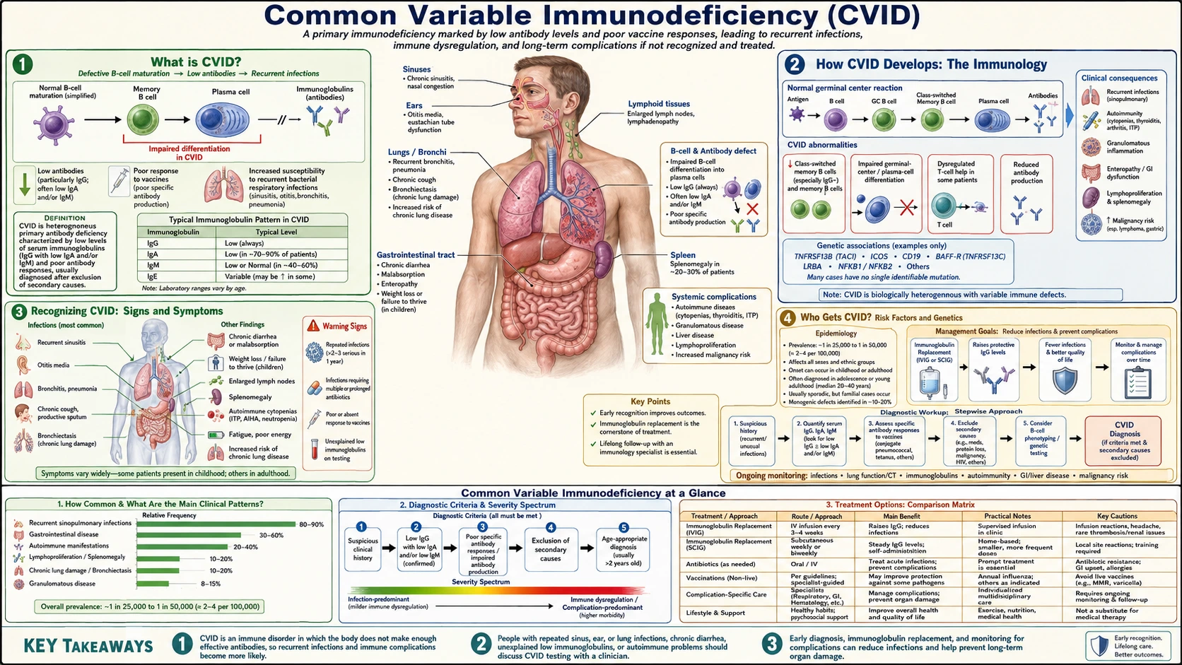
Table of Contents
- How CVID Is Defined
- How CVID Develops: The Immunology
- Recognizing CVID: Signs and Symptoms
- Risk Factors and Genetics: Who Gets CVID
- Diagnosing CVID
- Infections Associated with CVID
- Long-Term Complications
- Treatment: Immunoglobulin Replacement
- Living with CVID: Vaccines, Travel, and Daily Life
- Key Research Papers
- Connections
- Featured Videos
How CVID Is Defined
Common Variable Immunodeficiency (CVID) is the most common symptomatic primary antibody deficiency in adults, affecting approximately 1 in 25,000 people. Despite its name suggesting it is "common," CVID is still a rare disease — the word reflects its status as the most frequently encountered form among the primary immunodeficiencies that actually require treatment. The disease is defined by a failure of the immune system to produce adequate amounts of immunoglobulin (antibodies), particularly immunoglobulin G (IgG), leaving patients unable to mount effective defenses against a wide range of bacterial and other infections.
At its core, CVID represents a breakdown in B-cell maturation. B cells — the white blood cells responsible for making antibodies — are present in relatively normal numbers in most patients with CVID, but they fail to complete their developmental journey from naive B cells into antibody-secreting plasma cells. The result is hypogammaglobulinemia: abnormally low levels of circulating antibodies. Without the protective shield of adequate immunoglobulin G, patients experience recurrent, severe, or unusual infections that would not trouble a person with a normal immune system.
What makes CVID particularly challenging is its diagnostic delay. The average time from first symptoms to confirmed diagnosis is approximately 10 years. Most patients are told repeatedly that their infections are "just bad luck" or attributed to other causes before a clinician measures immunoglobulin levels and recognizes the pattern. During that decade, preventable organ damage — particularly to the lungs — accumulates steadily. Early diagnosis and treatment with immunoglobulin replacement therapy can halt or slow most of this damage.
The delay matters for survival as well. In a New York cohort of 248 patients followed for up to 25 years (Cunningham-Rundles and Bodian, 1999), 64% of men and 67% of women were still alive 20 years after diagnosis, compared with the 92% and 94% expected for people of the same age and sex. Lower immunoglobulin G levels, weaker T-cell responses and a smaller share of B cells in the blood were the features linked to death in that period.
CVID affects men and women equally and is diagnosed across a wide age range, but it has a bimodal onset: one peak in early childhood (ages 1–5) and a larger peak in young adulthood (ages 18–25). In the same 1999 cohort (patients aged 3 to 79), the median age at which symptoms began was 23 years for men and 28 years for women, and diagnosis came on average at 29 years for men and 33 years for women. In a larger New York cohort of 473 patients (Resnick and colleagues, 2012), the median age at diagnosis was 30 years for men and 33.5 years for women. The timing of symptom onset influences the pattern of organ damage seen by the time of diagnosis.
How CVID Develops: The Immunology
The immune system's antibody-making machinery depends on a precisely choreographed series of developmental steps. Understanding where this process goes wrong in CVID helps explain both the symptoms and the treatment logic.
Normal B-cell development begins in the bone marrow, where immature B cells learn to recognize foreign invaders through their surface receptors. After leaving the bone marrow, naive B cells circulate until they encounter an antigen — a fragment of a bacterium, virus, or other pathogen. When a B cell meets the right antigen together with help from T helper cells, it enters a germinal center reaction inside lymph nodes and the spleen. Inside the germinal center, B cells undergo two critical transformations: somatic hypermutation (fine-tuning their antibody to bind the antigen more tightly) and class-switch recombination (changing their antibody class from immunoglobulin M (IgM) to immunoglobulin G, immunoglobulin A (IgA), or immunoglobulin E (IgE) depending on the type of threat). Cells that complete this process successfully become long-lived plasma cells that secrete antibodies for months or years, and memory B cells that respond rapidly to future encounters with the same pathogen.
In CVID, this germinal center process is defective. Most patients have a dramatically reduced number of class-switched memory B cells — the cells that have completed the immunoglobulin M-to-IgG transition. B cells are present, they can start the process, but they cannot finish it efficiently. The result is a B-cell population that is largely stuck at the naive or transitional stage, unable to generate the durable immunoglobulin G responses needed for long-term protection.
T-cell dysfunction also plays a contributing role. Regulatory T cells are often abnormally expanded in CVID, and the signaling molecules that coordinate B-T cell cooperation — including CD40-CD40L interactions and cytokines like IL-21 — are disrupted in many patients. This means the problem is not confined to B cells alone; the entire adaptive immune architecture is dysregulated.
The consequence of this bottleneck is threefold: immunoglobulin G levels fall below the threshold needed to opsonize (coat) bacteria for destruction by neutrophils; immunoglobulin A levels — the antibody class that guards mucosal surfaces like the respiratory and gastrointestinal tracts — drop precipitously; and immunoglobulin M, while sometimes relatively preserved, cannot compensate for the loss of the other classes. Vaccines, which work by training B cells to produce long-lived immunoglobulin G memory responses, fail to generate meaningful protection in most CVID patients.
Recognizing CVID: Signs and Symptoms
The clinical picture of CVID is dominated by infections, but the range of possible manifestations is broad enough that the diagnosis is frequently missed for years. Clinicians who are not specifically thinking about primary immunodeficiency may attribute the pattern to smoking, asthma, allergy, or "just being prone to infections."
Recurrent Respiratory Infections
The most consistent and early presentation is recurrent sinopulmonary infections — sinusitis, otitis media, bronchitis, and pneumonia occurring more frequently, more severely, and with more unusual organisms than expected. The responsible pathogens are typically encapsulated bacteria — particularly Haemophilus influenzae and Streptococcus pneumoniae — because opsonizing antibodies are the primary mechanism for clearing these organisms. Without adequate immunoglobulin G, the immune system cannot efficiently tag these bacteria for destruction. Patients may have 3–5 pneumonias per year, or chronic sinusitis that never fully resolves despite multiple antibiotic courses.
Gastrointestinal Symptoms
Chronic diarrhea, malabsorption, and weight loss affect a significant minority of CVID patients. The most important pathogen to consider is Giardia lamblia, a parasitic protozoan that colonizes the small intestine and causes chronic, watery, foul-smelling diarrhea with fat malabsorption. Giardia is normally cleared by secretory immunoglobulin A in the gut mucosa; without it, the organism establishes a persistent infection that responds poorly to standard single-course treatment and requires repeated antibiotic therapy. Other gastrointestinal manifestations include a Crohn's-like inflammatory bowel picture, celiac-like villous atrophy (without the anti-tissue transglutaminase antibodies seen in true celiac disease), and nodular lymphoid hyperplasia of the gut.
Inflammatory and Autoimmune Features
Paradoxically, a dysregulated immune system that cannot make effective antibodies against bacteria can still produce autoantibodies against the body's own tissues. Approximately 25% of CVID patients develop autoimmune complications, the most common of which are immune thrombocytopenic purpura (ITP, low platelets from autoantibody-mediated destruction) and autoimmune hemolytic anemia. Pernicious anemia (autoantibodies against intrinsic factor, impairing B12 absorption) is also seen. These autoimmune manifestations can precede or follow the infectious phenotype and sometimes dominate the clinical picture.
Lymphoproliferative Features
Enlarged lymph nodes (lymphadenopathy) and an enlarged spleen (splenomegaly) are found in a substantial proportion of CVID patients. The lymphoid tissue is trying — and failing — to mount germinal center reactions, resulting in chronically stimulated, enlarged nodes. Splenomegaly can cause hypersplenism (excessive destruction of blood cells by the spleen) and left-sided abdominal discomfort.
Risk Factors and Genetics: Who Gets CVID
CVID is predominantly a sporadic disease — meaning most patients have no affected first-degree relatives and no identifiable inherited mutation. However, genetic factors clearly contribute to susceptibility, and a subset of patients have identifiable single-gene defects.
Identified Genetic Mutations
Only about 10–20% of CVID patients have a currently identifiable monogenic cause. The most important single-gene defects include:
- TACI (TNFRSF13B): Mutations in this B-cell receptor gene are the most commonly found genetic variant in CVID, present in roughly 8–10% of patients. TACI helps regulate B-cell survival and class-switch recombination. However, TACI variants are also found in healthy relatives of CVID patients, suggesting they act as susceptibility modifiers rather than definitive causes in most cases.
- ICOS: Inducible co-stimulator, a T-cell surface protein critical for providing help to B cells during germinal center reactions. ICOS deficiency causes a relatively severe CVID phenotype with marked T-cell dysfunction.
- CD19, CD21, CD81: Components of the B-cell co-receptor complex that fine-tune B-cell activation thresholds. Loss of these proteins impairs B-cell responses to T-cell help.
- NFKB1 and NFKB2: Transcription factors regulating B-cell and T-cell survival and function. NFKB1 haploinsufficiency is one of the more commonly identified dominant mutations and can be associated with autoimmune features.
- LRBA (Lipopolysaccharide-Responsive Beige-Like Anchor): LRBA deficiency causes a particularly severe CVID phenotype with prominent autoimmunity and CTLA4 pathway disruption. Importantly, these patients may respond to abatacept (a CTLA4-Ig fusion protein) and are candidates for hematopoietic stem cell transplantation.
- CTLA4 haploinsufficiency: Causes dysregulated T-cell activation and a severe immune dysregulation syndrome overlapping with CVID, with prominent autoimmunity and lymphocytic infiltration of multiple organs.
Familial CVID
Approximately 10–20% of CVID patients have a first-degree relative with CVID or selective immunoglobulin A deficiency (a milder antibody deficiency affecting only immunoglobulin A). The co-occurrence of these two conditions in families suggests they exist on a spectrum of the same underlying genetic susceptibility. For relatives who have frequent infections, a blood test of immunoglobulin levels is the test that shows whether they share the antibody deficiency.
No Identifiable Cause
For the majority of patients — roughly 80–90% — no causative mutation is found with current genetic testing panels. This does not mean the disease is not genetic; it means the relevant variants are likely polygenic (involving many small-effect genetic differences) or involve genes not yet discovered. Research in this area is active and the proportion of patients with identified mutations continues to grow as sequencing panels expand.
Diagnosing CVID
CVID diagnosis requires meeting a specific set of criteria and carefully excluding other causes of low immunoglobulins. The most widely used diagnostic criteria require all four of the following:
- Immunoglobulin G below 4 g/L (at least 2 standard deviations below the mean for age) on at least two separate measurements
- Immunoglobulin A below 0.7 g/L and/or immunoglobulin M below normal for age
- Age greater than 4 years (to exclude transient hypogammaglobulinemia of infancy, a self-resolving condition)
- Absent or poor antibody response to vaccines — failure to mount a protective antibody titer after immunization with protein antigens (tetanus, diphtheria) and/or polysaccharide antigens (pneumococcal polysaccharide vaccine). This is the most functionally critical criterion.
Additionally, secondary causes of hypogammaglobulinemia must be excluded. These include protein-losing conditions (nephrotic syndrome, protein-losing enteropathy), hematologic malignancies (chronic lymphocytic leukemia, lymphoma), medications (rituximab, mycophenolate, carbamazepine), and thymoma-associated hypogammaglobulinemia.
The Vaccine Challenge Test
The vaccine response test is arguably the most important diagnostic step and yet the most frequently omitted in non-specialist settings. The standard approach is to measure baseline antibody titers to pneumococcal serotypes and tetanus, then administer both 23-valent pneumococcal polysaccharide vaccine and tetanus toxoid, and recheck titers 4–8 weeks later. A protective response is defined as a fourfold rise in tetanus titer and/or a protective rise (to ≥1.3 μg/mL) in at least 70% of measured pneumococcal serotypes. CVID patients typically show minimal or no titer increase despite vaccination.
B-Cell Phenotyping
Flow cytometry of peripheral blood B cells helps characterize the specific maturation block and can distinguish CVID from X-linked agammaglobulinemia (XLA), in which B cells are virtually absent. In CVID, total B-cell numbers are often near-normal but switched memory B cells (CD19+CD27+IgD-IgM-) are markedly reduced. The EUROclass classification system uses B-cell and T-cell phenotyping to stratify CVID patients into subgroups with different complication risks — patients with very low switched memory B cells and elevated transitional B cells have higher rates of splenomegaly, granulomatous disease, and lymphoma.
Infections Associated with CVID
The infectious complications of CVID reflect the specific gap in immune defense created by antibody deficiency. Understanding which organisms are particularly dangerous helps clinicians and patients recognize early warning signs.
Encapsulated Bacteria: The Primary Threat
Bacteria with polysaccharide capsules — Streptococcus pneumoniae, Haemophilus influenzae, Neisseria meningitidis — are the organisms for which opsonizing immunoglobulin G antibodies are most critical. The capsule makes these bacteria invisible to phagocytes (neutrophils and macrophages) unless coated with antibody. Without adequate immunoglobulin G, these organisms cause unusually severe and recurrent infections: pneumonia, meningitis, septicemia, and otitis media. Two bouts of pneumococcal pneumonia, or one of meningococcal meningitis, are among the patterns that prompt testing for antibody deficiency.
Giardia lamblia: Chronic Gastrointestinal Infection
Giardia is so frequently associated with CVID that the combination of recurrent respiratory infections and chronic diarrhea is a recognised signal to test for both conditions at the same time. Giardia trophozoites attach to the duodenal mucosa and disrupt brush-border enzyme function, causing fat and carbohydrate malabsorption, bloating, flatulence, and weight loss. In immunocompetent individuals, Giardia infection is usually self-limited or responds to a single course of metronidazole or tinidazole. In CVID, eradication is much harder — the parasite recurs repeatedly because the secretory immunoglobulin A that normally prevents recolonization is absent. Multiple treatment courses with extended durations are often needed.
Enterovirus: A Rare but Serious Threat
Non-polio enteroviruses (echovirus, coxsackievirus) are normally controlled by neutralizing immunoglobulin G antibodies. In CVID patients, these viruses can cause chronic meningoencephalitis — a progressive, fatal inflammatory brain and spinal cord infection that is extremely difficult to treat. This complication underscores the importance of achieving adequate immunoglobulin G trough levels with immunoglobulin replacement therapy. High-dose IVIG (which contains high titers of neutralizing anti-enterovirus antibodies) is the primary treatment, sometimes with the addition of pleconaril (when available).
Respiratory Viruses and Atypical Organisms
CVID patients are also at increased risk from influenza (which can cause severe pneumonia), Mycoplasma pneumoniae (which can cause persistent respiratory tract infection), and Campylobacter (which can cause chronic or recurrent bacteremic diarrhea). Opportunistic infections — the fungi and parasites that afflict patients with T-cell deficiencies — are not typical of CVID; if they occur, this suggests a more complex combined immunodeficiency rather than pure CVID.
Long-Term Complications
Even with immunoglobulin replacement therapy, CVID is associated with significant organ complications that account for much of the morbidity and mortality beyond infection. The cumulative disease burden from these non-infectious complications is increasingly recognized as the dominant long-term management challenge.
Bronchiectasis: Lasting Airway Damage
In the 473-patient New York cohort followed over four decades (Resnick and colleagues, 2012), bronchiectasis was recorded in 11.2% of patients and chronic lung disease of any kind in 28.5%; in a 2024 UK registry study limited to 129 patients with "complex" CVID (those who already had non-infectious complications), bronchiectasis was found in 64%. Bronchiectasis is permanent, irreversible dilation and scarring of the bronchial walls caused by repeated infections and chronic inflammation. In bronchiectasis, the normal mucociliary clearance mechanism is disrupted, creating pockets where bacteria pool and breed. The condition becomes self-perpetuating: bacterial colonization drives inflammation, which causes more structural damage, which impairs clearance, which enables more infection. Computed tomography (CT) chest scanning is the gold standard for detecting bronchiectasis; specialist centres typically scan at diagnosis and at intervals afterwards. Management includes aggressive antibiotic treatment of exacerbations, airway clearance physiotherapy, and optimizing immunoglobulin G trough levels.
Non-Infectious Complications and Survival
In the same 473-patient cohort, 94% of patients had a history of infections and 68% also had at least one non-infectious complication. Over the follow-up, 19.6% of patients died, a shorter survival than people of the same age and sex. The risk of death was 11 times higher for patients with non-infectious complications (hazard ratio 10.95). Deaths were linked to lymphoma, any form of hepatitis, lung impairment and gut disease with or without malabsorption, but not to bronchiectasis, autoimmunity, other cancers, granulomatous disease or earlier removal of the spleen.
Autoimmunity: 25% of Patients
The autoimmune complications of CVID are paradoxical but well-documented. Immune thrombocytopenic purpura (ITP) is the most common, causing easy bruising and bleeding from platelet counts that may fall below 10 × 10&sup9;/L. Autoimmune hemolytic anemia (AIHA) follows a similar mechanism — antibodies against red blood cell surface antigens trigger their premature destruction. Pernicious anemia (autoantibodies against intrinsic factor or parietal cells impairing vitamin B12 absorption) is also seen at higher rates than in the general population. Management of these conditions in CVID is complicated by the fact that the usual first-line treatments (corticosteroids, rituximab) further suppress an already compromised immune system — so they are used cautiously, with careful monitoring for infectious complications.
Granulomatous-Lymphocytic Interstitial Lung Disease (GLILD)
GLILD is a severe, progressive pulmonary complication affecting an estimated 10–20% of CVID patients, characterized by non-caseating granulomas and lymphocytic infiltrates in the lung parenchyma. Clinically, it resembles sarcoidosis and causes progressive breathlessness, restrictive lung physiology, and hypoxemia. GLILD is often associated with splenomegaly, lymphadenopathy, and liver involvement. It is not caused by infection — cultures and stains are negative — but rather reflects the dysregulated immune activation characteristic of CVID. GLILD can progress, and the drugs used against it are immunosuppressive (steroids, azathioprine, mycophenolate, rituximab) despite the concern about suppressing an already weak immune system. Experts do not agree on whether to treat at once or first watch while optimising immunoglobulin replacement; when treatment is given, corticosteroids come first in the UK consensus described below.
A 2017 UK expert consensus (British Lung Foundation and the UK Primary Immunodeficiency Network, a three-round Delphi survey of specialists) agreed a definition: GLILD is a distinct lung disease in people with CVID, with lymphocytes and/or granulomas in the lung, diagnosed after other conditions have been considered and where possible excluded. The experts agreed the work-up includes a chest CT scan and lung function tests (including gas transfer). On treatment there was no consensus on whether watching and optimising immunoglobulin first is acceptable (67% agreed, 25% disagreed); 90% agreed that when treatment is needed, corticosteroids alone are the first-line choice. A 2023 Italian series of six patients given rituximab as their first treatment reported better symptoms and quality of life, improved lung function and improved CT scans, with no severe side effects; it had no randomised comparison group.
In 2026 the European Respiratory Society, working with the European Society for Immunodeficiencies, published the first clinical practice guideline on diagnosing GLILD. It recommends screening all adults with CVID for GLILD, preferably with high-resolution CT, with evaluation by a team from several specialties. Lung infection is excluded and lymphocytes are looked for in lung washings (bronchoalveolar lavage); a lung biopsy is not routinely required but is used for atypical cases, and lung function including gas transfer is measured. Most of its recommendations are conditional because the certainty of the evidence is low.
Lymphoma: A Raised Risk
Lymphoma is more common in CVID than in the general population, with non-Hodgkin's B-cell lymphomas predominating. In the 473-patient New York cohort, lymphoma occurred in 8.2% of patients, women were more likely than men to develop it, and lymphoma was one of the complications linked to death. The mechanisms are not fully understood but likely involve chronic antigenic stimulation of B cells, viral oncogenesis (Epstein-Barr virus in particular), and the background of immune dysregulation. Usual CVID care includes baseline lymphocyte subset testing and follow-up for growing lymph nodes, "B symptoms" (unexplained fever, night sweats, weight loss) and an enlarging spleen. Early recognition matters because treatment is possible.
Liver and Gastrointestinal Complications
Nodular regenerative hyperplasia (NRH) of the liver — a non-cirrhotic form of portal hypertension — occurs in a subset of CVID patients, often without obvious symptoms until portal hypertension has developed. Liver function tests may be only mildly abnormal, which is why specialist centres use periodic liver imaging. Gastrointestinal (GI) complications including inflammatory bowel-like disease and chronic Giardia can cause malabsorption leading to micronutrient deficiencies (iron, B12, folate, fat-soluble vitamins).
Treatment: Immunoglobulin Replacement
The cornerstone of CVID management is lifelong immunoglobulin replacement therapy — providing the immunoglobulin G antibodies the patient's own immune system cannot produce. This treatment, developed in the 1950s and progressively refined since, transforms CVID from a rapidly fatal disease into a manageable chronic condition.
How Immunoglobulin Replacement Works
Commercial immunoglobulin preparations (IVIG or SCIG) are pooled from the plasma of thousands of healthy donors and contain a broad, representative spectrum of immunoglobulin G antibodies against virtually all common pathogens. When infused into a CVID patient, these donor antibodies provide passive protection: they coat bacteria, neutralize viruses, and support phagocytic killing just as the patient's own antibodies would if the patient could make them. The protection is real but temporary — immunoglobulin G has a half-life of approximately 3–4 weeks in the bloodstream, which is why replacement must be given indefinitely and regularly.
Intravenous Immunoglobulin (IVIG)
Standard IVIG dosing is 400–600 mg/kg every 3–4 weeks, administered as an intravenous infusion typically taking 2–4 hours. The goal is to maintain an immunoglobulin G trough level above 5–7 g/L — the minimum associated with meaningful reduction in infection frequency. In patients with established bronchiectasis, higher targets (above 8 g/L) are often pursued. Dose and frequency adjustments are guided by trough immunoglobulin G levels measured just before each infusion, and by clinical response — when a patient is still having breakthrough infections at a given trough, the usual response is a higher dose. Side effects of IVIG include headache, flushing, nausea, and (rarely) aseptic meningitis or thromboembolic events. Pre-medication with acetaminophen and antihistamines reduces infusion reactions in susceptible patients.
Subcutaneous Immunoglobulin (SCIG)
An increasingly popular alternative, SCIG is administered as a slow infusion into subcutaneous tissue — typically the abdomen or thigh — using a small pump. Dosing is weekly or twice-weekly, at a fraction of the monthly IVIG dose (roughly 130–150% of the monthly IVIG dose divided across weekly administrations, to account for lower subcutaneous bioavailability). The advantages of SCIG are significant for many patients: it can be done at home, the smaller more frequent doses produce more stable immunoglobulin G levels with smaller peaks and troughs, and systemic side effects (headache, fatigue) are less common because the rate of absorption is slower. Patient satisfaction rates with SCIG are generally high. The main limitation is the volume required — patients with large monthly doses may need multiple simultaneous infusion sites.
Monitoring and Targets
Management of CVID requires ongoing monitoring beyond immunoglobulin levels. Key monitoring components include:
- Immunoglobulin G trough levels at each infusion visit — the single most actionable laboratory parameter
- Pulmonary function tests (spirometry and diffusion capacity) annually, or more frequently if respiratory symptoms develop
- Computed tomography chest at baseline and every 3–5 years (or immediately if clinical deterioration occurs) to monitor for bronchiectasis and GLILD
- Abdominal imaging (ultrasound or computed tomography) for spleen and liver size annually
- Lymphocyte subsets (B-cell and T-cell phenotyping) at baseline and periodically to track immunological progression
- Complete blood count (CBC) and liver function tests at regular intervals for autoimmune and hepatic complications
- Vitamin B12, iron, and folate levels if gastrointestinal symptoms suggest malabsorption
Additional Treatments
For patients with bronchiectasis, regular airway clearance physiotherapy (e.g., oscillating positive expiratory pressure devices, active cycle of breathing) is used to clear pooled mucus. Antibiotic prophylaxis with azithromycin or doxycycline is used in patients with frequent exacerbations despite optimal immunoglobulin G replacement. For autoimmune complications refractory to first-line immunosuppression, newer agents including rituximab, abatacept (particularly effective in LRBA/CTLA4 deficiency), and sirolimus are used. Hematopoietic stem cell transplantation remains investigational for CVID but has been successfully performed in patients with identified LRBA deficiency or CTLA4 haploinsufficiency with severe autoimmunity.
Living with CVID: Vaccines, Travel, and Daily Life
Living with CVID requires a different relationship with the healthcare system, with vaccines, and with everyday infection-risk situations. The practical adjustments are manageable, but they require clear knowledge of what is safe and what is dangerous.
Vaccines: What Is Allowed and What Is Not
This is one of the most critical pieces of information for any CVID patient: live vaccines are listed as contraindicated. Live vaccines contain attenuated (weakened) but still replication-competent organisms. In a person with normal immunity, the organism replicates briefly and is then cleared by the immune response it stimulates. In a CVID patient with impaired humoral immunity, these organisms can replicate unchecked and cause the actual disease the vaccine was meant to prevent.
Live vaccines not given in CVID:
- MMR (measles-mumps-rubella)
- Varicella (chickenpox) and zoster (shingles) live vaccine
- Live oral typhoid vaccine (Ty21a)
- Live attenuated influenza vaccine (nasal spray flu vaccine, LAIV)
- BCG (tuberculosis vaccine used in some countries)
- Yellow fever vaccine (live, required for entry to some countries)
- Oral polio vaccine (not used in the US but still administered in some countries)
Non-live vaccines (inactivated or subunit) used in CVID:
- Inactivated influenza vaccine (injectable, not nasal spray) — given yearly in CVID even though the response is poor, as partial protection may occur in some patients
- Non-live COVID-19 vaccines (mRNA and protein-subunit types; responses are reduced)
- Recombinant zoster vaccine — the shingles vaccine used in CVID, as it is not live
- Tetanus, diphtheria, pertussis (Tdap/Td booster) — safe but typically generates poor response
- Vi polysaccharide typhoid vaccine (injection) — the non-live alternative to oral typhoid
- Hepatitis A and B vaccines — non-live and given as a baseline, even if responses are suboptimal
Travel Considerations
Travel takes planning for CVID patients, and travel medicine clinics familiar with immunodeficiency handle these trips. The usual considerations include: an adequate supply of immunoglobulin for the duration of the trip (and a plan for infusion if the trip is extended), documentation from the treating immunologist explaining the diagnosis and current treatment, knowing the medical facilities at the destination, the fact that the live yellow fever vaccine cannot be given (some countries that require it accept a medical waiver), and the fact that even safe vaccines may produce diminished responses in CVID.
Infection Prevention in Daily Life
With well-maintained immunoglobulin G levels, many CVID patients live largely normal lives. The precautions described for reducing infection risk include frequent handwashing, limiting close contact with people who have received live vaccines (particularly oral polio vaccine, if traveling to regions where it is used), prompt treatment of respiratory symptoms rather than "waiting to see," a standby course of antibiotics prescribed for sinopulmonary exacerbations when access to medical care may be delayed, and household contacts receiving inactivated (not live) flu vaccines.
Mental Health and Quality of Life
The psychological burden of CVID — the decade-long diagnostic odyssey, the lifelong infusion schedule, the uncertainty about future complications, and the fatigue of managing a chronic illness — is substantial and often underappreciated. Non-profit patient organisations for primary immunodeficiency offer peer support networks, educational resources, and help with insurance and treatment access, and many newly diagnosed patients find them useful.
Key Research Papers
- Bonilla FA, Khan DA, Ballas ZK, et al. Practice parameter for the diagnosis and management of primary immunodeficiency. J Allergy Clin Immunol. 2015;136(5):1186-1205. — PubMed PMID: 26371839
- Cunningham-Rundles C. The many faces of common variable immunodeficiency. Hematology Am Soc Hematol Educ Program. 2012;2012:301-305. — PubMed PMID: 23233596
- Ameratunga R, Brewerton M, Slade C, et al. Comparison of diagnostic criteria for common variable immunodeficiency disorder. Front Immunol. 2014;5:415. — PubMed PMID: 25309532
- Cunningham-Rundles C, Bodian C. Common variable immunodeficiency: clinical and immunological features of 248 patients. Clin Immunol. 1999;92(1):34-48. — PubMed PMID: 10413651
- Bogaert DJ, Dullaers M, Lambrecht BN, et al. Genes associated with common variable immunodeficiency: one diagnosis to rule them all? J Med Genet. 2016;53(9):575-590. — PubMed PMID: 27250108
- Resnick ES, Moshier EL, Godbold JH, Cunningham-Rundles C. Morbidity and mortality in common variable immune deficiency over 4 decades. Blood. 2012;119(7):1650-1657. — PubMed PMID: 22180439
- Hurst JR, Verma N, Lowe D, et al. British Lung Foundation/United Kingdom Primary Immunodeficiency Network consensus statement on the definition, diagnosis, and management of granulomatous-lymphocytic interstitial lung disease in common variable immunodeficiency disorders. J Allergy Clin Immunol Pract. 2017;5(4):938-945. — PubMed PMID: 28351785
- Quinti I, Soresina A, Guerra A, et al. Effectiveness of immunoglobulin replacement therapy on clinical outcome in patients with primary antibody deficiencies: results from a multicenter prospective cohort study. J Clin Immunol. 2011;31(3):315-322. — PubMed PMID: 21365217
- Bintalib HM, Grigoriadou S, Patel SY, et al. Investigating pulmonary and non-infectious complications in common variable immunodeficiency disorders: a UK national multi-centre study. Front Immunol. 2024;15:1451813. — PubMed PMID: 39318627
- Tessarin G, Baronio M, Gazzurelli L, et al. Rituximab monotherapy is effective as first-line treatment for granulomatous lymphocytic interstitial lung disease (GLILD) in CVID patients. J Clin Immunol. 2023;43(8):2091-2103. — PubMed PMID: 37755605
- Bintalib HM, van de Ven AAJM, Rojas-Reyes MX, et al. Clinical practice guideline for the diagnosis of granulomatous-lymphocytic interstitial lung disease in patients with common variable immunodeficiency disorders: an ERS Clinical Research Collaboration. Eur Respir J. 2026;68(3). — PubMed PMID: 42350065
Search PubMed for more: Common Variable Immunodeficiency on PubMed
Connections
- Immunology
- Bronchiectasis
- Thrombocytopenia (ITP)
- Giardiasis
- Anemia
- X-Linked Agammaglobulinemia
- Chronic Granulomatous Disease
- Immunoglobulin Lab Tests
- All Conditions
- Edelman & Porter — the structure of the antibody that is missing, and how IVIG replaces it
- Genetic Susceptibility to Vaccine Adverse Reactions — live-vaccine caution and vaccine failure from absent antibody response