Phenylketonuria
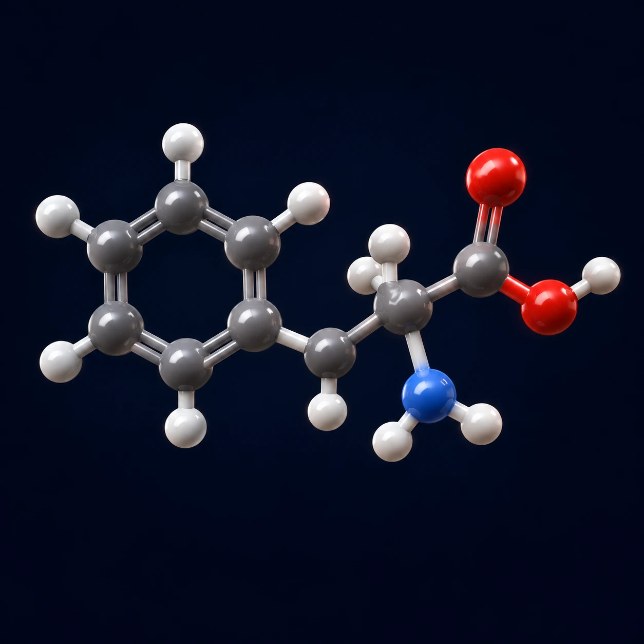
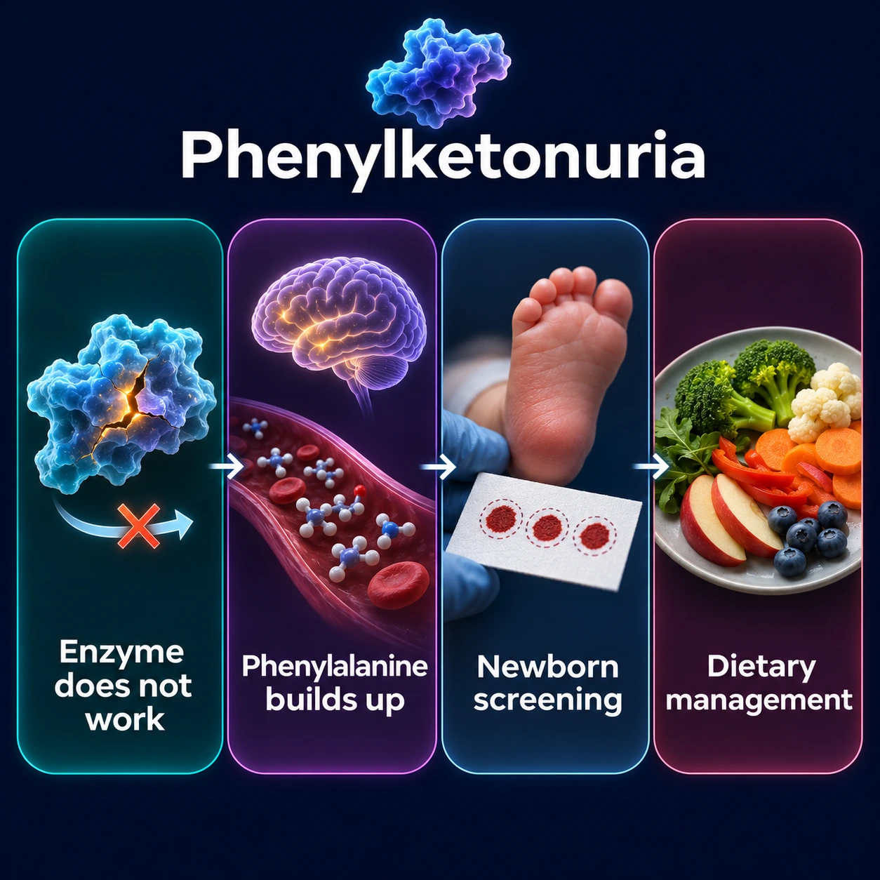

Overview and Epidemiology
Phenylketonuria (PKU) is an autosomal recessive inborn error of amino acid metabolism caused by mutations in the PAH gene, which encodes the enzyme phenylalanine hydroxylase (PAH), located on chromosome 12q23.2. PAH normally catalyzes the irreversible hydroxylation of the essential amino acid phenylalanine (Phe) to tyrosine, a reaction requiring tetrahydrobiopterin (BH4) as an essential cofactor. When PAH activity is severely reduced or absent, phenylalanine accumulates to toxic concentrations in blood and brain — a condition termed hyperphenylalaninemia. Left untreated, the resulting neurotoxicity causes profound and irreversible intellectual disability. Detected at birth and treated promptly, children with PKU can achieve completely normal cognitive development.
PKU affects approximately 1 in 10,000–1 in 15,000 newborns in populations of predominantly Caucasian ancestry. Prevalence varies substantially by ethnicity and geography: Ireland has one of the highest rates (~1 in 4,500), Turkey ~1 in 4,000–1 in 6,000, and parts of Eastern Europe are also heavily affected. Conversely, prevalence is considerably lower in African and East Asian populations. A 2020 worldwide genetic study (16,092 affected people in 51 countries) estimated that about 0.45 million people have PKU, a global prevalence of 1 in 23,930 live births, ranging from 1 in 4,500 in Italy to 1 in 125,000 in Japan; 62% had classic PKU, 22% mild PKU and 16% mild hyperphenylalaninemia. Within Europe classic PKU is most common in the east, and the share of classic PKU rises from Europe (56%) to the Middle East (71%) and Australia (80%). More than 950 PAH gene variants have been identified in people with PKU (Blau 2016), catalogued in ClinVar and the PAHvdb database; missense mutations predominate, but nonsense, frameshift, splice-site, and large deletion variants are all well represented.
The disease was first described in 1934 by Norwegian physician Asbjørn Følling, who detected phenylpyruvic acid — an abnormal metabolite — in the urine of two intellectually disabled siblings. Følling's astute biochemical investigation established the link between a single enzyme defect and a specific clinical syndrome, making PKU one of the founding examples of an inborn error of metabolism. The disease was historically called "Følling disease" in his honor. A watershed moment came in 1963 when Robert Guthrie developed a simple, inexpensive newborn screening test using dried blood spots, enabling early detection and dietary intervention before brain damage occurs — one of public health's most impactful achievements in preventing intellectual disability.
Genetics and Molecular Basis
The PAH gene spans approximately 90 kilobases on chromosome 12q23.2, comprising 13 exons. More than 950 variants have been identified in people with PKU, with missense mutations accounting for the majority. PKU is inherited in an autosomal recessive pattern: an individual must inherit two defective copies of the gene — one from each parent — to develop the disease. Compound heterozygosity (two different pathogenic alleles, one inherited from each parent) is extremely common — about 76% of patients (Blau 2016) — and accounts for the majority of affected individuals outside of consanguineous populations. Carriers of a single pathogenic allele are typically healthy with normal phenylalanine levels.
A clinically critical concept in PKU genetics is the strong genotype-phenotype correlation determined by residual PAH enzyme activity. Mutations that abolish enzyme activity entirely (PAH activity <1% of normal) produce classic PKU with severely elevated blood Phe. Mutations allowing 1–5% residual activity produce mild PKU. Mutations preserving >5% activity result in mild hyperphenylalaninemia (HPA), the mildest phenotype, where blood Phe is elevated above normal but may not require full dietary treatment. Understanding this correlation guides treatment decisions and prognosis.
Tetrahydrobiopterin (BH4) is the essential cofactor for PAH catalysis. BH4 deficiency — caused by mutations in genes encoding enzymes of BH4 synthesis (GCH1, PTS, SPR) or regeneration (QDPR encoding dihydropteridine reductase) — produces a distinct and clinically more complex form of hyperphenylalaninemia requiring additional treatment beyond dietary Phe restriction. These BH4 synthesis or recycling defects are excluded in every patient with confirmed HPA, as they require treatment with neurotransmitter precursors (levodopa (L-DOPA), 5-hydroxytryptophan) in addition to BH4 itself, and their neurological deterioration can be severe even with normal Phe levels. Urine pterin profiling and DHPR enzyme assay are the diagnostic tests of choice.
BH4-responsive PKU, distinct from BH4 deficiency disorders, occurs in approximately 25–30% of people with PKU. In these patients, residual PAH enzyme is structurally present but misfolded or kinetically impaired; pharmacological doses of BH4 act as a molecular chaperone — stabilizing the misfolded protein, preventing its premature degradation, and increasing enzyme activity enough to substantially raise dietary Phe tolerance. This form is identified by a standardized BH4 loading test and underlies the rationale for sapropterin pharmacotherapy.
Pathophysiology: From Enzyme Deficiency to Neurotoxicity
When PAH activity is severely reduced, dietary phenylalanine cannot be converted to tyrosine at sufficient rates, and plasma Phe rises to toxic levels. The central mechanism of brain injury in PKU involves phenylalanine's competition with other large neutral amino acids (LNAAs) — including tyrosine, tryptophan, leucine, isoleucine, valine, and methionine — for transport across the blood-brain barrier via the LAT1 (large neutral amino acid transporter 1, encoded by SLC7A5). In the context of hyperphenylalaninemia, phenylalanine saturates LAT1, dramatically reducing the brain uptake of all other LNAAs. This has cascading consequences for neurotransmitter synthesis: reduced tyrosine availability impairs dopamine (via DOPA) and norepinephrine production, while reduced tryptophan availability impairs serotonin synthesis (via 5-hydroxytryptophan).
The neurotoxic consequences extend beyond neurotransmitter deficiency. Adequate LNAA supply is essential for protein synthesis during the critical period of rapid postnatal brain growth — synaptic formation, axonal elaboration, and myelination all depend on continuous amino acid availability. Reduced LNAA delivery to the developing brain impairs myelination, a process requiring abundant lipid and protein synthesis in oligodendrocytes, contributing to the white matter abnormalities detectable on brain magnetic resonance imaging (MRI) in insufficiently treated patients. The resulting cognitive impairment is not purely functional (neurotransmitter) but also structural (myelin and synapse formation).
Excess phenylalanine is diverted to alternative metabolic pathways under conditions of PAH deficiency. Transamination of phenylalanine by aminotransferases produces phenylpyruvic acid — the compound Følling detected in 1934 — which is further metabolized to phenylacetic acid and its glutamine conjugate phenylacetylglutamine. These organic acids are excreted in high concentrations in the urine and sweat of untreated PKU patients, producing the characteristic musty or mousy odor that was historically a clinical clue before biochemical testing was available. Phenylethylamine, another minor Phe metabolite, contributes to some of the behavioral symptoms.
Tyrosine becomes a conditionally essential amino acid in PKU because it can no longer be synthesized adequately from phenylalanine. Reduced tyrosine availability decreases melanin synthesis in melanocytes — melanin synthesis requires tyrosine as its precursor — producing the characteristic hypopigmentation of untreated PKU: fair skin, blond hair, and blue eyes even in children from darker-skinned ancestry. This phenotypic feature directly reflects the biochemical block and normalizes partially with adequate tyrosine supplementation. Tyrosine supplementation is incorporated into PKU medical formulas.
PKU Phenotypes: Classic, Mild, and Hyperphenylalaninemia
Classic PKU represents the most severe end of the phenylketonuria spectrum, defined by PAH enzyme activity below 1% of normal and sustained blood phenylalanine concentrations exceeding 1,200 μmol/L (reference range <120 μmol/L) on an unrestricted diet. Without newborn screening and early dietary intervention, classic PKU reliably causes severe intellectual disability (IQ typically 20–50), seizures, and profound neurological impairment. Classic PKU generally does not respond to BH4 loading — the underlying mutations have abolished enzyme function to a degree that pharmacological BH4 cannot compensate — and requires lifelong low-phenylalanine dietary management, with or without adjunctive pharmacotherapy.
Mild PKU occupies an intermediate position, with PAH activity of 1–5% of normal and blood Phe levels of 600–1,200 μmol/L on a normal diet. Dietary treatment is typically required to prevent neurocognitive consequences, though the degree of restriction may be less severe than in classic PKU. A substantial proportion of mild PKU patients respond to BH4 loading and may be candidates for sapropterin therapy, which can significantly increase their dietary Phe tolerance.
Mild hyperphenylalaninemia (HPA) — sometimes called benign hyperphenylalaninemia — is characterized by PAH activity above 5% of normal and blood Phe in the range of 120–600 μmol/L on a normal diet, below the threshold for classic PKU. International guidelines vary on whether mild HPA requires dietary treatment; the 2017 European guidelines recommend treating untreated Phe levels of 360–600 μmol/L during the first 12 years of life, and state that patients aged 12 or older with untreated Phe below 600 μmol/L do not require treatment, while some programs in other countries monitor at lower levels without initiating dietary restriction. Virtually all patients with mild HPA are BH4-responsive, which BH4 loading testing identifies; sapropterin can often normalize their Phe levels entirely without dietary restriction.
BH4-responsive PKU is identified by the BH4 loading test: after a baseline blood Phe measurement, the patient ingests BH4 at 20 mg/kg body weight orally; blood Phe is then measured at 8 and 24 hours. A reduction of Phe by 30% or more from baseline at either time point constitutes a positive response, indicating the presence of residual, pharmacologically stabilizable PAH enzyme. Approximately 25–30% of all people with PKU are responsive, with the highest rates in mild PKU and mild HPA. Identifying BH4-responsive patients is clinically critical because sapropterin therapy can substantially expand dietary freedom, reducing reliance on Phe-restricted medical formulas and improving quality of life.
Clinical Features of Untreated PKU
In countries without newborn screening, or historically before the Guthrie test was introduced, PKU presented in infancy and early childhood with a characteristic and devastating neurodevelopmental syndrome. Newborns with PKU appear clinically normal at birth; phenylalanine begins accumulating after feeding is established, and neurological consequences unfold over weeks to months. The window between birth and the onset of irreversible brain damage underscores why newborn screening is so critically important — every day of delay in diagnosis and treatment narrows the margin for a normal outcome.
Intellectual disability is the central and most devastating feature of untreated classic PKU. Without treatment, IQ typically falls in the range of 20–50, representing severe intellectual disability. The disability is progressive in the first years of life as the developing brain is exposed to continuously elevated Phe; after early childhood, the baseline damage is largely fixed. Seizures occur in a majority of untreated patients and include a wide spectrum: infantile spasms, tonic-clonic seizures, absence seizures, and focal seizures. Electroencephalogram (EEG) characteristically shows diffuse slowing and epileptiform discharges. Behavioral and psychiatric manifestations are prominent and include hyperactivity, anxiety, depression, aggression, and features overlapping with autism spectrum disorder. These may be present even in treated patients with suboptimal Phe control in later life.
Microcephaly develops progressively as Phe toxicity impairs normal brain growth. Motor abnormalities, including increased muscle tone, tremor, and hyperreflexia, reflect ongoing neurological dysfunction. The characteristic mousy or musty odor of urine and sweat, caused by excretion of phenylacetic acid, was historically a bedside diagnostic clue — an odor so distinctive that experienced clinicians could recognize the diagnosis on entering an untreated patient's room. Hypopigmentation — lighter skin, hair, and eye color relative to family members — results from impaired melanin synthesis and was another historical clinical pointer.
Eczema occurs in a significant proportion of untreated patients, though the precise mechanism linking phenylalanine excess to skin inflammation is not fully established. Brain magnetic resonance imaging abnormalities are well-documented and follow a characteristic pattern: T2-hyperintense signal in the periventricular and subcortical white matter, reflecting demyelination or dysmyelination. These changes correlate with metabolic control and can partially reverse with tightening of dietary management, particularly in younger patients — a finding with important implications for long-term monitoring. Children diagnosed and treated from birth by newborn screening programs can achieve entirely normal intellectual development, a testament to the effectiveness of metabolic intervention when started early.
Maternal PKU Syndrome
Maternal PKU syndrome is among the most urgent challenges in PKU management — a preventable teratogenic catastrophe that occurs when women with PKU carry pregnancies without achieving adequate metabolic control. Phenylalanine crosses the placenta freely and concentrates in the fetal circulation at levels 1.5–2 times maternal levels, because the fetus lacks the PAH capacity to metabolize the transferred Phe. Even when the fetus is merely a heterozygous carrier (not PKU-affected), fetal brain, heart, and somatic development are profoundly damaged by the high intrauterine Phe environment. Phenylalanine acts as a direct teratogen at high concentrations.
The clinical consequences form a specific syndrome. Microcephaly is the most common structural defect, occurring in approximately 73% of pregnancies with uncontrolled maternal Phe. Intellectual disability affects the offspring in over 90% of cases when maternal Phe is uncontrolled throughout pregnancy — striking because the infant may have inherited only one copy of the PAH mutation and would otherwise be a healthy carrier. A 1980 international survey of 524 pregnancies in 155 women found that 95% of mothers with blood Phe of 20 mg/dL or higher had at least one child with intellectual disability. Congenital heart defects: in the Maternal PKU Collaborative Study, 34 of 235 children (14%) born to mothers with a basal Phe of 900 μmol/L or more whose Phe was not under control by the 8th week of pregnancy had congenital heart disease, compared with 1% of control children; a basal maternal Phe above 1,800 μmol/L significantly raised the risk, and narrowing of the aorta (coarctation) and an underdeveloped left heart (hypoplastic left heart syndrome) were over-represented. Defects described include conotruncal defects (tetralogy of Fallot, truncus arteriosus) and septal defects (ventricular septal defect (VSD), atrial septal defect (ASD)). Intrauterine growth restriction (IUGR), low birth weight, and facial dysmorphia complete the syndrome. The risk correlates directly with maternal Phe levels during organogenesis and throughout fetal brain development.
Prevention requires intensive pre-conception counseling and metabolic management. The target maternal blood Phe is below 360 μmol/L ideally throughout pregnancy, with many guidelines recommending 120–240 μmol/L as the optimal range. The 2017 European guidelines note that in the Maternal PKU Collaborative Study good child outcomes were associated with well-treated pregnancies with blood Phe between 120 and 360 μmol/L, and that no heart defects were described when maternal Phe was 120–360 μmol/L during the first 8 weeks of pregnancy. Crucially, metabolic control must be established before conception — organogenesis begins in the first weeks after fertilization, often before the woman knows she is pregnant. Women with PKU of reproductive age therefore need support in maintaining good metabolic control year-round, not only after a positive pregnancy test. Monitoring requires very frequent blood Phe measurements (at least weekly, sometimes more often) and dietitian-supervised formula adjustment throughout the pregnancy. Sapropterin may be used in BH4-responsive women to ease the dietary burden, though its safety in pregnancy continues to be evaluated. Maternal PKU syndrome is entirely preventable with appropriate pre-conception and antenatal management.
Newborn Screening and Diagnosis
The introduction of newborn screening for PKU transformed the disease from a leading cause of preventable intellectual disability into a manageable chronic condition with a normal prognosis when detected and treated early. The Guthrie test, developed by Robert Guthrie and Ada Susi in 1963, used a bacterial inhibition assay on dried blood spots collected from a heel prick to detect elevated phenylalanine. This simple, inexpensive method, applicable to large populations of newborns, became the template for all subsequent expanded newborn screening programs. Guthrie's innovation is estimated to have prevented intellectual disability in hundreds of thousands of individuals worldwide.
Modern newborn screening programs have replaced the Guthrie bacterial inhibition assay with tandem mass spectrometry (MS/MS), which simultaneously quantifies phenylalanine and hundreds of other metabolites from a single dried blood spot, enabling detection of dozens of metabolic disorders in addition to PKU. MS/MS measures both absolute blood Phe concentration and the Phe:Tyr ratio — a ratio above 2.5 is highly suggestive of PAH deficiency and reduces false-positives from non-specific Phe elevation. Blood is collected at 24–48 hours of life (after feeding is established, to allow Phe accumulation). PKU newborn screening is now universal in all 50 US states, across the European Union, and in most high-income countries globally.
A positive or borderline newborn screen requires prompt confirmatory workup. Plasma quantitative amino acid analysis (fasting) measures Phe and Tyr precisely; Phe above 120 μmol/L on two separate samples confirms hyperphenylalaninemia. The critical next step is exclusion of BH4 deficiency disorders: urine pterins profile (biopterin, neopterin, primapterin) and erythrocyte dihydropteridine reductase (DHPR) enzyme assay are standard in every newly diagnosed HPA patient. BH4 deficiency disorders require different treatment (neurotransmitter precursors, BH4 itself) and carry worse neurological prognosis if not identified immediately. Once BH4 deficiency is excluded, PAH gene sequencing confirms the molecular diagnosis, identifies specific mutations, guides genotype-phenotype prediction, enables family carrier testing, and determines BH4-responsiveness likelihood based on known mutation data.
The BH4 loading test (20 mg/kg sapropterin orally; Phe measured at baseline, 8 h, and 24 h) is considered for newly diagnosed patients under the 2017 European guidelines, except those whose genotype is already known to be non-responsive (patients with two known BH4-responsive variants may go straight to a treatment trial). Long-term monitoring of treated PKU requires regular blood Phe measurements — self-collected dried blood spot cards sent to the laboratory. Target Phe in treated patients: under the 2017 European guidelines, 120–360 μmol/L up to age 12 and 120–600 μmol/L after age 12; <360 μmol/L (US guidelines). Frequency: weekly in infancy and early childhood, less frequently in stable adults; increases during pregnancy and illness.
Treatment: Dietary Management and Pharmacotherapy
The cornerstone of PKU management for over 60 years has been a low-phenylalanine diet. Because phenylalanine is an essential amino acid present in all natural proteins, the dietary strategy involves severely restricting or eliminating high-protein natural foods — meat, fish, poultry, eggs, dairy products, legumes, and most grains — which together provide the bulk of dietary Phe. Small, carefully measured amounts of natural protein are permitted, individually titrated to each patient's blood Phe tolerance (which varies by residual PAH activity, age, and growth demands). The bulk of protein, energy, and micronutrients must be supplied by specialized Phe-free amino acid medical formulas, which provide all essential amino acids (with ample tyrosine supplementation), vitamins, and minerals without phenylalanine. These formulas are unpalatable to many patients and adherence, particularly in adolescence and adulthood, is a major clinical challenge.
A critical shift in the field over recent decades has been the recognition that PKU is a lifelong condition requiring lifelong dietary management — not a childhood-only disease that can be relaxed in adulthood. Studies consistently show that adults with PKU who relax dietary control experience cognitive deterioration, executive function impairment, psychiatric symptoms, and measurable white matter changes on brain magnetic resonance imaging. Adult PKU management remains challenging; many patients struggle with the demanding restrictions, and transition from pediatric to adult services often leads to metabolic deterioration. Products sweetened with aspartame (aspartame sweeteners) are a hidden Phe source — aspartame is a dipeptide of aspartate and phenylalanine. The 2017 European guidelines note that a 360 ml can of diet cola holds about 130 mg of aspartame and one teaspoon of the table-top sweetener 15–20 mg, that small studies in the 1980s and 1990s showed a small but consistent rise in blood Phe with aspartame, and that sucralose, saccharin and acesulfame potassium contain no Phe; food labeling laws require "PHENYLKETONURICS: CONTAINS PHENYLALANINE" warnings on all aspartame-containing products.
Sapropterin dihydrochloride is a synthetic form of BH4, approved by the U.S. Food and Drug Administration (FDA) in 2007 for PKU patients who have demonstrated BH4 responsiveness. It was the first drug approved for PKU. In its phase III trial (89 patients, 6 weeks), blood Phe fell by a mean of 236 μmol/L on sapropterin 10 mg/kg vs a 3 μmol/L rise on placebo, and 44% vs 9% had a fall of 30% or more. Sapropterin acts as a pharmacological chaperone for the PAH enzyme — it stabilizes the misfolded or kinetically impaired enzyme, preventing its degradation and increasing its residual activity. In responsive patients, treatment allows substantially higher dietary Phe tolerance, often reducing or partially eliminating the need for medical formula and expanding the range of acceptable foods. Dose is 10–20 mg/kg/day orally (tablet or dissoluble formulation). Sapropterin does not benefit patients with classic PKU caused by null or severely dysfunctional PAH alleles — the underlying enzyme must be present, even if misfolded, for chaperone therapy to work.
Sepiapterin is an oral powder with an FDA label (initial U.S. approval 2025) for the treatment of hyperphenylalaninemia in adults and children 1 month of age and older with sepiapterin-responsive PKU, used together with a Phe-restricted diet. In the international phase 3 APHENITY trial (Lancet 2024), 114 of 156 people with PKU (73%) responded to sepiapterin in a 14-day test (a fall in blood Phe of at least 15%); among responders, 6 weeks of sepiapterin lowered blood Phe by 63% on average versus 1% on placebo. Most side effects were mild, short-lived stomach and gut upsets, and there were no serious or severe adverse events.
Pegvaliase was approved by the U.S. Food and Drug Administration in 2018; its current label covers adults and children 12 years of age and older with PKU whose blood Phe stays above 600 μmol/L on existing management. It represents a fundamentally different mechanism: rather than supporting the endogenous PAH enzyme, pegvaliase is a PEGylated recombinant phenylalanine ammonia lyase (PAL) derived from the cyanobacterium Anabaena variabilis. PAL metabolizes phenylalanine through an alternative pathway to ammonia and trans-cinnamic acid, completely bypassing the dysfunctional PAH enzyme. Administered by subcutaneous injection, pegvaliase achieves dramatically reduced blood Phe — many patients reach near-normal levels (<120 μmol/L) after dose titration. Significant adverse effects include systemic hypersensitivity and immune-mediated reactions: the label carries a boxed warning for anaphylaxis, which may occur at any time during treatment; the drug is available only through a Risk Evaluation and Mitigation Strategy [REMS] program; the first dose is given under medical supervision with observation for at least 60 minutes after injection; epinephrine is prescribed to every patient, to be carried at all times; and the label says to "consider premedication" with an H1-receptor antagonist, H2-receptor antagonist and/or fever reducer. Other common effects are arthralgia, fatigue, and injection site reactions. In the final results of its phase 3 program (261 adults treated for a mean of 36.6 months), 71.3%, 65.1% and 59.4% reached blood Phe of ≤600, ≤360 and ≤120 μmol/L; the most common side effects were joint pain, injection-site reactions and headache, and allergic reactions were most frequent in the first 6 months. A slow, individualized dose escalation protocol over many months is required.
Large neutral amino acid (LNAA) supplementation provides an alternative strategy for adult PKU patients who cannot adhere to dietary treatment or who are not candidates for other therapies. Supplementing LNAA competes with phenylalanine at the LAT1 transporter on the blood-brain barrier, reducing Phe entry into the brain even without lowering systemic blood Phe levels. While LNAA supplements do not normalize blood Phe, they can reduce brain phenylalanine and alleviate some neurological and psychiatric symptoms. Looking ahead, PAH gene therapy trials using adeno-associated virus (AAV) vectors are in active clinical development. Early-phase results are promising, with sustained reductions in blood Phe following a single AAV administration, potentially offering a durable cure-like approach for classic PKU.
Key Research Papers
- Blau N, et al. Phenylketonuria. Lancet. 2010;376(9750):1417-1427. — PubMed PMID: 20971365
- van Wegberg AMJ, et al. The complete European guidelines on phenylketonuria. Orphanet J Rare Dis. 2017;12(1):162. — PubMed PMID: 29025426
- Vockley J, et al. Phenylalanine hydroxylase deficiency. Genet Med. 2014;16(2):188-200. — PubMed PMID: 24385074
- Blau N. Genetics of Phenylketonuria: Then and Now. Hum Mutat. 2016;37(6):508-515. — PubMed PMID: 26919687
- Levy HL, et al. Efficacy of sapropterin dihydrochloride (tetrahydrobiopterin, 6R-BH4) for reduction of phenylalanine concentration in patients with phenylketonuria: a phase III randomised placebo-controlled study. Lancet. 2007;370(9586):504-510. — PubMed PMID: 17693179
- Longo N, et al. Single-dose, subcutaneous recombinant phenylalanine ammonia lyase conjugated with polyethylene glycol in adult patients with phenylketonuria. Lancet. 2014;384(9937):37-44. — PubMed PMID: 24743000
- Waisbren SE, et al. Phenylalanine blood levels and clinical outcomes in phenylketonuria: a systematic literature review and meta-analysis. Mol Genet Metab. 2007;92(1-2):63-70. — PubMed PMID: 17591452
- Lenke RR, Levy HL. Maternal phenylketonuria and hyperphenylalaninemia. An international survey of the outcome of untreated and treated pregnancies. N Engl J Med. 1980;303(21):1202-1208. — PubMed PMID: 7421947
- Levy HL, et al. Congenital heart disease in maternal phenylketonuria: report from the Maternal PKU Collaborative Study. Pediatr Res. 2001;49(5):636-642. — PubMed PMID: 11328945
- Muntau AC, et al. Tetrahydrobiopterin as an alternative treatment for mild phenylketonuria. N Engl J Med. 2002;347(26):2122-2132. — PubMed PMID: 12501224
- Guthrie R, Susi A. A simple phenylalanine method for detecting phenylketonuria in large populations of newborn infants. Pediatrics. 1963;32:338-343. — PubMed PMID: 14063511
- Lichter-Konecki U, Vockley J. Phenylketonuria: Current Treatments and Future Developments. Drugs. 2019;79(5):495-500. — PubMed PMID: 30864096
- Hillert A, et al. The Genetic Landscape and Epidemiology of Phenylketonuria. Am J Hum Genet. 2020;107(2):234-250. — PubMed PMID: 32668217
- Muntau AC, et al. Effects of oral sepiapterin on blood Phe concentration in a broad range of patients with phenylketonuria (APHENITY): results of an international, phase 3, randomised, double-blind, placebo-controlled trial. Lancet. 2024;404(10460):1333-1345. — PubMed PMID: 39368841
- Harding CO, et al. Pegvaliase for the treatment of phenylketonuria: Final results of a long-term phase 3 clinical trial program. Mol Genet Metab Rep. 2024;39:101084. — PubMed PMID: 38694233
- U.S. National Library of Medicine, DailyMed. Pegvaliase-pqpz injection, prescribing information (revised 2/2026). — DailyMed label
- U.S. National Library of Medicine, DailyMed. Sepiapterin oral powder, prescribing information (initial U.S. approval 2025). — DailyMed label