⚠ Evidence warning: Clinical trials or other evidence do not support the claims that a liver transplant restores 85-90% of the body's enzyme activity, that more than 95% of recipients have no metabolic crises, and that living donors can give to several relatives over time at lower surgical risk. This page keeps it as a report of what people, websites or other media say.
Maple Syrup Urine Disease
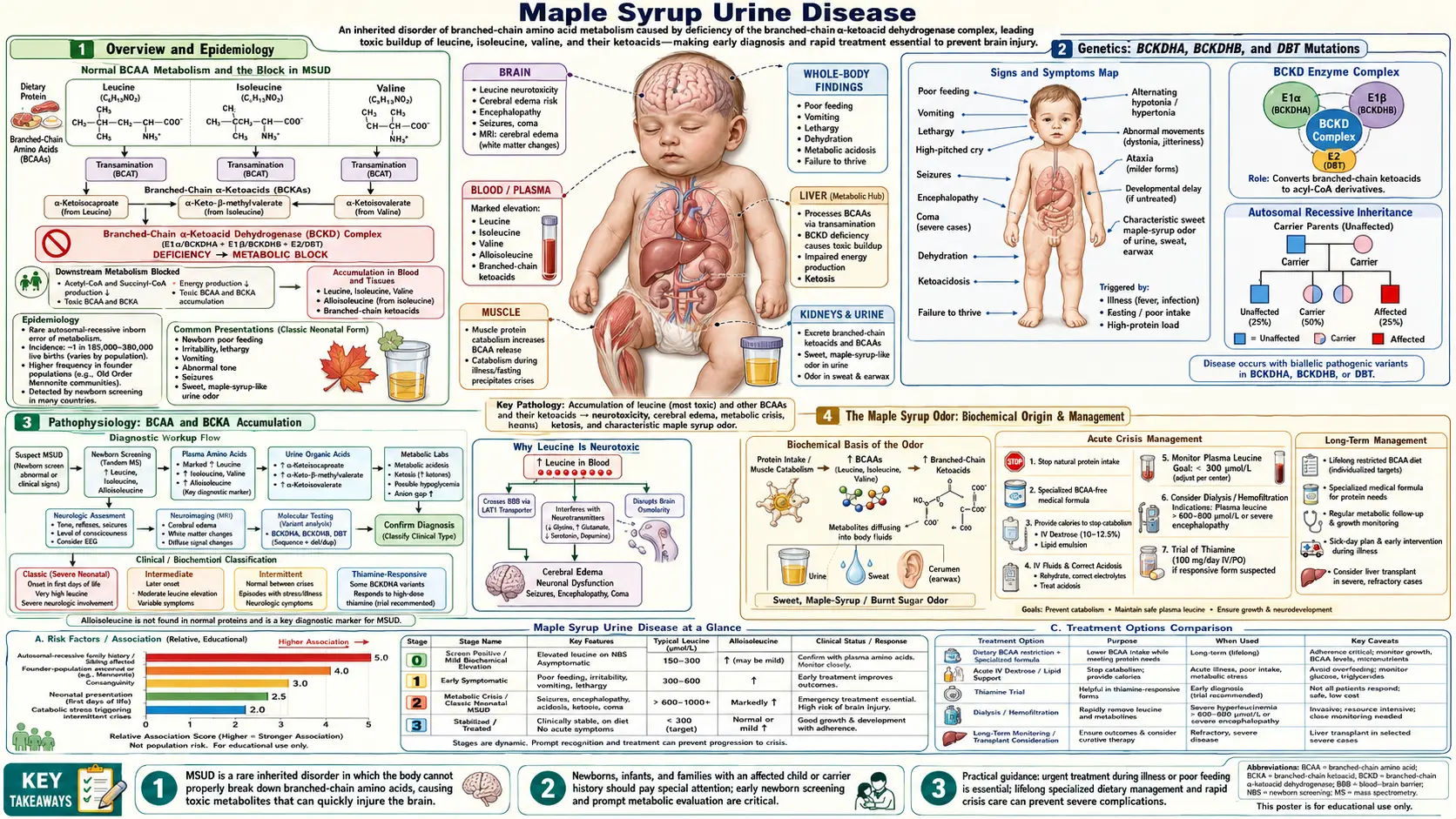
Overview and Epidemiology
Maple Syrup Urine Disease (MSUD) is an autosomal recessive inborn error of branched-chain amino acid (BCAA) catabolism caused by deficiency of the branched-chain alpha-keto acid dehydrogenase (BCKAD) complex. The three branched-chain amino acids (BCAAs) — leucine, isoleucine, and valine — accumulate in blood and tissue along with their respective branched-chain keto acid (BCKA) metabolites. Without treatment, classic MSUD presents as a neonatal neurological emergency leading to cerebral edema and death.
The disease is named for the characteristic sweet maple syrup or caramel odor of urine, sweat, and earwax caused by accumulating BCKA metabolites. First described by Menkes et al. in 1954 in four siblings who died of a rapidly progressive neurological illness in infancy, MSUD is now included on the Recommended Uniform Screening Panel (RUSP) for U.S. newborn screening programs.
Prevalence: approximately 1:185,000 live births in the general population. Dramatically higher in Old Order Mennonite and Amish communities — approximately 1:380 live births — due to a founder mutation (Y393N in the BCKDHA gene). More than 200 pathogenic variants across three causal genes have been documented worldwide.
Genetics: BCKDHA, BCKDHB, and DBT Mutations
The BCKAD complex is a mitochondrial multi-enzyme complex with four subunit types. Three genes account for the vast majority of MSUD cases:
- BCKDHA (chromosome 19q13.2): encodes the E1α subunit. This is the most commonly mutated gene. The Mennonite founder mutation Y393N resides here and accounts for a large proportion of MSUD cases in Old Order Mennonite and Amish populations.
- BCKDHB (chromosome 6q14.1): encodes the E1β subunit. Mutations here are the second most common cause of MSUD.
- DBT (chromosome 1p21.2): encodes the E2 subunit (dihydrolipoyl transacylase). Thiamine-responsive forms of MSUD are often associated with DBT mutations.
A fourth subunit, E3 (dihydrolipoamide dehydrogenase, encoded by the DLD gene on chromosome 7q31.1), is shared with the pyruvate dehydrogenase complex and the alpha-ketoglutarate dehydrogenase complex. E3 deficiency therefore causes a combined organic acidemia affecting all three complexes simultaneously, producing a more severe and metabolically complex phenotype.
MSUD follows strict autosomal recessive inheritance. Residual BCKAD enzyme activity correlates directly with clinical severity: less than 2% residual activity produces classic MSUD; 3–30% produces the intermediate form; greater than 30% with stress-triggered crises produces the intermittent subtype. More than 200 pathogenic variants have been documented across the three main genes.
Pathophysiology: Branched-Chain Amino Acid and BCKA Accumulation
Under normal physiology, dietary leucine, isoleucine, and valine are first transaminated by the branched-chain aminotransferase (BCAT) enzyme — predominantly in muscle and brain — to produce their respective branched-chain keto acids (BCKAs): alpha-ketoisocaproic acid (KIC) from leucine, alpha-keto-beta-methylvaleric acid (KMV) from isoleucine, and alpha-ketoisovaleric acid (KIV) from valine. These BCKAs then undergo oxidative decarboxylation by the BCKAD complex to yield acyl-CoA intermediates that enter the TCA cycle for energy production.
In MSUD, the BCKAD complex is deficient. BCKAs cannot be decarboxylated and therefore accumulate in plasma, urine, and tissues alongside their parent branched-chain amino acids.
Leucine and KIC are the most neurotoxic species:
- Leucine competes at the LAT1 large neutral amino acid transporter at the blood-brain barrier, displacing tyrosine, tryptophan, and phenylalanine. This reduces brain uptake of these precursors and impairs synthesis of dopamine, serotonin, and norepinephrine.
- KIC directly inhibits glutamate dehydrogenase, disrupting the balance of gamma-aminobutyric acid (GABA) and glutamate neurotransmitters.
- Leucine and KIC cause cerebral edema through intracellular mechanisms — astrocyte swelling is particularly prominent — contributing to the neonatal encephalopathy.
- Both leucine and KIC impair mitochondrial function, causing neuronal energy failure that accelerates the cascade toward irreversible brain injury.
A diagnostically important byproduct of the blocked pathway is alloisoleucine, which forms non-enzymatically from the accumulated KMV (2-oxo-3-methylvalerate). Healthy people have only small amounts of alloisoleucine in plasma (averages of 1.3 to 1.9 μmol/L in a 1999 reference study). A plasma level above 5 μmol/L on quantitative amino acid analysis was the most specific and most sensitive diagnostic marker for all forms of MSUD in that study, and alloisoleucine is considered pathognomonic for MSUD.
The Maple Syrup Odor: Biochemical Origin
The hallmark sweet, caramel-like maple syrup odor that gives the disease its name arises from the accumulation of branched-chain keto acids, particularly through the production of sotolone (4,5-dimethyl-3-hydroxy-2[5H]-furanone), a lactone metabolite derived from the isoleucine keto acid pathway. Sotolone is also the primary compound responsible for the scent of fenugreek seeds and artificial maple flavoring used in food products.
The odor is detectable in urine, in cerumen (earwax — often the first location where parents or nurses notice it), and in sweat. Babies with untreated classic MSUD are born without symptoms; the odor can be detected in earwax at 12–24 hours of life, before the neurological crisis becomes apparent, and becomes apparent in urine at 4–6 days. It can also be detected by clinicians examining diapers during a metabolic crisis.
Before the era of tandem mass spectrometry newborn screening, the maple syrup odor served as a critical early diagnostic clue that prompted urgent investigation. Even today, the odor in an acutely ill neonate is a sign hospitals treat as an emergency, prompting immediate plasma amino acid testing. Parents of diagnosed children are counseled to recognize changes in the intensity of the odor as an early warning sign of metabolic decompensation during illness.
MSUD Subtypes: Classic, Intermediate, Intermittent, Thiamine-Responsive, and E3-Deficient
MSUD is not a single uniform disorder. Five clinically and biochemically distinct subtypes are recognized, each associated with different levels of residual BCKAD enzyme activity and different clinical trajectories.
Classic MSUD
The most common and most severe subtype. Residual BCKAD activity is less than 2% of normal. Presents with neonatal metabolic crisis at 3–5 days of life. The maple syrup odor can be detected in earwax from 12–24 hours of life. All three branched-chain amino acids are markedly elevated, and alloisoleucine is invariably detected on plasma amino acid analysis. Without urgent treatment, classic MSUD is fatal in the newborn period.
Intermediate MSUD
Residual BCKAD activity is 3–30% of normal. Patients do not experience acute neonatal crises but have chronic elevation of branched-chain amino acids leading to intellectual disability, poor growth, and developmental delay presenting during infancy or childhood. The diagnosis is often made later than classic MSUD.
Intermittent MSUD
Residual BCKAD activity is 5–15% of normal — sufficient for normal branched-chain amino acid metabolism under basal conditions. Patients are asymptomatic and develop normally between episodes. However, catabolic stress from intercurrent infection, surgery, fasting, or excessive protein intake can trigger an acute crisis clinically indistinguishable from classic MSUD. Diagnosis is frequently delayed because routine newborn screening may miss patients with this subtype; the first identified crisis may occur in older childhood.
Thiamine-Responsive MSUD
Often associated with DBT or BCKDHA mutations. High-dose thiamine supplementation (50–300 mg/day) improves residual BCKAD complex activity, leading to measurable reductions in plasma leucine, isoleucine, and valine levels. Clinical response is variable and does not eliminate the need for dietary management entirely in most patients. GeneReviews describes a trial of thiamine by mouth as a way to find out whether a person has the thiamine-responsive form.
E3-Deficient MSUD (Combined)
Caused by mutations in the DLD gene encoding the E3 (dihydrolipoamide dehydrogenase) subunit, which is shared by three mitochondrial enzyme complexes. E3 deficiency therefore simultaneously impairs the BCKAD complex, the pyruvate dehydrogenase complex, and the alpha-ketoglutarate dehydrogenase complex. The result is a combined organic acidemia with features of MSUD plus lactic acidosis and alpha-ketoglutaric aciduria. This subtype is often severe and metabolically complex to manage, requiring attention to multiple metabolic pathways simultaneously.
Clinical Presentation: Neonatal Crisis and Long-Term Complications
The timeline of the classic neonatal presentation is characteristic and closely tied to the accumulation of dietary leucine after feeding is established:
- Days 1–2: apparently normal. Colostrum and early breast milk contain relatively low leucine compared with formula, so leucine loading begins gradually after feeding is well established.
- Days 2–3: poor feeding, irritability, lethargy — early nonspecific signs that may be dismissed as normal newborn behavior.
- Days 4–6: progressive encephalopathy — stupor transitioning to coma, alternating hypotonia and rigidity, stereotyped movement patterns including "bicycling" or "fencing" dystonic posturing, opisthotonus, and a bulging fontanelle signaling rising intracranial pressure.
- Days 7–10 if untreated: critical brain swelling (cerebral edema) and coma, brainstem herniation, central respiratory failure, and death.
The maple syrup odor is detectable throughout the neonatal period in classic disease.
Magnetic resonance imaging (MRI) findings in acute MSUD are nearly pathognomonic: cytotoxic edema with diffusion-weighted imaging (DWI) restricted diffusion preferentially affecting the deep brain structures — cerebellar white matter, brainstem, cerebral peduncles, posterior limb of the internal capsule, and globus pallidus. This distribution mirrors the myelination pattern of the newborn brain, because myelinated regions accumulate branched-chain amino acid metabolites preferentially. This distinctive magnetic resonance imaging appearance strongly suggests MSUD even before biochemical confirmation.
Long-term complications in treated survivors include:
- Intellectual disability — severity correlates with the duration and intensity of the neonatal crisis and with the quality of subsequent metabolic control throughout childhood.
- Spastic quadriplegia following severe neonatal encephalopathy.
- Seizures, often refractory to standard antiepileptic treatment.
- Psychiatric disorders — depression, anxiety, and attention-deficit/hyperactivity disorder (ADHD) occur at elevated rates even in patients with excellent metabolic control, suggesting that chronic low-level branched-chain amino acid dysregulation or neurotransmitter imbalance contributes independently of acute crises.
- Recurrent pancreatitis — mechanism is not fully established.
- Metabolic crises throughout life, triggered by any catabolic state such as infection, fever, surgery, or prolonged fasting.
A study of 184 people followed over three decades concluded that strict diet keeps blood levels within acceptable limits but does not fully prevent thinking and psychiatric problems, and that liver transplant removes the need for a prescription diet but brings predictable medical problems of its own and does not reverse earlier neurological damage. The authors describe a critical unmet need for safe, effective treatments that can start early in life, and see the disease as a guide for designing therapies based on gene replacement or editing.
Diagnosis: Newborn Screening and Confirmatory Testing
Newborn screening in the United States uses tandem mass spectrometry (MS/MS) on dried blood spots collected at 24–48 hours of age. Key markers include:
- Elevated leucine plus isoleucine (these two amino acids co-elute at the same mass of 132 Da and cannot be distinguished from each other by standard MS/MS).
- Elevated valine.
- Alloisoleucine, the pathognomonic marker, cannot be told apart from leucine and isoleucine by standard MS/MS. It is measured by a second-tier test on the same blood spot or by quantitative plasma amino acid analysis.
- GeneReviews names whole-blood ratios of (leucine + isoleucine) to alanine and to phenylalanine, above the screening laboratory's cutoff, as the suggestive screening findings.
- Elevated leucine/(isoleucine + valine) ratio.
- Elevated leucine/phenylalanine ratio to reduce false positives.
Sensitivity for classic MSUD exceeds 99%. Intermittent MSUD may be missed if sampled during a period of normal branched-chain amino acid levels.
Confirmatory testing includes:
- Quantitative plasma amino acids: markedly elevated leucine, isoleucine, and valine. The presence of alloisoleucine (greater than 5 μmol/L) is pathognomonic — healthy people have only small amounts of this amino acid in plasma (averages of 1.3 to 1.9 μmol/L).
- Urine organic acids: elevated BCKA metabolites including 2-oxoisocaproic acid (from leucine), 2-oxo-3-methylvaleric acid (from isoleucine), and 2-oxoisovaleric acid (from valine).
- BCKAD enzyme activity measured in leukocytes or cultured skin fibroblasts — quantifies residual activity and helps classify the subtype.
- Molecular gene sequencing of BCKDHA, BCKDHB, and DBT — confirms diagnosis, identifies the specific mutation for family studies, and helps predict thiamine responsiveness.
Monitoring during treatment: plasma branched-chain amino acids via dried blood spot cards at home. Target plasma leucine is 75–300 μmol/L during treatment (normal less than 200 μmol/L). Leucine is the primary target for monitoring and dietary adjustments.
Treatment: Acute Crisis Management and Chronic Dietary Therapy
The fundamental goal of acute treatment is to rapidly reduce plasma leucine — the most neurotoxic branched-chain amino acid — while simultaneously maintaining anabolism to prevent the breakdown of endogenous protein that would release additional leucine from muscle.
Acute Crisis Management
- High-rate intravenous 10% dextrose at a glucose infusion rate of 8–10 mg/kg/min or higher, combined with insulin at 0.05–0.1 units/kg/hour. This combination drives a strongly anabolic state, shifting branched-chain amino acids into protein synthesis and causing rapid reduction of plasma leucine, isoleucine, and valine.
- Stopping all exogenous branched-chain amino acid intake — protein intake and branched-chain amino acid-containing formula are held for 24–48 hours during the acute phase.
- Branched-chain amino acid-free amino acid solution administered intravenously or orally. This provides all essential amino acids minus leucine, isoleucine, and valine, preventing catabolism of endogenous protein stores and accelerating the leucine reduction.
- Dialysis — hemodialysis is preferred over peritoneal dialysis for speed of effect — when plasma leucine exceeds 1,500–2,000 μmol/L, or when neurological deterioration continues despite maximal medical management. Dialysis is the most rapid method available for reducing plasma leucine.
- Plasma amino acids are monitored every 4–6 hours during the crisis. Dietary protein is not reintroduced until plasma leucine falls below 300 μmol/L.
- Thiamine 100–300 mg/day is given to patients presenting with MSUD while awaiting determination of thiamine responsiveness.
Chronic Dietary Management
Long-term treatment is built around a specialized medical formula that provides all essential amino acids, calories, vitamins, and minerals while omitting leucine, isoleucine, and valine. Small, carefully measured amounts of natural protein are added — titrated to plasma leucine levels and the patient's growth requirements. A 2017 review lists leucine intake in infancy as 40–100 mg per kilogram of body weight per day at 0–6 months and 40–75 mg per kilogram per day at 7–12 months, from natural protein, changing with age and adjusted based on home dried blood spot monitoring results.
Frequent monitoring is essential: dried blood spot testing 2–3 times per week in infancy and weekly in older patients during stable periods. An emergency sick-day protocol means increasing formula, reducing natural protein, and contacting the metabolic team at the first sign of intercurrent illness or catabolism, because a missed meal or a febrile illness can trigger a life-threatening crisis within hours in classic and intermediate MSUD.
In a 2010 study of a specially designed formula, 15 Old Order Mennonite children started on it between birth and 34 months of age all grew and developed normally over 14–33 months. Hospital stays fell from 0.35 to 0.14 per patient per year, and the large majority of illnesses that could set off a crisis were managed at home with sick-day formula and frequent amino acid monitoring.
Liver Transplantation in MSUD
The rationale stated for liver transplantation in MSUD is that the BCKAD enzyme complex is expressed predominantly in hepatic tissue, with a successful liver transplant said to restore approximately 85–90% of total body BCKAD enzyme activity and to be essentially curative with respect to metabolic crises. No source for these statements was found. A 2017 review says the first step of branched-chain amino acid breakdown mostly takes place in skeletal muscle rather than the liver, and that neither deceased unrelated nor living related livers can restore enzyme activity to normal levels, so acute metabolic intoxication can still occur after transplant. The authors of a 2012 transplant series state that enzyme activity restored to under 30% of liver cells is under 5% of whole-body activity. In one patient described in a 2000 report, whole-body leucine breakdown was normal with the breakdown confined to the new liver, while blood levels of branched-chain amino acids stayed at 2 to 3 times those of healthy controls.
Papers on liver transplantation in MSUD appeared in 1999 and 2000, and a 2006 series reported elective transplantation in 10 further children. Subsequent case series have consistently demonstrated that liver transplantation in classic MSUD resolves recurrent metabolic crises, allows near-normal dietary intake including natural protein, and substantially improves quality of life. A figure of more than 95% of transplanted patients experiencing zero metabolic crises post-transplant is also stated, with no source found; the three-decade study of 184 people says transplant safeguards patients from life-threatening metabolic crises. Plasma leucine, isoleucine, and valine normalize on a regular diet in the majority of recipients.
In a series of 35 children transplanted between 2004 and 2009, patient and graft survival were 100% at about 4.5 years, and leucine tolerance rose more than 10-fold. One-third had an IQ of 70 or below before surgery, with no statistically significant change a year later; the authors concluded that transplant "may arrest brain damage, but will not reverse it." Liver transplantation does not reverse pre-existing neurological damage — intellectual disability and other deficits acquired before or during the procedure are not recovered. The best outcomes are achieved when transplantation is performed early, before irreversible injury accumulates from repeated crises or chronic branched-chain amino acid elevation.
A particularly important consideration is that heterozygous donor livers — from carriers with one normal and one pathogenic allele — provide sufficient BCKAD activity (approximately 50% of normal, which far exceeds the threshold needed to handle a normal diet with no crisis risk). This has made living-related liver donation feasible in Mennonite and Amish communities where carriers are common. It is also stated, with no source found, that this allows donors to contribute to multiple family members over time with relatively lower surgical risk than obtaining cadaveric organs. A 2017 review says living related donor liver can be used when deceased donor tissue is limited.
Because other organs still hold the working enzyme, the liver removed from a child with MSUD can be given to someone who does not have the disease (a "domino" transplant). In the same 2012 series, six patients donated their livers this way, and all recipients were alive and well with normal branched-chain amino acid balance at the time of the report.
Transplantation is increasingly considered standard of care for classic MSUD, particularly where the burden of dietary management and metabolic crises is high, and where suitable living-related donors are available.
Key Research Papers
- Strauss KA, Puffenberger EG, Carson VJ. Maple Syrup Urine Disease. GeneReviews [Internet]. Seattle: University of Washington; 2006 (updated 2020) — PubMed PMID: 20301495
- Strauss KA, et al. Classical maple syrup urine disease and brain development: principles of management and formula design. Mol Genet Metab. 2010;99(4):333-45. — PubMed PMID: 20061171
- Barschak AG, et al. Oxidative stress in plasma from maple syrup urine disease patients during treatment. Metab Brain Dis. 2008;23(1):71-80. — PubMed PMID: 18026828
- Mazariegos GV, et al. Liver transplantation for classical maple syrup urine disease: long-term follow-up in 37 patients and comparative United Network for Organ Sharing experience. J Pediatr. 2012;160(1):116-21.e1. — PubMed PMID: 21839471
- Chuang DT, et al. Lessons from genetic disorders of branched-chain amino acid metabolism. J Nutr. 2006;136(1 Suppl):243S-249S. — PubMed PMID: 16365091
- Morton DH, et al. Diagnosis and treatment of maple syrup disease: a study of 36 patients. Pediatrics. 2002;109(6):999-1008. — PubMed PMID: 12042535
- Bodner-Leidecker A, et al. Branched-chain L-amino acid metabolism in classical maple syrup urine disease after orthotopic liver transplantation. J Inherit Metab Dis. 2000;23(8):805-18. — PubMed PMID: 11196106
- Snyderman SE. The therapy of maple syrup urine disease. Am J Dis Child. 1967;113(1):68-73. — PubMed PMID: 6015908
- Blackburn PR, Gass JM, Vairo FPE, Farnham KM, Atwal HK, Macklin S, Klee EW, Atwal PS. Maple syrup urine disease: mechanisms and management. Appl Clin Genet. 2017;10:57-66. — PubMed PMID: 28919799
- Strauss KA, Mazariegos GV, Sindhi R, et al. Elective liver transplantation for the treatment of classical maple syrup urine disease. Am J Transplant. 2006;6(3):557-64. — PubMed PMID: 16468966
- Wendel U, Saudubray JM, Bodner A, Schadewaldt P. Liver transplantation in maple syrup urine disease. Eur J Pediatr. 1999;158 Suppl 2:S60-4. — PubMed PMID: 10603101
- Strauss KA, Carson VJ, Soltys K, et al. Branched-chain α-ketoacid dehydrogenase deficiency (maple syrup urine disease): treatment, biomarkers, and outcomes. Mol Genet Metab. 2020;129(3):193-206. — PubMed PMID: 31980395
- Schadewaldt P, Bodner-Leidecker A, Hammen HW, Wendel U. Significance of L-alloisoleucine in plasma for diagnosis of maple syrup urine disease. Clin Chem. 1999;45(10):1734-40. — PubMed PMID: 10508118
- Oglesbee D, Sanders KA, Lacey JM, et al. Second-tier test for quantification of alloisoleucine and branched-chain amino acids in dried blood spots to improve newborn screening for maple syrup urine disease (MSUD). Clin Chem. 2008;54(3):542-9. — PubMed PMID: 18178665
- Podebrad F, Heil M, Reichert S, Mosandl A, Sewell AC, Böhles H. 4,5-dimethyl-3-hydroxy-2[5H]-furanone (sotolone)--the odour of maple syrup urine disease. J Inherit Metab Dis. 1999;22(2):107-14. — PubMed PMID: 10234605