Small Bowel Obstruction
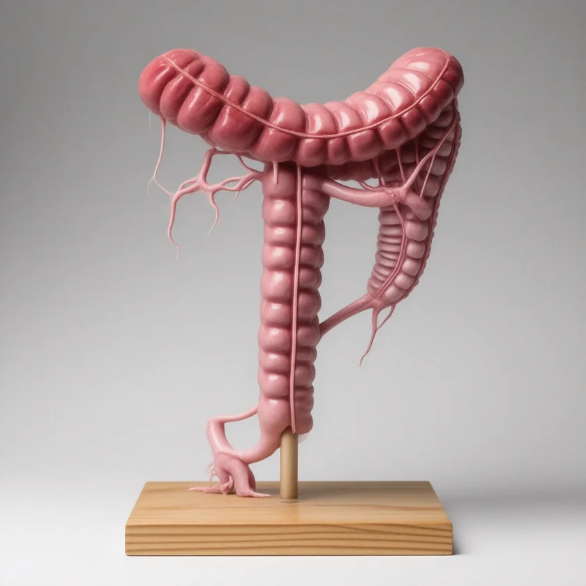
- Overview
- Causes and Pathophysiology
- Strangulation and Closed-Loop Obstruction
- Adhesive SBO — The Most Common Cause
- Clinical Presentation
- Diagnosis — Imaging
- Non-Operative Management
- Surgical Management
- Special Causes — Hernias, Gallstone Ileus, Intussusception
- Prognosis and Prevention
- Key Research Papers
- Connections
- Featured Videos
Overview
In plain words: cramping belly pain, vomiting, a swollen belly and no gas or stool are the signs of a blocked small bowel. Steady severe pain, fever or a rigid belly are signs of dying bowel that hospitals treat as an emergency.
Small bowel obstruction (SBO) is a mechanical impediment to the normal transit of intestinal contents through the small intestine. It is one of the most common and consequential emergencies in general surgery, accounting for as many as 16% of surgical admissions and more than 300,000 operations annually in the United States, according to the Eastern Association for the Surgery of Trauma guideline. SBO generates an estimated $3 billion in healthcare costs each year.
The small intestine — spanning roughly 6 to 7 meters from the pylorus to the ileocecal valve — handles the bulk of nutrient absorption and fluid regulation. When a mechanical block develops, the bowel proximal to the obstruction dilates as luminal contents, swallowed air, and bacterially produced gas accumulate. The bowel distal to the blockage decompresses as residual contents are absorbed or pass through. This asymmetric distension drives the cardinal symptoms: colicky abdominal pain, nausea, vomiting, and cessation of flatus and bowel movements.
If untreated, the sequence is predictable and dangerous. Progressive distension leads to venous congestion within the bowel wall. Mucosal blood flow decreases, impairing the gut's protective barrier function. Bacterial translocation across a compromised mucosa seeds the portal circulation and mesenteric lymphatics. In the worst cases, arterial supply is cut off — strangulation — and full-thickness bowel necrosis follows within hours. Perforation releases fecal contents into the peritoneal cavity, triggering peritonitis and septic shock. Mortality from strangulated SBO, even with prompt surgery, ranges from 20 to 35%.
Early recognition, accurate imaging, appropriate fluid resuscitation, and judicious timing of surgical intervention are the pillars of management. The central clinical dilemma is distinguishing the majority of cases that will resolve without an operation from the minority that require urgent surgery.
Causes and Pathophysiology
SBO results from any process that physically narrows or compresses the bowel lumen, kinks the bowel, or traps it in a way that prevents onward passage of intestinal contents. Causes are conventionally grouped by their relationship to the bowel wall: extrinsic (outside the wall), intrinsic (within the wall), or intraluminal (inside the lumen).
Major Causes by Frequency
- Adhesions (60–70%): Peritoneal scar bands forming after prior abdominal or pelvic surgery are by far the leading cause. The inflammatory response to any surgical incision, peritoneal handling, or spillage of intestinal contents triggers fibroblast proliferation. Fibrinous deposits that normally resolve instead organize into fibrous adhesions that tether loops of bowel to each other, the abdominal wall, or the pelvic organs. Appendectomy, hysterectomy, and colorectal resection carry the highest subsequent SBO risk, but any laparotomy or laparoscopy can produce adhesions.
- Hernias (10–15%): A portion of bowel herniates through a defect in the abdominal wall (inguinal, femoral, umbilical, incisional) or through an internal peritoneal opening (Petersen's space after Roux-en-Y bypass, foramen of Winslow, paraduodenal fossa). If the hernia neck is tight, the bowel becomes incarcerated (trapped, non-reducible). Progressive edema and venous congestion lead to strangulation (arterial compromise).
- Malignancy (10–15%): Primary small bowel tumors (adenocarcinoma, gastrointestinal stromal tumor (GIST), lymphoma, carcinoid) can obstruct intrinsically. More commonly, extrinsic compression from metastatic disease — particularly peritoneal carcinomatosis from ovarian, gastric, or colorectal primaries — creates multiple partial or complete obstruction points. Malignant SBO in the setting of advanced cancer carries a grave prognosis and requires careful goals-of-care discussion before surgical intervention.
- Crohn's Disease (5%): Transmural inflammation and repeated cycles of ulceration and healing generate strictures — fixed, fibrostenotic narrowings that no amount of anti-inflammatory therapy will reverse. Crohn's strictures most commonly affect the terminal ileum. Distinguishing fibrotic (surgical) from inflammatory (medical) strictures on magnetic resonance (MR) enterography guides treatment decisions.
- Intussusception (<5% in adults): Telescoping of a proximal bowel segment (intussusceptum) into the lumen of an adjacent distal segment (intussuscipiens). In children, most cases are idiopathic and reducible with air enema. In adults, a lead-point lesion — a polyp, gastrointestinal stromal tumor, lymphoma, or lipoma — is present in the vast majority of cases, mandating resection even after successful reduction.
- Gallstone Ileus (1–4%): A large gallstone (typically >2.5 cm) erodes through an inflamed gallbladder wall into the adjacent duodenum or colon, creating a cholecystoenteric fistula. The stone migrates distally until it impacts, most commonly at the narrow terminal ileum. The classic radiographic finding is Rigler's triad: pneumobilia (air in the biliary tree), small bowel obstruction pattern, and an ectopic calcified density. Bouveret syndrome is the rare variant where the stone lodges in the duodenum or pylorus, presenting as gastric outlet obstruction.
Pathophysiology in Detail
Once the bowel is mechanically blocked, several cascades unfold simultaneously. The proximal bowel distends with luminal gas — primarily swallowed air (nitrogen-rich) plus carbon dioxide and hydrogen from bacterial fermentation. Simultaneously, the intestine continues secreting 6 to 8 liters of fluid per day (saliva, gastric juice, bile, pancreatic secretions, intestinal secretions) that cannot be reabsorbed distally. This fluid pools in the obstructed lumen — a process called third spacing — and is functionally lost from the intravascular compartment.
The result is profound hypovolemia, often underappreciated on initial presentation. Vomiting compounds fluid and electrolyte loss. Early in the course, loss of hydrochloric acid from the stomach drives a hypochloremic, hypokalemic metabolic alkalosis. Later, if ischemia supervenes, lactic acid from ischemic bowel drives a metabolic acidosis — an ominous shift indicating the need for urgent surgical intervention.
Bowel wall edema from venous congestion disrupts the tight-junction integrity of the intestinal epithelium. Normal commensal bacteria — primarily gram-negative enterics and anaerobes — translocate across this leaky barrier into the mesenteric lymphatics and portal circulation, priming the systemic inflammatory response. This bacterial translocation, rather than frank perforation, is often the mechanism by which SBO patients develop fever, leukocytosis, and early sepsis physiology even before gross bowel necrosis occurs.
Strangulation and Closed-Loop Obstruction
Strangulation — impairment of blood supply to the obstructed bowel segment — transforms a manageable surgical problem into a life-threatening emergency. It occurs in approximately 10 to 15% of all SBOs but accounts for the majority of SBO mortality. Even with prompt surgical intervention, mortality from strangulated SBO ranges from 20 to 35%, compared to less than 5% for simple (non-strangulated) SBO.
The mechanism begins with venous obstruction. Elevated intraluminal pressure compresses mesenteric veins, which have lower wall tension and pressure than arteries. Venous outflow obstruction leads to engorgement and edema of the bowel wall. As wall tension rises, arterial inflow eventually becomes compromised, producing full-thickness ischemia and necrosis within hours. The necrotic segment becomes a reservoir for gram-negative bacteria and endotoxin, which escape into the peritoneal cavity and systemic circulation.
Clinical Clues to Strangulation
The challenge is that no single clinical finding reliably identifies strangulation preoperatively, and some patients with strangulation initially appear deceptively well. However, surgeons treat the following constellation of findings as grounds for urgent operative planning:
- Fever above 38.5°C
- Peritoneal signs on exam — guarding, rebound tenderness, rigidity
- Leukocytosis above 15,000 cells/mm³
- Metabolic acidosis — elevated serum lactate, base deficit on arterial blood gas
- Tachycardia and hemodynamic instability
- Constant, non-colicky pain replacing the earlier crampy, intermittent pattern (indicates ischemia; the bowel stops peristalsing against a dead segment)
Computed tomography (CT) findings highly suggestive of strangulation include mesenteric fat stranding, free peritoneal fluid (especially if high-density, indicating hemorrhage), pneumatosis intestinalis (gas within the bowel wall), portal venous gas, and absent or diminished bowel wall enhancement after intravenous (IV) contrast — the imaging correlate of transmural ischemia.
Closed-Loop Obstruction
A closed-loop obstruction occurs when both ends of a bowel segment are blocked simultaneously, creating an isolated, non-decompressable loop. Unlike a simple obstruction where distension can distribute proximally, the closed loop has nowhere to decompress and rapidly develops ischemia from pressure-related vascular compromise. Common mechanisms include a single adhesive band trapping a loop at two points, an internal hernia, or a volvulus (torsion around a fixed mesenteric axis).
Computed tomography findings specific to closed-loop obstruction include the whirl sign (swirling of mesenteric vessels and fat around a twisted mesenteric pedicle), the beak sign (tapered ends of the obstructed loop pointing toward the obstruction site), and a U-shaped or C-shaped configuration of a distended, isolated bowel loop. Closed-loop obstruction is a surgical emergency — there is no role for a non-operative trial. Strangulation can develop within 6 hours of onset, and the physical exam often lags behind the computed tomography findings.
Adhesive SBO — The Most Common Cause
Adhesive SBO deserves individual attention because it is so prevalent, because most episodes resolve without surgery, and because the decision of when to operate versus observe is the central management challenge surgeons face daily. Adhesions form in response to peritoneal injury — any surgery, infection, or inflammation that disrupts the mesothelial lining. The peritoneum attempts to seal the injured surface with a fibrinous exudate. If fibrinolytic activity is insufficient to dissolve this exudate, fibroblasts invade and the fibrinous layer matures into a permanent fibrous adhesion within 5 to 7 days.
Between 10 and 20% of patients who undergo abdominal surgery will experience at least one adhesion-related SBO during their lifetime. The risk is not limited to the immediate postoperative period — adhesions can mature, contract, and trap bowel loops years or decades after the inciting surgery. The Bologna guideline notes that younger patients have a higher lifetime risk.
Water-Soluble Contrast (Diatrizoate) Challenge
The water-soluble contrast agent meglumine diatrizoate has emerged as both a diagnostic and therapeutic tool for adhesive SBO. When 100 mL is administered orally or via nasogastric tube and a plain abdominal X-ray is obtained 4 to 24 hours later, the presence of contrast in the colon predicts resolution without surgery. Pooled figures for contrast reaching the colon within 4 to 24 hours are a sensitivity of 96% and a specificity of 98% (Branco 2010, 14 prospective studies); a later pooled analysis gives a sensitivity of 92% and a specificity of 93% (Ceresoli 2016). This allows early identification of patients who can continue non-operative management with confidence.
Beyond its predictive value, diatrizoate has been proposed to have a direct therapeutic effect. Its high osmolarity (~1900 mOsm/L) draws fluid into the bowel lumen from the bowel wall and surrounding tissues, reducing edema and potentially physically breaking the adhesive band by increasing luminal pressure proximal to the obstruction. Older pooled analyses reported shorter hospital stays and fewer operations (Branco 2010: odds ratio 0.62; Ceresoli 2016: odds ratio 0.55, and time to resolution about 28 hours shorter). Newer pooled analyses of randomized trials are less favorable. In 11 randomized trials with 817 patients (Klingbeil 2022), hospital stay in non-surgical patients fell by 1.95 days, but operation rates did not change significantly (19.8% vs 21.4%), and the authors note considerable design limitations. A 2025 pooled analysis, described under Non-Operative Management, found no significant reduction in operations.
Recurrence After Non-Operative Management
The Bologna guideline reports that 12% of patients treated without surgery for adhesive SBO are readmitted within 1 year, rising to 20% after 5 years, and that after surgery the figures are 8% and 16%. In a Norwegian series of 500 patients operated on for adhesive obstruction (Fevang 2004), cumulative recurrence after one operation was 18% at 10 years and 29% at 30 years, reaching 81% for patients with 4 or more admissions. After a second episode managed non-operatively, recurrence rates rise further. Surgeons weigh this cumulative recurrence risk against the risk of creating new adhesions with each operative intervention — a paradox surgeons call the "adhesion cycle." Laparoscopic adhesiolysis is associated with fewer new adhesions than open surgery, but it does not eliminate the underlying tendency to form adhesions.
Clinical Presentation
The classic tetrad of SBO — colicky abdominal pain, nausea and vomiting, abdominal distension, and obstipation — is taught in every medical school, but the relative prominence of each symptom varies significantly with the level and completeness of the obstruction.
Pain
Early in the course, pain is colicky — crampy and intermittent, peaking every 3 to 4 minutes as peristaltic waves build against the obstruction and then subside. Patients often describe feeling waves of pain that make them writhe, followed by brief periods of relative relief. The periumbilical location reflects the midgut origin of most small bowel. A critical red flag is the transition from colicky to constant pain, which suggests that the bowel has stopped peristalsing against a dead or ischemic segment — a harbinger of strangulation requiring urgent reassessment and imaging.
Vomiting
Vomiting occurs early in proximal SBO (jejunal) and may temporarily relieve the distension. The vomitus is initially bilious (yellow-green bile-stained fluid). As obstruction persists, bacterial overgrowth in the stagnant proximal bowel produces increasingly malodorous, feculent vomiting — a late and ominous sign indicating that the obstructed bowel has been colonized by colonic flora. Feculent vomiting can also indicate a gastrocolic or enterocolic fistula.
Distension
Abdominal distension is proportional to the length of bowel proximal to the obstruction and the duration of the obstruction. Proximal (high jejunal) obstruction produces minimal distension because so little bowel is involved. Distal (ileal) obstruction — which represents the majority of cases — produces massive distension, a tympanitic abdomen, and the characteristic radiographic ladder pattern.
Obstipation
True obstipation — the complete absence of flatus and stool — indicates a complete obstruction. Patients with partial SBO may continue passing small amounts of gas or loose stool as contents squeeze past the obstruction. Eliciting whether the patient has passed any flatus since symptom onset is one of the most important questions in the history.
Bowel Sounds
Early in SBO, bowel sounds are characteristically high-pitched and tinkling — the result of gas and fluid under pressure sloshing through dilated loops against the obstruction. As the bowel fatigues and peristalsis decreases, bowel sounds diminish. A silent abdomen in the context of SBO suggests advanced obstruction, ileus, or strangulation with peritonitis. Audible peristaltic rushes coinciding with crampy pain episodes are a supportive finding.
Why a Calm-Looking Belly Does Not Rule Out Strangulation
According to the Bologna guideline, physical examination detects a strangulated bowel only 48% of the time, even in experienced hands. Adhesive obstruction averages 8 days in hospital and carries a 3% in-hospital death rate per episode.
Diagnosis — Imaging
The diagnosis of SBO is established by clinical history and physical examination but confirmed and characterized by imaging. The choice and sequence of imaging depend on clinical urgency.
Plain Abdominal X-Ray
Upright and supine abdominal radiographs are obtained first as a rapid, inexpensive screen. Classic findings include:
- Dilated small bowel loops greater than 3 cm in diameter (normal small bowel is less than 2.5–3 cm)
- Absence of distal colon gas in complete obstruction
- Step-ladder pattern — multiple air-fluid levels of differing heights in adjacent loops on the upright film
- String-of-pearls sign — a row of small gas bubbles trapped in the valvulae conniventes (circular folds of the small bowel mucosa), indicating fluid-filled distended loops with only small gas collections
Plain films have moderate sensitivity (60–70%) and poor specificity for SBO. They cannot reliably identify the cause, level, or presence of strangulation. A normal plain film does not exclude SBO, particularly in early or proximal obstruction where fluid-filled loops may not contain enough gas to be visible.
Computed Tomography Abdomen and Pelvis with Intravenous Contrast
Computed tomography is the gold standard for evaluating suspected SBO, with sensitivity of 93–96% and specificity of 94–96%. It provides information unavailable from plain films:
- Transition point — the precise location where dilated bowel meets collapsed bowel, identifying the site and sometimes the cause of obstruction
- Cause — adhesive band (abrupt cutoff, no mass), hernia (bowel through fascial defect), tumor (soft tissue mass), Crohn's (mural thickening, fat wrapping, stricture), gallstone (calcified density in bowel lumen with pneumobilia)
- Closed-loop findings — whirl sign (swirling mesenteric vessels), beak sign (tapered obstructed ends), U- or C-shaped isolated loop
- Ischemia indicators — mesenteric fat stranding, free peritoneal fluid (particularly if high-density/hemorrhagic), pneumatosis intestinalis, portal venous gas, absent bowel wall enhancement
- Completeness — presence of contrast or gas in the colon suggests partial rather than complete obstruction
Computed tomography is performed with intravenous contrast to assess bowel wall perfusion. Oral contrast is generally omitted in SBO (the obstructed bowel itself provides sufficient intraluminal contrast) and can delay diagnosis.
Water-Soluble Contrast Follow-Through
The water-soluble contrast challenge has been used in both diagnostic and therapeutic roles (detailed in the Adhesive SBO section). Briefly: 50–100 mL of diatrizoate administered orally or via nasogastric tube, followed by abdominal X-ray at 4 and 24 hours. Contrast reaching the colon at 24 hours predicts non-operative resolution with high accuracy. Contrast not reaching the colon at 24 hours predicts the need for surgery, allowing timely but non-emergency operative planning rather than waiting an additional 24–48 hours of futile observation.
Non-Operative Management
The majority of adhesive SBOs — between 65 and 85% — resolve without surgical intervention. Non-operative management is the initial approach for hemodynamically stable patients without signs of strangulation, peritonitis, or closed-loop obstruction.
Initial Resuscitation
Patients with SBO are almost universally volume-depleted from vomiting, third-spacing of fluids into the obstructed lumen and bowel wall, and inadequate oral intake. Fluid deficits of 3 to 6 liters are common at presentation. Aggressive intravenous fluid resuscitation with isotonic crystalloid (Lactated Ringer's or normal saline) is the cornerstone of initial management. Resuscitation adequacy is monitored with urine output (target 0.5 mL/kg/hour), heart rate, and blood pressure trends. Serum electrolytes (particularly sodium, potassium, chloride, and bicarbonate) and renal function require serial monitoring and correction.
Nasogastric Decompression
A nasogastric tube (NGT) placed on continuous low-wall suction decompresses the stomach and proximal bowel, removing the gas and fluid that drive the distension-pain-vomiting cycle. NGT decompression relieves vomiting, reduces abdominal distension and pain, and lowers the risk of aspiration. The tube is kept in place until bowel sounds return and the obstruction resolves. NGT output provides a continuous readout of ongoing third-space losses to be replaced.
Serial Clinical Assessment
The most critical element of non-operative management is repeated clinical reassessment every 4 to 6 hours. The clinician looks for evolving signs of strangulation: new or worsening fever, tachycardia, peritoneal signs, rising white blood cell count, or a metabolic acidosis on repeat labs. Any of these findings ends the non-operative trial and mandates urgent surgery. Repeated bedside examination remains central, but it has limits: the Bologna guideline puts the sensitivity of physical examination for strangulation at only 48%, even in experienced hands, so a single exam is insufficient for dynamic surgical decision-making, and computed tomography is used alongside it.
Duration of Non-Operative Trial
The Bologna guideline states that a trial of non-operative management for adhesive SBO can be continued safely for 72 hours. It adds that evidence for the optimal duration is absent, that most authors and the panel consider 72 hours safe and appropriate, and that going beyond 72 hours when a decompression tube keeps draining heavily but nothing else has worsened remains debated. The water-soluble contrast challenge at 24 hours is used to stratify patients: those with contrast reaching the colon continue non-operative management confidently; those without contrast in the colon at 24 hours are scheduled for early elective surgery before they deteriorate further. Waiting beyond 72 hours without progress is associated with increased complications and mortality from strangulation that was initially occult.
Whether the contrast itself treats the obstruction is disputed. A 2025 pooled analysis of 11 randomized trials (Gowell 2025) found no significant reduction in operations (odds ratio 0.63, 95% confidence interval 0.39 to 1.01; P = 0.053), bowel death, bowel removal or deaths with water-soluble contrast, and only one of those trials used computed tomography to confirm the diagnosis. Its authors state that the review does not support therapeutic use of these agents and that further adequately powered trials are needed.
Surgical Management
Surgery is required when non-operative management fails or when clinical features mandate urgent intervention. The fundamental principle is the same regardless of approach: relieve the obstruction, resect non-viable bowel, and restore intestinal continuity when conditions allow.
Indications for Surgery
- Signs of strangulation (fever, peritoneal signs, metabolic acidosis, tachycardia)
- Closed-loop obstruction on computed tomography
- SBO that fails to resolve during a trial of non-operative management (the Bologna guideline considers up to 72 hours safe)
- Peritonitis or perforation
- Hemodynamic instability attributed to the SBO
- Malignant obstruction requiring tissue diagnosis or palliative bypass
- Obstructed hernia not reducible by external manipulation
Laparoscopic Adhesiolysis
The Bologna guideline says a laparoscopic approach "may be beneficial for selected cases of simple" adhesive SBO. Compared to open laparotomy, it is reported to offer faster return of bowel function, shorter hospital stay, less postoperative pain, and fewer new adhesions, although the guideline adds that a lower risk of later adhesive obstruction could not be confirmed in randomized trials. A pooled analysis of 14 comparative studies (Sajid 2016) favored laparoscopy on complications and deaths and found enterotomy rates equal, and the guideline notes strong selection bias in those series toward less severe cases. In the LASSO randomized trial (2019), 104 selected patients likely to have a single adhesive band were assigned to keyhole or open surgery: hospital stay after surgery was 4.2 vs 5.5 days, complications within 30 days were 31% vs 43% (not significant, p = 0.23), and one patient died in each group. However, laparoscopic management of SBO is technically demanding. Massively distended loops of bowel crowd the operative field, obscure the anatomy, and are at risk of enterotomy with trocar insertion or instrument manipulation. Reported enterotomy rates in laparoscopic SBO surgery range from 3 to 6%, with most occurring during trocar insertion into a distended abdomen.
Conversion to open laparotomy is necessary in approximately 20 to 30% of laparoscopic attempts — most commonly for dense adhesions, unrecognized ischemia requiring bowel resection, or enterotomy. Conversion is not a failure; it is sound surgical judgment.
Open Laparotomy
Open surgery remains the standard for complex cases: multiple prior laparotomies, massive distension precluding laparoscopic visualization, suspicion of strangulation requiring resection, or hemodynamic instability. A midline incision provides maximal exposure. Adhesiolysis is carried out sharply to minimize bleeding and serosal trauma. All adhesions are divided, not just those immediately at the obstruction — incomplete adhesiolysis leaves proximal constriction points that may cause recurrence.
Bowel Resection and Anastomosis
If a segment of bowel is frankly necrotic — uniformly black or grey, non-peristalsing, without mesenteric pulsation after a period of warm observation — it is resected. Whether to perform a primary anastomosis immediately or create a temporary ostomy (and reconnect in a second operation) depends on the degree of fecal contamination, the patient's hemodynamic stability, and the surgeon's assessment of anastomotic healing conditions. The "damage control" strategy — resection, ostomy, abdominal packing, and intensive care unit (ICU) optimization before second-look laparotomy — is appropriate for critically ill patients in whom attempting a tenuous anastomosis could be fatal.
Special Causes — Hernias, Gallstone Ileus, Intussusception
Hernias
Hernia-related SBO demands particular attention because the diagnosis requires a thorough physical examination of all potential hernia sites — inguinal rings, femoral canal, umbilicus, and all prior surgical incision sites. A small incarcerated femoral hernia in an obese patient is notoriously easy to miss on a cursory exam. An important variant is the Richter's hernia, in which only the anti-mesenteric wall of the bowel is trapped in the hernia defect. Because the lumen is not completely occluded, the patient may not have obvious obstipation and the diagnosis is frequently delayed — yet strangulation of the trapped wall segment can still produce bowel necrosis and perforation.
Internal hernias are a specific concern after Roux-en-Y gastric bypass: the Petersen's space (between the Roux limb and the transverse mesocolon), the jejunojejunostomy mesenteric defect, and the transverse mesocolic defect created at bypass are all potential sites for internal herniation — producing a closed-loop obstruction that is often missed on computed tomography and requires high clinical suspicion in any bypass patient with abdominal pain.
Gallstone Ileus
Gallstone ileus accounts for 1 to 4% of all SBOs but a disproportionately high share of SBO mortality — largely because it affects predominantly elderly women with multiple comorbidities, and because the diagnosis is frequently delayed. Rigler's triad — pneumobilia, small bowel obstruction, and ectopic calcified gallstone — is pathognomonic but present in only 30 to 35% of plain films; computed tomography identifies the triad more reliably. Treatment has traditionally been emergency laparotomy to extract the impacted stone (enterolithotomy) with or without simultaneous cholecystectomy and fistula repair. Given the high operative risk in this population, a staged approach — initial enterolithotomy only, with interval elective cholecystectomy once the patient recovers — is increasingly favored. The fistula itself rarely causes symptoms and may be left to close spontaneously.
Intussusception in Adults
Adult intussusception is rare (<5% of SBOs) but almost always requires surgery because a pathological lead point — most commonly a lipoma, gastrointestinal stromal tumor, or lymphoma — is identified in 70 to 90% of cases. Even after successful radiologic or laparoscopic reduction, the lead-point segment is resected to exclude malignancy and prevent recurrence. The ileocolic variant (ileum telescoping into the ascending colon) is the most common in adults and is typically caused by an ileal tumor. Small bowel–small bowel (enteroenteric) intussusception is more often idiopathic or caused by a benign polyp.
Prognosis and Prevention
Prognosis
Overall mortality for SBO has declined dramatically over the past 50 years, from approximately 25% in the pre-CT era to under 5% for simple, non-strangulated SBO today. This improvement reflects earlier diagnosis, better fluid resuscitation, and advances in anesthesia and critical care rather than fundamental changes in operative technique. Strangulated SBO carries a mortality of 20 to 35% even with prompt surgery — a figure that has not improved substantially because the pathophysiology of massive bacterial translocation and multiorgan failure proceeds faster than surgical rescue. Delay in diagnosing strangulation remains the most preventable cause of SBO death.
Long-term outcomes after adhesive SBO depend on recurrence. After non-operative treatment, the Bologna guideline reports readmission for 20% of patients within 5 years. After a second non-operative episode, the rate of a third episode is even higher. After surgical adhesiolysis, recurrence rates of 10 to 15% at 5 years have been reported, and the Bologna guideline gives 16%, reflecting the formation of new adhesions from the operation itself.
A large observational study points the other way on surgery. Among 27,904 Ontario patients with a first adhesive obstruction, propensity-matched, recurrence was 13.0% after surgery vs 21.3% without surgery (hazard ratio 0.62). The 5-year chance of another recurrence rose with each episode until surgery, then fell by about half.
Prevention
Several strategies reduce adhesion formation without eliminating it entirely. Laparoscopic approaches produce fewer adhesions than equivalent open operations, though the difference narrows for complex or re-operative procedures. Meticulous peritoneal technique — minimizing serosal trauma, avoiding foreign body contamination, maintaining tissue hydration — reduces the inflammatory trigger for adhesion formation. Bioresorbable adhesion barriers (e.g., a hyaluronate-carboxymethylcellulose film) applied at the time of abdominal surgery have been shown in randomized trials to reduce adhesion density and the incidence of SBO requiring surgery. In a trial of 1,701 patients having intestinal resection (Fazio 2006), overall bowel obstruction did not differ, while adhesive obstruction requiring reoperation was 1.8% vs 3.4%. The Bologna guideline reports a relative risk of 0.49 for reoperation across three colorectal trials. Their benefit is most pronounced after colectomy and intestinal resection. The same guideline says younger patients have a higher lifetime risk of recurrent obstruction and "might therefore benefit" from these barriers.
Key Research Papers
The following peer-reviewed studies form the evidence base for the diagnosis, risk stratification, and management of small bowel obstruction.
- Miller G, Boman J, Shrier I, Gordon PH. Natural history of patients with adhesive small bowel obstruction. Br J Surg. 2000;87(9):1240-7. — PubMed PMID: 10971435
- Biondo S et al. Randomized clinical study of Gastrografin administration in patients with adhesive small bowel obstruction. Br J Surg. 2003;90(5):542-6. — PubMed PMID: 12734858
- Fevang BT et al. Long-term prognosis after operation for adhesive small bowel obstruction. Ann Surg. 2004;240(2):193-201. — PubMed PMID: 15273540
- Maung AA et al. Evaluation and management of small-bowel obstruction: an Eastern Association for the Surgery of Trauma practice management guideline. J Trauma Acute Care Surg. 2012. — PubMed PMID: 23114494
- Fevang BT, Jensen D, Svanes K, Viste A. Early operation or conservative management of patients with small bowel obstruction? Eur J Surg. 2002;168(8-9):475-81. — PubMed PMID: 12549688
- Ten Broek RPG et al. Bologna guidelines for diagnosis and management of adhesive small bowel obstruction (ASBO): 2017 update of the evidence-based guidelines from the world society of emergency surgery ASBO working group. World J Emerg Surg. 2018. — PubMed PMID: 29946347
- Paulson EK, Thompson WM. Review of small-bowel obstruction: the diagnosis and when to worry. Radiology. 2015. — PubMed PMID: 25906301
- Lopez-Kostner F, Hool GR, Lavery IC. Management and causes of acute large-bowel obstruction. Surg Clin North Am. 1997;77(6):1265-90. — PubMed PMID: 9431339
- Sajid MS et al. A systematic review comparing laparoscopic vs open adhesiolysis in patients with adhesional small bowel obstruction. Am J Surg. 2016;212(1):138-50. — PubMed PMID: 27162071
- Branco BC et al. Systematic review and meta-analysis of the diagnostic and therapeutic role of water-soluble contrast agent in adhesive small bowel obstruction. Br J Surg. 2010;97(4):470-8. — PubMed PMID: 20205228
- Ceresoli M et al. Water-soluble contrast agent in adhesive small bowel obstruction: a systematic review and meta-analysis of diagnostic and therapeutic value. Am J Surg. 2016;211(6):1114-25. — PubMed PMID: 26329902
- Klingbeil KD et al. The Effect of Hyperosmolar Water-Soluble Contrast for the Management of Adhesive Small Bowel Obstruction: A Systematic Review and Meta-Analysis. Ann Surg. 2022;276(6):981-988. — PubMed PMID: 35837888
- Gowell M et al. Water-soluble contrast agents in adhesional small bowel obstruction: meta-analysis and PRECIS-2 assessment of trials. BJS Open. 2025;9(3):zraf049. — PubMed PMID: 40341790
- Sallinen V et al. Laparoscopic versus open adhesiolysis for adhesive small bowel obstruction (LASSO): an international, multicentre, randomised, open-label trial. Lancet Gastroenterol Hepatol. 2019;4(4):278-286. — PubMed PMID: 30765264
- Behman R et al. Association of Surgical Intervention for Adhesive Small-Bowel Obstruction With the Risk of Recurrence. JAMA Surg. 2019;154(5):413-420. — PubMed PMID: 30698610
- Fazio VW et al. Reduction in adhesive small-bowel obstruction by Seprafilm adhesion barrier after intestinal resection. Dis Colon Rectum. 2006;49(1):1-11. — PubMed PMID: 16320005
Connections
- Gastroenterology
- Crohn's Disease
- Gallstones
- Appendicitis
- Diverticulitis
- Cholangitis
- Peritonitis
- Hernia