Eosinophilic Gastroenteritis
- Overview
- Classification — Klein's Three Layers
- Epidemiology and Atopic Background
- Pathophysiology — IL-5 and IL-13 Eosinophil Recruitment
- Clinical Presentation
- Diagnosis — Endoscopy, Biopsy, and Exclusions
- Differential Diagnosis
- Treatment — Steroids, Diet, and Emerging Biologics
- Prognosis and Long-Term Course
- Practical Guidance for Patients
- Key Research Papers
- Connections
Overview
Eosinophilic gastroenteritis (EGE) is a rare inflammatory disorder in which eosinophils — white blood cells normally absent from most of the gastrointestinal tract — infiltrate the wall of the stomach and small intestine in large numbers. It belongs to the family of eosinophilic gastrointestinal diseases (EGIDs), which includes eosinophilic esophagitis (EoE, esophagus), eosinophilic gastritis (EoG, stomach alone), eosinophilic enteritis (small intestine alone), and eosinophilic colitis (colon). EGE most often involves more than one of these segments simultaneously.
A 2016 estimate from a United States national insurance-claims database (people aged 0–64) put EGE at about 8 in 100,000 people, though this figure likely underestimates true prevalence because the disease is frequently misdiagnosed as irritable bowel syndrome, Crohn's disease, or food intolerance before the correct diagnosis is established. It can occur at any age — from infancy through late adulthood; in that database it was most common in children under 5 years old.
The unifying theme across EGIDs is a Th2-driven immune response to food antigens (and, in some patients, aeroallergens), which recruits eosinophils into gut tissue via key cytokines — particularly IL-5, IL-13, and the eosinophil-specific chemoattractant eotaxin-3 (CCL26). Unlike the esophagus, the stomach and intestine normally contain small numbers of resident eosinophils (particularly in the right colon), so distinguishing pathological eosinophilia from normal variation requires careful biopsy interpretation.
Because EGE is rare and biologically heterogeneous, clinical trials are scarce. Most treatment evidence comes from case series and small cohort studies.
Classification — Klein's Three Layers
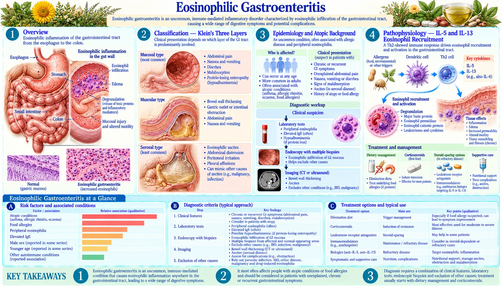
The most widely used classification system for EGE was proposed by Klein and colleagues in 1970 and divides the disease by which layer of the bowel wall is predominantly infiltrated by eosinophils. Each layer produces a strikingly different clinical syndrome.
1. Mucosal EGE (most common, approximately 60% of cases)
Eosinophils infiltrate the innermost lining of the gut — the mucosa and lamina propria. This causes villous atrophy (damage to the absorptive villi of the small intestine), crypt hyperplasia, and disruption of enterocyte function. The result is a malabsorption syndrome that closely mimics celiac disease: chronic diarrhea (sometimes with steatorrhea in severe cases), protein-losing enteropathy (hypoalbuminemia, edema), iron deficiency anemia, and weight loss. Food is not absorbed normally, and proteins leak out of the inflamed mucosa.
2. Muscularis EGE (10–30% of cases)
Eosinophilic infiltration extends into the muscular wall of the bowel (muscularis propria). This causes bowel wall thickening, rigidity, and dysmotility — the gut cannot contract and relax properly. Patients develop a picture of mechanical or functional bowel obstruction: severe cramping abdominal pain, nausea, postprandial vomiting, and abdominal distension. On computed tomography (CT) imaging, affected segments show diffuse wall thickening. Rarely, this layer of disease leads to obstruction severe enough to require surgical intervention.
3. Serosal EGE (about 12% to 39% of cases, depending on the series)
Eosinophils infiltrate the outermost layer of the bowel (serosa) and spill into the peritoneal cavity. The hallmark is eosinophilic ascites — accumulation of peritoneal fluid that is rich in eosinophils (typically >10% eosinophils among ascitic fluid cells). It is the most dramatic presentation of EGE, and its share varies widely between case series: 5 of 40 patients (about 12%) had subserosal disease in a 1990 Mayo Clinic series, while a 2011 French series of 43 patients classified 39% as subserosal and only 12% as muscular. Patients present with abdominal distension, a shifting dullness on percussion, and confirmed ascites on imaging. Analysis of the ascitic fluid distinguishes eosinophilic ascites from ascites caused by cirrhosis, malignancy, or infection. Peripheral blood eosinophilia is usually very high in serosal EGE. The serosal form responds well to corticosteroids but can recur.
These three layers can occur in isolation or in combination. Pure mucosal disease is by far the most common presentation encountered in clinical practice.
Epidemiology and Atopic Background
EGE is genuinely rare. A 2016 analysis of a United States national insurance-claims database (more than 75 million people, ages 0–64) estimated the prevalence of eosinophilic gastroenteritis at 8.4 per 100,000, eosinophilic gastritis at 6.3 per 100,000 and eosinophilic colitis at 3.3 per 100,000; the authors put the total number of people affected by these three conditions in the United States at fewer than 50,000. The disease appears to be more common in Western, industrialized countries, consistent with the "hygiene hypothesis" that reduced early-life microbial exposure promotes atopic disease.
Age and sex: EGE can occur at any age. Children are affected, with EGE presenting as failure to thrive, protein-losing enteropathy, or protein-wasting diarrhea in infants and toddlers. In the 2016 United States claims analysis, eosinophilic gastroenteritis was most common in children under 5 years old, while eosinophilic gastritis was more common in older age groups. Unlike EoE (which is strongly male-predominant), EGE has a more equal sex distribution, with some series showing a slight female predominance.
Atopic background: Approximately 50–70% of EGE patients have a history of atopic disease — asthma, allergic rhinitis, atopic dermatitis, or food allergy. This atopic association is one of the strongest clues that food antigens and Th2 immune dysregulation drive the disease. Patients often have elevated serum immunoglobulin E (IgE) and peripheral blood eosinophilia. In the 2016 United States claims analysis, 45.6% of people with eosinophilic gastroenteritis also had an allergic condition, most often children. In the 1990 Mayo Clinic series, 20 of 40 patients reported a history of allergy, and 9 of 40 had no raised eosinophil count in the blood. A personal or family history of allergy is one of the clues that points toward EGE in a person with unexplained gastrointestinal (GI) symptoms.
Food allergen associations: Unlike immunoglobulin E-mediated food allergy (in which standard allergy testing reliably identifies triggers), EGE triggering foods are identified through empirical elimination and reintroduction with endoscopic monitoring. Common implicated foods overlap with EoE triggers: dairy, wheat, egg, soy, and seafood. Systematic food elimination can induce remission in many patients, although the evidence base is thinner than for EoE.
Seasonal variation in symptom severity — with worsening during high-pollen seasons — has been reported in some patients, paralleling observations in EoE and suggesting that aeroallergens (not just food) contribute to eosinophil recruitment in the gut.
Pathophysiology — IL-5 and IL-13 Eosinophil Recruitment
The central immune abnormality in EGE is a Th2-polarized adaptive immune response that drives eosinophil production, recruitment, and tissue residence in the stomach and intestine. The same cytokine axes that drive EoE operate in EGE, but the target tissue differs.
IL-5 — the master eosinophil cytokine: IL-5, produced by Th2 CD4+ T cells, ILC2 innate lymphoid cells, and mast cells, is the primary driver of eosinophil differentiation in the bone marrow and release into the bloodstream. Elevated IL-5 in EGE accounts for the prominent peripheral blood eosinophilia seen in most patients. Anti-IL-5 therapies (mepolizumab, benralizumab) have been used in hypereosinophilic syndrome (HES) — a systemic eosinophilic disorder that can overlap with EGE — with proven efficacy, providing proof of concept that this pathway is the key therapeutic target.
IL-13 and eotaxin-3 (CCL26): IL-13, another Th2 cytokine, acts on gastrointestinal epithelial cells to upregulate eotaxin-3, the key tissue chemoattractant for eosinophils. IL-13 also disrupts epithelial barrier function, allowing food antigens to penetrate the mucosa and perpetuate the inflammatory cycle. In EoE, IL-13 blockade with dupilumab (anti-IL-4Rα, which also blocks IL-4) is U.S. Food and Drug Administration-approved; in the phase 2 DEGAS trial in eosinophilic gastritis (published 2026), dupilumab lowered stomach eosinophil counts by 50%, against 4% on placebo, over 12 weeks.
Immunoglobulin E-independent mechanisms: Most food-triggered EGE is not immunoglobulin E-mediated — standard allergy skin tests and serum food-specific immunoglobulin E levels do not reliably identify trigger foods in EGE. Instead, the underlying mechanism appears to involve T-cell-mediated delayed hypersensitivity, non-IgE-dependent mast cell activation, and potentially ILC2-driven type 2 inflammation. This is why patch testing (which detects T-cell-mediated reactions) may be more informative than skin-prick tests for food triggers in some patients.
Normal eosinophil distribution in the gut: A key diagnostic challenge is that small numbers of eosinophils are normally present in the stomach, small intestine, and particularly the right colon. The diagnosis of EGE requires pathologically elevated counts — generally accepted thresholds are ≥30 eosinophils per high-power field (eos/hpf) in the stomach and ≥50 eos/hpf in the small intestine, with no universally agreed standard. Context matters: the same count that is diagnostic in an inflamed-appearing stomach is less meaningful in the normal-appearing right colon.
Clinical Presentation
EGE produces non-specific symptoms that vary enormously depending on the layer of bowel wall affected (Klein classification), the segment of gastrointestinal tract involved, and disease severity. There is no single symptom or sign that is pathognomonic for EGE — the diagnosis requires biopsy. Patients often go years before the correct diagnosis is made.
Most common presenting symptoms:
- Abdominal pain — the most common symptom overall; typically colicky, epigastric or periumbilical, worsened by eating.
- Nausea and vomiting — common in gastric involvement; postprandial, bilious if obstruction is partial.
- Diarrhea — watery or bulky (steatorrhea in mucosal type with fat malabsorption); may be chronic or episodic.
- Weight loss — from malabsorption, poor intake, or both; can be significant (5–15 kg) before diagnosis.
- Bloating and early satiety — particularly with gastric involvement.
Signs of malabsorption (mucosal type):
- Hypoalbuminemia — protein-losing enteropathy; serum albumin may fall below 2.5 g/dL in severe cases, causing dependent edema.
- Iron deficiency anemia — from occult gastrointestinal blood loss and impaired iron absorption.
- Steatorrhea — fatty, floating, foul-smelling stools from fat malabsorption.
- Micronutrient deficiencies — vitamins D, B12, zinc, calcium (with prolonged active disease).
Obstruction (muscularis type): Episodic or persistent nausea, vomiting, cramping, constipation, and abdominal distension. May mimic small bowel obstruction from adhesions or Crohn's disease stricture.
Eosinophilic ascites (serosal type): Painless progressive abdominal distension, shifting dullness, and confirmed ascites on imaging. May be the sole presenting feature or accompany mucosal or muscularis disease. Paracentesis reveals a striking eosinophil-rich fluid.
Laboratory clues:
- Peripheral blood eosinophilia (absolute eosinophil count >500/µL) — present in approximately 70–80% of EGE patients; levels often correlate with disease activity.
- Elevated serum immunoglobulin E — reflecting atopic background; not diagnostic but supportive.
- Elevated erythrocyte sedimentation rate (ESR)/C-reactive protein (CRP) — in some but not all patients; less reliable than in Crohn's disease.
- Low albumin, low prealbumin — in mucosal/protein-losing EGE.
- Normal peripheral eosinophil count does not exclude EGE — up to 20–30% of patients have counts within the normal range.
Diagnosis — Endoscopy, Biopsy, and Exclusions
There is no single diagnostic test for EGE. The diagnosis rests on three pillars: compatible symptoms + histological eosinophilia on biopsy + exclusion of other causes of gut eosinophilia. The diagnostic workup is systematic and is usually carried out by gastroenterologists.
Step 1: Exclude secondary causes of gastrointestinal eosinophilia
Several conditions cause gut eosinophilia that can mimic EGE; they are ruled out before the eosinophilia is attributed to a primary EGID:
- Parasitic infections — most importantly: Strongyloides stercoralis (can be latent for decades; dissemination in immunosuppressed patients is life-threatening), Toxocara canis/cati (visceral larva migrans), Anisakis simplex (ingested in raw fish), hookworm (Ancylostoma, Necator). Stool ova and parasite examination × 3 samples, stool antigen tests, and serology (Strongyloides antibody, Toxocara antibody) are the usual first tests. When a parasite is found, treating it can clear the eosinophilia, and the EGE diagnosis then no longer applies.
- Drug reactions — numerous medications cause gut eosinophilia (nonsteroidal anti-inflammatory drugs (NSAIDs), proton pump inhibitors (PPIs), carbamazepine, clozapine, gold salts, rifampicin). This is why the medication history matters in the workup.
- Inflammatory bowel disease — Crohn's disease can cause prominent mucosal eosinophilia, particularly in the small bowel and stomach. The distinction requires clinical context + pathology (granulomas favor Crohn's; pure eosinophilia without architectural distortion favors EGE).
- Celiac disease — villous atrophy with eosinophilic infiltrate can mimic EGE; anti-tTG immunoglobulin A (IgA) and total immunoglobulin A are the standard blood tests for it.
- Hypereosinophilic syndrome (HES) — systemic eosinophil overproduction (absolute eosinophil count (AEC) >1,500/µL for >1 month + end-organ damage including gastrointestinal, heart, skin, central nervous system). EGE may be a manifestation of HES or a distinct condition; cardiac involvement (Löffler's endocarditis) distinguishes HES from pure EGE.
- Connective tissue diseases — eosinophilic granulomatosis with polyangiitis (EGPA, formerly called Churg-Strauss syndrome), systemic lupus erythematosus.
- Malignancy — lymphoma, mastocytosis, myeloproliferative neoplasms can cause reactive eosinophilia.
Step 2: Endoscopy with multiple biopsies
Upper endoscopy (esophagogastroduodenoscopy, EGD) is the primary diagnostic tool. Key principles:
- Biopsy multiple segments — gastric antrum, gastric body, duodenal bulb, and second/third portion of duodenum. Normal-appearing mucosa can harbor diagnostic eosinophilia. In screening data from a lirentelimab trial (published 2022), eosinophils were patchy: on average only 2.6 of 8 stomach biopsies and 2.2 of 4 duodenal biopsies per person met the threshold, and the authors concluded that counting eosinophils in at least 8 gastric and 4 duodenal biopsies is required to identify every case. A 1990 Mayo Clinic series found that endoscopic biopsies missed the diagnosis in 5 of 40 patients, presumably because of patchy disease.
- Endoscopic appearances are non-specific — erythema, nodularity, edema, erosions, and fold thickening may be present, but the mucosa can look completely normal even with high eosinophil counts in biopsies.
- Colonoscopy with ileal intubation if symptoms suggest colonic involvement.
- Capsule endoscopy or enteroscopy (push/balloon-assisted) if mid-small bowel is implicated (high suspicion of muscularis or serosal disease with normal EGD/colonoscopy).
Step 3: Histology — eosinophil counts
Eosinophil counts per high-power field (eos/hpf) at the peak infiltrated area are the diagnostic measure. General guiding thresholds (no universal consensus exists):
- Gastric antrum or body: ≥30 eos/hpf
- Duodenum (small bowel): ≥50 eos/hpf
- Eosinophilic abscesses, degranulation, and subepithelial fibrosis increase diagnostic confidence.
Step 4: Ascitic fluid analysis (serosal EGE)
In patients with ascites of uncertain cause, paracentesis reveals: eosinophil-rich fluid (>10% of nucleated cells are eosinophils), typically exudative, with a low SAAG (serum-ascites albumin gradient), no bacteria on culture, and no malignant cells. This picture is essentially diagnostic of serosal EGE in the right clinical context.
Imaging:
- Computed tomography abdomen — bowel wall thickening (particularly muscularis type), ascites, mesenteric lymphadenopathy; helps define extent of disease.
- Ultrasound — first-line for confirming ascites; guides paracentesis.
- MR enterography — superior soft tissue contrast; preferred for evaluating small bowel wall thickness and mesentery in pediatric patients.
Allergy testing: Standard skin-prick testing and serum food-specific immunoglobulin E are of limited utility for identifying EGE trigger foods (non-IgE mechanism). Patch testing (atopy patch test, APT) may identify T-cell-mediated food triggers in some patients and is used in conjunction with dietitian-guided elimination.
Differential Diagnosis
EGE is distinguished from a range of conditions that produce overlapping symptoms and, in many cases, overlapping histological eosinophilia. The differential is broad because gut eosinophilia is a histological finding, not a diagnosis in itself.
- Eosinophilic esophagitis (EoE) — esophageal eosinophilia only; dysphagia and food impaction rather than abdominal pain and diarrhea; EGE and EoE can coexist (pan-EGID).
- Crohn's disease — key distinctions: transmural granulomatous inflammation, skip lesions, perianal disease, elevated fecal calprotectin, C-reactive protein; eosinophilia can be present but is not the primary histological finding.
- Celiac disease — villous atrophy, intraepithelial lymphocytes (not eosinophils), positive anti-tTG immunoglobulin A antibodies, dramatic HLA-DQ2/DQ8 association; gluten-free diet diagnostic and therapeutic.
- Parasitic infections — most important to exclude first; may produce identical histological eosinophilia; stool studies and serology.
- Hypereosinophilic syndrome (HES) — AEC >1,500/µL for >1 month, multi-organ involvement (cardiac Löffler's endocarditis is hallmark), possibly FIP1L1-PDGFRA rearrangement (imatinib-sensitive).
- Drug-induced gut eosinophilia — nonsteroidal anti-inflammatory drugs, proton pump inhibitors, antibiotics, antidepressants; resolution after drug discontinuation.
- Mast cell disease / mastocytosis — tryptase elevation, cutaneous lesions (urticaria pigmentosa), bone marrow involvement.
- Lymphoma — T-cell lymphoma can cause eosinophil recruitment in the gut; biopsy with immunohistochemistry distinguishes.
- Irritable bowel syndrome — no histological eosinophilia; normal endoscopy and biopsies (though subtle mucosal eosinophilia has been described in some irritable bowel syndrome (IBS) patients).
Treatment — Steroids, Diet, and Emerging Biologics
EGE treatment is based on a small evidence base of case series and expert consensus. The primary goals are symptom relief, induction and maintenance of histological remission, correction of nutritional deficiencies, and avoidance of long-term steroid toxicity.
1. Corticosteroids — the mainstay of therapy
Systemic corticosteroids (prednisolone/prednisone 20–40 mg/day orally for 2–4 weeks) produce dramatic responses in the vast majority of patients — approximately 90% or more experience significant symptom relief and histological improvement within 2–4 weeks. This rapid steroid responsiveness is one of the most distinctive features of EGE and helps confirm the diagnosis. The main limitation is relapse after steroids are tapered in a share of patients: in a French series of 43 patients followed for a median of 13 years, 42% had a single flare without relapse, while 37% had recurring flares and 21% had chronic disease. A 2019 review notes that some patients have no recurrences, while a few have symptoms return during or right after stopping corticosteroids.
In serosal EGE with eosinophilic ascites, steroids are equally effective — ascites typically resolves within days to weeks of starting prednisolone.
Budesonide (oral; enteric-coated formulations designed for small bowel release) is increasingly used as a steroid-sparing alternative, particularly for mucosal small bowel disease. Its high first-pass hepatic metabolism (90%) reduces systemic side effects compared to prednisolone. Experience is from case series, not randomized trials.
2. Dietary therapy
Based on the evidence from EoE and the known atopic/food-trigger pathophysiology of EGE, dietary elimination is used as a first-line or add-on strategy:
- Six-food elimination diet (6FED) — removing milk, wheat/gluten, eggs, soy, peanuts/tree nuts, and seafood. Success has been reported in case series; foods are reintroduced sequentially with endoscopic confirmation of continued remission.
- Elemental diet — an amino acid-based formula that eliminates all intact food antigens. Highly effective for inducing remission (analogous to its use in EoE and Crohn's disease) but poor palatability and high cost limit use in adults; more feasible in infants via nasogastric tube.
- Targeted elimination — based on patch testing or clinical history; less systematic but more practical when a specific trigger is suspected (e.g., wheat or dairy).
Elimination diets are usually carried out with a gastrointestinal dietitian experienced in elimination protocols and nutritional monitoring.
3. Mast cell stabilizers and antihistamines (steroid-sparing)
- Cromolyn sodium — stabilizes mast cells; used in case series for steroid-dependent EGE with variable results; oral formulation (not well absorbed, acts locally).
- Ketotifen — H1-antihistamine with mast cell-stabilizing properties; has shown benefit in several small case series; common side effects: sedation, weight gain.
- Montelukast — leukotriene receptor antagonist; limited evidence in EGE; some improvement in symptoms reported in case reports.
4. Emerging biologics
- Dupilumab (anti-IL-4Rα, blocks IL-4 and IL-13) — U.S. Food and Drug Administration-approved for EoE, atopic dermatitis, asthma; case reports and small series in EGE with promising results; the phase 2 DEGAS trial in eosinophilic gastritis was completed and published in 2026 (below).
- Mepolizumab (anti-IL-5) — proven in HES; used off-label in cases of EGE overlapping with HES or steroid-dependent EGE; limited case reports.
- Cendakimab (anti-IL-13) and etrasimod (S1P receptor modulator) — in clinical trials for EGID spectrum disorders.
- Dupilumab trial (DEGAS, 2026) — in a phase 2 trial of 41 people aged 12–59 with eosinophilic gastritis, stomach eosinophil counts fell by 50% over 12 weeks with dupilumab, against 4% with placebo. Side effects were about as common in both groups (81% vs 85%), and no serious adverse events were reported. The authors note that eosinophilic gastritis currently has no approved treatments.
- Lirentelimab (ENIGMA trial, 2020) — an anti-Siglec-8 antibody that removes eosinophils and calms mast cells. In this phase 2 trial of 65 adults with eosinophilic gastritis and/or duodenitis, gut eosinophil counts fell by 86% with the drug, against a 9% rise with placebo, and symptom scores fell by 48% versus 22%. Mild-to-moderate infusion reactions were more common on the drug (60% vs 23%).
- Benralizumab trial (2023) — an eosinophil-removing antibody against the interleukin-5 receptor. In a single-centre phase 2 trial of 26 people aged 12–60 with eosinophilic gastritis, 10 of 13 on benralizumab and 1 of 13 on placebo reached histological remission (no tissue eosinophilia) at 12 weeks. Endoscopy scores, tissue-structure scores and patient-reported symptoms did not improve more than with placebo, and the authors concluded that a mechanism independent of eosinophils also drives the disease.
5. Supportive care and nutritional management
- Iron supplementation (intravenous (IV) iron if oral not tolerated in severe mucosal disease).
- Vitamin D and calcium supplementation — especially important given fat malabsorption and long-term steroid use (both cause bone loss).
- Albumin infusions in severe protein-losing enteropathy (short-term bridge while disease control is established).
- Enteral nutrition support (nasojejunal feeds) in patients with severe disease impairing oral intake.
6. Surgical management (rare)
Surgery is occasionally required for muscularis EGE presenting with complete or near-complete bowel obstruction not responding rapidly to steroids. Laparoscopic adhesiolysis, stricturoplasty, or limited bowel resection may be needed. Ascites in serosal EGE can be drained for comfort but systemic steroid treatment is the definitive approach.
Prognosis and Long-Term Course
EGE follows a relapsing-remitting or chronic course in about half of patients. In a French series of 43 patients followed for a median of 13 years, spontaneous remission occurred in 40%, and the authors concluded that about 40% of patients resolve the disease without relapse, while about 50% have unpredictable relapses or a chronic course. A 2019 review likewise reports spontaneous remission in around 30% to 40% of cases, with most patients needing ongoing treatment. Severe complications such as perforation, major hemorrhage, or malignant transformation are uncommon.
Long-term course and steroid dependence: In the 13-year French follow-up, 18 of 43 patients (42%; 9 of them with the subserosal, ascites-forming type) had a single flare and never relapsed, 16 (37%) had flares separated by periods of full remission, and 9 (21%) had chronic disease. For patients whose disease returns as steroids are tapered or stopped, steroid dependence is the central management challenge. Long-term steroid use carries well-known risks: osteoporosis, glucose intolerance, adrenal suppression, cataracts, and susceptibility to infection. This drives the need for steroid-sparing strategies (dietary therapy, budesonide, biological agents).
Nutritional complications: Patients with mucosal EGE may develop lasting nutritional consequences from prolonged protein-losing enteropathy — low bone density, muscle wasting, and micronutrient depletion. Careful dietitian follow-up and periodic laboratory monitoring (albumin, ferritin, vitamin D, B12, zinc) are important parts of long-term care.
Malignancy risk: There is no established increased risk of gastrointestinal cancer in EGE (unlike inflammatory bowel disease), and eosinophilic infiltration itself has not been shown to be premalignant. Long-term surveillance endoscopy intervals are guided by clinical need rather than oncological surveillance.
Quality of life: EGE profoundly impairs quality of life through dietary restriction, meal-related anxiety, unpredictable diarrhea or pain, and the physical and psychological burden of steroid dependence. Validated patient-reported outcome measures specific to EGID are under development. Psychological support and patient advocacy organizations (e.g., the American Partnership for Eosinophilic Disorders, APFED) offer support to patients and families.
Practical Guidance for Patients
Living with eosinophilic gastroenteritis can feel overwhelming, particularly because the disease is rare, many general practitioners are unfamiliar with it, and treatment decisions are individualized. The points below summarise how care is usually organised and what published case series report.
Specialist care: EGE is usually managed by a gastroenterologist experienced with eosinophilic disorders, often at an academic or referral center; some centers have dedicated EGID clinics. Gastrointestinal dietitians who specialize in elimination diets and malabsorption are part of many care teams, and allergists/immunologists are often involved when asthma, eczema or food allergy are also present.
The subtype matters: The Klein layer that is mainly involved — mucosal, muscularis, or serosal — predicts the main symptoms and shapes treatment priorities (malabsorption workup for mucosal; obstruction management for muscularis; paracentesis and drainage for serosal).
Food elimination: Even without positive allergy testing, a structured elimination diet (starting with the six most common triggers) has induced remission in some patients in case series. A follow-up endoscopy is used to confirm a histological response, because symptom improvement alone does not show that the diet is working. Calcium, vitamin D and iron are the nutrients most watched during elimination.
Steroids as a short-term tool: Prednisone works fast and reliably, but long-term use carries the risks listed above. Treatment plans aim to control the disease with the lowest effective steroid dose while dietary and biological options are explored to reduce or end steroid dependence. Bone protection (calcium, vitamin D, and for some long-term steroid users bisphosphonates) is part of these plans.
Symptom diary: A record of foods eaten, symptoms, and blood eosinophil counts (where these are monitored) can show patterns and possible triggers, and gives the care team something concrete to review at follow-up visits.
Patient support: The American Partnership for Eosinophilic Disorders (APFED) at apfed.org and the Campaign Urging Research for Eosinophilic Disease (CURED) Foundation provide patient education, support communities, and research updates. Because EGE is rare, patient registries and clinical trials depend on the people who take part in them.
Key Research Papers
- Klein NC, Hargrove RL, Sleisenger MH, Jeffries GH. Eosinophilic gastroenteritis. Medicine (Baltimore). 1970;49(4):299–319. — PubMed PMID: 5426746
- Talley NJ, Shorter RG, Phillips SF, Zinsmeister AR. Eosinophilic gastroenteritis: a clinicopathological study of patients with disease of the mucosa, muscle layer, and subserosal tissues. Gut. 1990;31(1):54–58. — PubMed PMID: 2318432
- Caldwell JM, Collins MH, Stucke EM, et al. Histologic eosinophilic gastritis is a systemic disorder associated with blood and extragastric eosinophilia, TH2 immunity, and a unique gastric transcriptome. J Allergy Clin Immunol. 2014;134(5):1114–1124. — PubMed PMID: 25234644
- Furuta GT, Atkins FD, Lee NA, Lee JJ. Changing roles of eosinophils in health and disease. Ann Allergy Asthma Immunol. 2014;113(1):3–8. — PubMed PMID: 24795292
- Pesek RD, Reed CC, Muir AB, et al. Increasing rates of diagnosis, substantial co-occurrence, and variable treatment patterns of eosinophilic gastritis, gastroenteritis, and colitis based on 10-year data across a multicenter consortium. Am J Gastroenterol. 2019;114(6):984–994. — PubMed PMID: 31008735
- Dellon ES, Gonsalves N, Hirano I, et al. ACG clinical guideline: evidenced based approach to the diagnosis and management of esophageal eosinophilia and eosinophilic esophagitis. Am J Gastroenterol. 2013;108(5):679–692. — PubMed PMID: 23567357
- Uppal V, Kreiger P, Kutsch E. Eosinophilic gastroenteritis and colitis: a comprehensive review. Clin Rev Allergy Immunol. 2016;50(2):175–188. — PubMed PMID: 26054822
- Gonsalves N, Yang GY, Doerfler B, et al. Elimination diet effectively treats eosinophilic esophagitis in adults; food reintroduction identifies causative factors. Gastroenterology. 2012;142(7):1451–1459. — PubMed PMID: 22391333
- Lwin T, Melton SD, Genta RM. Eosinophilic gastritis: histopathological characterization and quantification of the normal gastric eosinophil content. Mod Pathol. 2011;24(4):556–563. — PubMed PMID: 21169993
- Straumann A, Bauer M, Fischer B, Blaser K, Simon HU. Idiopathic eosinophilic esophagitis is associated with a T(H)2-type allergic inflammatory response. J Allergy Clin Immunol. 2001;108(6):954–961. — PubMed PMID: 11742273
- van Rhijn BD, Verheij J, Smout AJ, Bredenoord AJ. Rapidly increasing incidence of eosinophilic esophagitis in a large cohort. Neurogastroenterol Motil. 2013;25(1):47–52.e5. — PubMed PMID: 22963642
- Sunkara T, Rawla P, Yarlagadda KS, Gaduputi V. Eosinophilic gastroenteritis: diagnosis and clinical perspectives. Clin Exp Gastroenterol. 2019;12:239–253. — PubMed PMID: 31239747
- Jensen ET, Martin CF, Kappelman MD, Dellon ES. Prevalence of eosinophilic gastritis, gastroenteritis, and colitis: estimates from a national administrative database. J Pediatr Gastroenterol Nutr. 2016;62(1):36–42. — PubMed PMID: 25988554
- Pineton de Chambrun G, Gonzalez F, Canva JY, et al. Natural history of eosinophilic gastroenteritis. Clin Gastroenterol Hepatol. 2011;9(11):950–956.e1. — PubMed PMID: 21806952
- Dellon ES, Gonsalves N, Rothenberg ME, et al. Determination of biopsy yield that optimally detects eosinophilic gastritis and/or duodenitis in a randomized trial of lirentelimab. Clin Gastroenterol Hepatol. 2022;20(3):535–545.e15. — PubMed PMID: 34089846
- Dellon ES, Peterson KA, Murray JA, et al. Anti-Siglec-8 antibody for eosinophilic gastritis and duodenitis. N Engl J Med. 2020;383(17):1624–1634. — PubMed PMID: 33085861
- Kliewer KL, Murray-Petzold C, Collins MH, et al. Benralizumab for eosinophilic gastritis: a single-site, randomised, double-blind, placebo-controlled, phase 2 trial. Lancet Gastroenterol Hepatol. 2023;8(9):803–815. — PubMed PMID: 37336228
- Gonsalves NP, Dellon ES, Kliewer KL, et al. Dupilumab versus placebo in adults and adolescents with eosinophilic gastritis (DEGAS): a double-blind, placebo-controlled, phase 2, multicentre, randomised controlled trial. Lancet Gastroenterol Hepatol. 2026;11(8):672–688. — PubMed PMID: 42341804