Coarctation of the Aorta
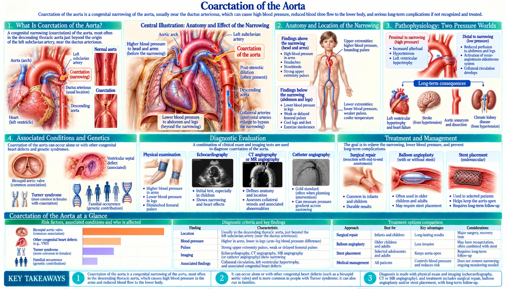
Table of Contents
- Coarctation of the Aorta Explained
- Anatomy and Location of the Narrowing
- Pathophysiology: Two Pressure Worlds
- Associated Conditions and Genetics
- Clinical Presentations by Age
- Physical Examination Signs
- Diagnosis: Imaging and Hemodynamics
- Treatment: Catheter-Based and Surgical Repair
- Lifelong Surveillance After Repair
- Long-Term Complications
- Key Research Papers
- Connections
- Featured Videos
Coarctation of the Aorta Explained
Coarctation of the aorta (CoA) is a congenital narrowing of the aorta — the body's main artery — that obstructs blood flow from the heart to the lower body. It accounts for 5–8% of all congenital heart defects and occurs in approximately 3–4 per 10,000 live births. Males are affected roughly twice as often as females, though the condition is strongly linked to Turner syndrome (45,X) in women.
The narrowing creates two pressure zones: dangerously high blood pressure in the arms and head (above the coarctation), and abnormally low pressure in the legs and abdominal organs (below it). Without repair, most people with significant CoA develop severe hypertension, left ventricular failure, aortic dissection, or stroke before the age of 50. With modern catheter-based or surgical repair, outcomes are excellent — but the condition requires lifelong cardiac follow-up because it is never truly "cured."
Anatomy and Location of the Narrowing
The aorta arises from the left ventricle, arches over the heart (the aortic arch), then descends through the chest and abdomen. The coarctation almost always occurs at the aortic isthmus — a short segment of the descending aorta just past the origin of the left subclavian artery, at the point where the ductus arteriosus (or its remnant, the ligamentum arteriosum) connects to the aorta.
The narrowing itself is typically a discrete fibromuscular ridge or shelf that protrudes into the aortic lumen, reducing its diameter by 50% or more. In severe or complex cases, the entire aortic arch may be underdeveloped — called a hypoplastic aortic arch — rather than a single focal narrowing. A long-segment or diffuse coarctation is more challenging to treat and may require more extensive surgery.
Why this location? During fetal development, the ductus arteriosus allows blood to bypass the fetal lungs. After birth, the ductus closes (normally within 24–48 hours). Ductal tissue extends into the adjacent aortic wall; as this tissue contracts when the ductus closes, it can constrict the neighboring aorta — producing the coarctation. This explains why critical CoA often becomes life-threatening only after the ductus closes.
Pathophysiology: Two Pressure Worlds
The coarctation acts as a fixed obstruction to left ventricular outflow:
- Proximal (upstream) effects: The left ventricle must pump against increased afterload. Over months and years, the LV wall thickens (left ventricular hypertrophy, LVH). The aorta, branches of the aortic arch, and the brain are exposed to chronically elevated pressure — causing early hypertension, accelerated coronary artery disease, and increased stroke risk. The ascending aorta may also dilate (aortopathy) even after successful repair.
- Distal (downstream) effects: Blood pressure below the coarctation is reduced. The kidneys respond to their perceived low perfusion pressure by activating the renin-angiotensin-aldosterone system (RAAS) — which raises blood pressure throughout the body. This creates a vicious cycle: the kidneys try to normalize their pressure by raising systemic pressure, which further increases the load on the left ventricle and the risk of hypertensive complications above the coarctation.
Collateral Circulation
Over months to years, the body attempts to bypass the obstruction by enlarging collateral arterial vessels. Blood flows from the internal mammary arteries through enlarged intercostal arteries around the narrowing to reach the descending aorta below. These expanded intercostal arteries erode the undersides of the ribs — producing the characteristic rib notching seen on chest X-ray (typically ribs 3 through 9, bilaterally, not present until late childhood because collaterals take years to develop).
While collaterals partially compensate for the obstruction, they do not normalize distal pressure and do not protect against the proximal hypertension and LVH. They also complicate surgical repair — the surgeon may need to tie off (ligate) or manage large collateral vessels.
Associated Conditions and Genetics
Coarctation of the aorta rarely occurs in isolation. The most important associated conditions are:
- Bicuspid aortic valve (BAV): Present in 50–70% of patients with CoA. Both CoA and BAV share a common developmental origin — abnormal migration of neural crest cells during embryogenesis. Patients with CoA and BAV face additional risks: aortic stenosis or regurgitation from the abnormal valve, and accelerated aortopathy (dilation and dissection risk) affecting the entire aortic root and ascending aorta, even after CoA repair.
- Turner syndrome (45,X): The most common chromosomal cause of CoA in females. In a heart-scan study using magnetic resonance imaging in 152 women with Turner syndrome (45,X), 19 (12.5%) had coarctation and 52 (34%) had a bicuspid aortic valve, the more common of the two. Coarctation can also be the first sign of Turner syndrome in a baby girl: in a Utah birth-defects registry of girls born from 1997 to 2011, 21 of 167 girls with coarctation (12.6%) had Turner syndrome confirmed by chromosome testing, all diagnosed within the first 4 months of life; because only 86 of the girls (51%) had chromosome studies, the authors put the figure at “at least 12.6%.” Turner syndrome patients also have an elevated risk of spontaneous aortic dissection, making careful imaging surveillance critical throughout life.
- Intracranial (circle of Willis) aneurysms: In a 2003 US study that scanned 100 adults with coarctation by magnetic resonance angiography (MRA), 10 had an intracranial aneurysm (10%, against about 2% predicted for the general population — approximately 5 times as often). A 2023 pooled analysis of 5 cohorts (442 patients) found a lower average of 3.8%, with high blood pressure the only risk factor linked to the aneurysms (odds ratio 3.1). These berry aneurysms are thought to result from the chronic hypertensive stress on the cerebral vasculature. Rupture causes subarachnoid hemorrhage (SAH, bleeding over the surface of the brain) — a devastating event. An undiagnosed coarctation is one of the causes looked for in young adults who have an SAH.
- Ventricular septal defect (VSD): The most common intracardiac lesion associated with CoA, found in ~30% of cases with complex congenital heart disease.
- Patent ductus arteriosus (PDA): In neonatal critical CoA, the PDA is life-sustaining — it supplies blood flow to the descending aorta while the coarctation blocks antegrade flow. Prostaglandin E1 (PGE1) is given intravenously to prevent ductal closure and stabilize the neonate before repair.
- Mitral valve anomalies and sub-aortic stenosis may also coexist, particularly in the Shone complex (a constellation of left-sided obstructive lesions including CoA, mitral stenosis, parachute mitral valve, and subaortic stenosis).
Clinical Presentations by Age
The presentation of CoA spans a wide spectrum, from cardiovascular collapse in the first days of life to incidental hypertension discovered in a teenager or adult. Severity depends on the degree of narrowing, whether a PDA is patent, and the presence of associated cardiac defects.
Neonatal Critical Coarctation
In severe CoA, the ductus arteriosus provides most of the blood supply to the lower body during fetal life. When the ductus closes (typically at 24–72 hours of age), the lower body suddenly loses its blood supply. The infant presents with:
- Sudden cardiovascular collapse: pallor, poor perfusion, metabolic acidosis
- Differential cyanosis — upper body is pink (from the left ventricle) while lower body is blue or pale (ductal blood from the right ventricle; or simply underperfused below the coarctation)
- Absent or very weak femoral pulses
- Rapid deterioration to multi-organ failure
Emergency management: Intravenous prostaglandin E1 (alprostadil) is given immediately to reopen or maintain the ductus arteriosus, restoring lower-body perfusion. This stabilizes the infant for diagnostic evaluation and surgical repair, typically within days. Without PGE1, critical neonatal CoA is rapidly fatal.
Childhood Presentation
Less severe coarctations may not cause symptoms in the neonatal period. Children may be diagnosed when hypertension is found on routine screening, when a murmur is noted, or when a large blood pressure difference between the arms and legs is detected. Some children develop exercise intolerance, headaches, nosebleeds (from hypertension), or leg fatigue (claudication from reduced perfusion). The classic presentation in an otherwise healthy child is upper extremity hypertension found at a routine visit.
Adult Presentation
Adults with undiagnosed or previously undetected CoA typically present with:
- Upper extremity hypertension — often severe and resistant to multiple medications, because the underlying obstruction is not being treated
- Headaches, epistaxis, or hypertensive urgency
- Angina or exertional dyspnea from LVH and reduced coronary reserve
- Stroke or intracranial hemorrhage from a ruptured circle of Willis aneurysm
- Aortic dissection (type A or B)
- Endocarditis (on a bicuspid aortic valve)
In some adults, CoA is discovered incidentally when an echocardiogram performed for another reason reveals LVH, or when an aortogram shows the characteristic narrowing.
Physical Examination Signs
A thorough physical examination can strongly suggest coarctation of the aorta before any imaging is obtained. The key findings are:
- Blood pressure differential between arms and legs: This is the cornerstone of CoA diagnosis. Systolic blood pressure (BP) in the right arm will be significantly higher than in either leg (femoral or popliteal cuff). A difference of >20 mmHg is considered abnormal and requires investigation. In young people with high blood pressure, clinicians check both arms and one leg at each visit for this reason. (Note: if the left subclavian artery arises near or at the coarctation, the left arm blood pressure may be falsely low — which is why the right arm is used as the reference.)
- Brachial-femoral pulse delay: Palpating the radial (wrist) and femoral pulses simultaneously, the femoral pulse is felt later and with reduced amplitude compared to the radial. This delay reflects the time it takes for the pressure wave to pass through or around the coarctation. In severe CoA, the femoral pulse may be barely palpable or absent.
- Systolic murmur: A systolic ejection murmur is heard over the left sternal border and left interscapular area (posterior chest, between the shoulder blades) — the sound of blood accelerating through the narrowed aortic segment. The murmur may be continuous if collateral vessels are prominent. A separate systolic ejection click and murmur may be present at the base if a bicuspid aortic valve is present.
- Palpable collateral vessels: In older children and adults with long-standing CoA, enlarged intercostal collateral arteries may be palpable along the posterior chest wall.
- Findings of left ventricular hypertrophy: Sustained, forceful apical impulse (apex beat displaced leftward and inferiorly). Fourth heart sound (S4) may be present if the LV is stiff from chronic pressure overload.
Diagnosis: Imaging and Hemodynamics
Four-Limb Blood Pressure Measurement
The first and most important step is measuring blood pressure in all four limbs with appropriately sized cuffs. A right arm-to-leg systolic gradient of >20 mmHg is diagnostic of significant CoA. This simple test can be performed in any clinic or emergency room.
Chest X-Ray Findings
The chest X-ray in CoA shows two classic findings:
- Rib notching: Bilateral erosion of the inferior margins of ribs 3–9, caused by enlarged intercostal collateral arteries rubbing against the rib undersurfaces over years. This is a late finding and is not present in neonates or young children, or in patients with short-segment CoA without significant collaterals.
- Figure-3 sign: The aortic knob (dilated aorta above the coarctation) and the post-stenotic dilation (dilated aorta below the coarctation) create the shape of the numeral "3" at the left upper mediastinal contour on the posteroanterior (PA, back-to-front) chest X-ray. On a barium esophagram, the impression of these vessels on the esophagus creates a reversed-3 (or "E" sign).
Echocardiography
Two-dimensional and Doppler echocardiography is the primary imaging modality in neonates and young children, and for initial evaluation in older patients. Echo demonstrates the coarctation site and can estimate the gradient (peak Doppler velocity >4 m/s across the narrowing corresponds to a peak gradient >64 mmHg — severe). Echo also evaluates: bicuspid aortic valve and its severity, left ventricular function and wall thickness, aortic arch anatomy, and other intracardiac lesions. However, echo has limited spatial resolution for the descending aorta in adults and may underestimate the gradient if significant collaterals decompress the gradient.
Computed Tomography and Magnetic Resonance Imaging Angiography
Cross-sectional imaging provides definitive anatomical detail before intervention:
- Computed tomography (CT) angiography (CTA): Excellent spatial resolution; demonstrates the exact location, length, and severity of the coarctation; shows collateral vessels; fast acquisition. Preferred in older children and adults for pre-procedural planning. Radiation exposure is a consideration for serial follow-up in younger patients.
- Cardiac MRI (CMR): No radiation; excellent for serial follow-up; can quantify aortic flow, measure gradients, and assess LV function. Phase-contrast magnetic resonance imaging (MRI) can measure the hemodynamic significance of the coarctation and collateral flow. Preferred for long-term surveillance in young patients.
Cardiac Catheterization
Catheter-based hemodynamic assessment (measuring the peak-to-peak gradient between the aorta above and below the coarctation) is the gold standard for defining severity. A resting peak-to-peak gradient of >20 mmHg, or >10 mmHg with imaging evidence of significant narrowing and collaterals, generally justifies intervention. Catheterization is routinely combined with balloon angioplasty and stent placement in a single procedure in older children and adults.
Treatment: Catheter-Based and Surgical Repair
When to Intervene
Intervention is recommended when:
- Resting peak-to-peak gradient across the coarctation is >20 mmHg at catheterization
- Upper extremity hypertension is present (systolic blood pressure >95th percentile for age)
- Significant LV hypertrophy or dysfunction
- Symptoms (headache, exertional dyspnea, claudication)
- Significant anatomical narrowing by imaging (>50% reduction in aortic diameter) even with lower measured gradient if extensive collaterals decompress the gradient
The optimal timing of repair depends on age and associated lesions. In neonates with critical CoA and cardiovascular collapse, emergency repair after PGE1 stabilization is performed in days. In asymptomatic older children, repair is typically planned electively at age 2–5 years before hypertension and LVH become established.
Catheter-Based Intervention (Transcatheter Approach)
Transcatheter treatment is preferred for native CoA in adolescents and adults, and for re-coarctation (recurrent narrowing after prior repair) at all ages where vessels are large enough to accept the stent delivery system.
- Balloon angioplasty alone: Historically used; it carries a risk of aortic aneurysm at the dilation site, and in a 36-centre comparison of the three treatments it gave poorer blood-pressure and imaging results than stenting or surgery at follow-up, and more aortic wall injury (see below). Rarely used alone today except in selected neonates/infants as a bridge to definitive repair.
- Stent placement: The current preferred transcatheter approach. A balloon-expandable stent is deployed across the narrowing under fluoroscopic and angiographic guidance. Covered stents (stent grafts) are strongly preferred in adults with native CoA — they line the aortic wall, prevent dissection and aneurysm formation at the treatment site, and provide excellent hemodynamic results. Results: <10 mmHg residual gradient in >90% of cases; relief of hypertension; 5-year patency >85%.
Transcatheter repair requires a vessel diameter large enough to accommodate a 12–14 French sheath (typically children >25 kg / 8–10 years, depending on anatomy). Stenting is avoided in infants because they will outgrow the stent — though some centers use small stents that can be redilated ("stent-within-stent" or staged redilation strategies).
What the Stent and Comparison Studies Found
- Stent, surgery or balloon in children: A 36-centre observational study of 350 children over 10 kg with native coarctation (217 stented, 72 operated on, 61 treated with a balloon alone) found fewer immediate complications with stents (2.3%) than with surgery (8.1%) or balloon alone (9.8%), and shorter hospital stays with stents than with surgery (2.4 vs 6.4 days). Stent patients were more likely to need a planned repeat procedure, and balloon-only patients had more aortic wall injury, both right after treatment and at follow-up. Because the treatments were not assigned at random, the authors state that the results are to be interpreted with caution.
- Bare stents (COAST trial): In the Coarctation of the Aorta Stent Trial (COAST), bare platinum stents were placed in 104 of 105 children and adults with native or recurrent coarctation, with no procedural deaths or serious adverse events and lasting relief of the arm-to-leg blood pressure difference to 2 years. Six aortic aneurysms were found (5 treated with a covered stent, 1 resolved without treatment); stent fractures were seen in 11 patients at 2 years without loss of stent integrity; and 9 repeat procedures were done in the first 2 years to re-expand stents or treat aneurysms.
- Covered stents (COAST II trial): A covered stent is a metal stent wrapped in a graft lining that seals the aortic wall. COAST II placed covered stents in 158 patients (median age 19) who already had aortic wall injury or were at increased risk of it. The pressure difference from the ascending to the descending aorta fell from an average of 27 to 4 mmHg; existing wall injury was fully covered in 76 of 83 patients (92%); 4 patients had an important injury to the artery where the catheter went in; there were no deaths, repeat procedures or new wall injuries in the short term.
- Stents in adults: A pooled analysis of 27 studies (705 adults, average age 34) found that stenting succeeded technically in 97% of cases, with 1% dying within 30 days. Over a median follow-up of 29 months, 68 repeat procedures (8%) were done, 95.5% of them again through a catheter.
Surgical Repair
Surgery is preferred for neonates, infants, and small children (too small for stent delivery systems), and for complex arch anatomy, long-segment coarctation, or major associated intracardiac lesions requiring simultaneous repair. Surgical approaches:
- Resection with end-to-end anastomosis (EEA): The coarcted segment is excised and the two ends of the aorta are sewn together directly. Preferred technique for focal native CoA in children. Long-term results are excellent with minimal re-coarctation when performed in infancy; however, if the aortic ends are mobilized under tension (common in long-segment CoA), re-coarctation risk rises.
- Extended end-to-end anastomosis: Cuts further back into the underside of the aortic arch to widen the reconstruction — useful when there is associated mild arch hypoplasia.
- Subclavian flap aortoplasty: The left subclavian artery is divided and turned down as a flap to patch the coarctation. Historically popular; avoids the risk of circumferential suture lines. Disadvantage: sacrifices the left subclavian artery, which may lead to a shorter/colder left arm, a persistent blood pressure discrepancy between arms, and inability to use the left arm for future dialysis access. Less commonly used today.
- Patch aortoplasty: A Dacron patch is sutured to the aorta to widen the narrowed segment. Carries a 20–30% risk of late aortic aneurysm at the patch site; largely abandoned in favor of EEA or stenting.
- Conduit bypass: A synthetic graft bypasses the coarctation (grafted from ascending to descending aorta). Used for complex anatomy (long-segment, multiple recurrences, kinking, or calcification after prior repairs) not amenable to direct reconstruction.
Lifelong Surveillance After Repair
One of the most important clinical messages about CoA is that repair does not cure the underlying disease. All repaired CoA patients — regardless of the repair type, age at repair, or apparent initial success — require lifelong follow-up. Key reasons:
- Re-coarctation (re-stenosis): Occurs in 5–10% of patients after surgical repair and 5–15% after balloon angioplasty alone. Residual narrowing as little as 20 mmHg can maintain enough hypertension to cause LVH and premature coronary artery disease (CAD). Re-coarctation is treatable by stenting.
- Persistent or recurrent hypertension: Even after anatomically successful repair, up to 35% of patients remain hypertensive — particularly those repaired after age 5–6 years. This is thought to reflect irreversible changes in baroreflex sensitivity and vascular compliance that develop from years of high-pressure exposure. These patients require antihypertensive medications (angiotensin-converting enzyme inhibitors (ACE inhibitors) or angiotensin receptor blockers (ARBs) are preferred given the RAAS activation; beta-blockers are added for aortopathy).
- Aortic aneurysm: Can develop at the repair site (especially after patch aortoplasty), or anywhere in the ascending aorta or aortic root (from aortopathy associated with bicuspid AV and CoA). Annual blood pressure measurement and magnetic resonance imaging/computed tomography every 3–5 years are standard.
- Intracranial aneurysms: Persistent hypertension increases the risk of growth and rupture. Guidelines disagree on routine MRA screening of the circle of Willis in adults with CoA: a 2023 review notes that the 2018 American Heart Association guideline lists it as “recommended” and the 2020 European Society of Cardiology guideline as “not recommended,” and the review’s authors judged routine screening “likely of low-value” given the 3.8% pooled prevalence.
- Bicuspid aortic valve progression: BAV stenosis or regurgitation may develop or worsen over decades and require valve intervention.
The American College of Cardiology / American Heart Association and the European Society of Cardiology recommend that all CoA patients be followed by an adult congenital heart disease (ACHD) specialist at a center experienced in ACHD throughout their lifetime.
Long-Term Complications
Despite successful repair, CoA patients face higher rates of several cardiovascular complications than the general population:
- Premature coronary artery disease: Years of hypertension before and, in some patients, after repair accelerate coronary atherosclerosis. Myocardial infarction (MI) risk is elevated 2–5 fold compared to age-matched controls.
- Aortic dissection: Risk is elevated by the combination of chronic hypertension, bicuspid AV aortopathy, and potential structural changes at repair sites. Type A dissection (ascending aorta) is particularly feared. Risk is highest in Turner syndrome patients with CoA.
- Stroke and transient ischemic attack (TIA): From hypertension, bicuspid AV-related thromboembolism, atrial fibrillation (which may develop from chronic LV pressure overload), or rupture of an intracranial aneurysm.
- Left ventricular dysfunction: Long-standing pressure overload leads to diastolic and eventually systolic dysfunction. Even patients with normal ejection fraction may have heart failure with preserved ejection fraction (HFpEF) physiology (diastolic dysfunction, elevated filling pressures with exercise) explaining exercise intolerance years after repair.
- Infective endocarditis: Primarily on the bicuspid aortic valve (if present) or at the coarctation repair site in earlier surgical eras. Standard endocarditis prophylaxis guidelines apply.
Key Research Papers
The following citations are from peer-reviewed literature on coarctation of the aorta — epidemiology, interventional outcomes, genetics, and long-term surveillance.
- Hoffman JI, Kaplan S. The incidence of congenital heart disease. J Am Coll Cardiol. 2002;39(12):1890-1900. — PubMed PMID: 12084585
- Forbes TJ, Garekar S, Amin Z, et al. Procedural results and acute complications in stenting native and recurrent coarctation of the aorta in patients over 4 years of age: a multi-institutional study. Catheter Cardiovasc Interv. 2007;70(2):276-85. — PubMed PMID: 17630670
- Warnes CA, Williams RG, Bashore TM, et al. ACC/AHA 2008 Guidelines for the Management of Adults with Congenital Heart Disease: a report of the American College of Cardiology/American Heart Association Task Force on Practice Guidelines (writing committee to develop guidelines on the management of adults with congenital heart disease). Circulation. 2008;118(23):e714-833. — PubMed PMID: 18997169
- Baumgartner H, De Backer J, Babu-Narayan SV, et al. 2020 ESC Guidelines for the management of adult congenital heart disease. Eur Heart J. 2021;42(6):563-645. — PubMed PMID: 32860028
- Oliver JM, Gallego P, Gonzalez A, et al. Risk factors for aortic complications in adults with coarctation of the aorta. J Am Coll Cardiol. 2004;44(8):1641-7. — PubMed PMID: 15489097
- Presbitero P, Demarie D, Villani M, et al. Long term results (15-30 years) of surgical repair of aortic coarctation. Br Heart J. 1987;57(5):462-7. — PubMed PMID: 3593616
- Meadows J, Minahan M, McElhinney DB, McEnaney K, Ringel R; COAST Investigators. Intermediate Outcomes in the Prospective, Multicenter Coarctation of the Aorta Stent Trial (COAST). Circulation. 2015;131(19):1656-64. — PubMed PMID: 25869198
- Stout KK, Daniels CJ, Aboulhosn JA, et al. 2018 AHA/ACC Guideline for the Management of Adults With Congenital Heart Disease: A Report of the American College of Cardiology/American Heart Association Task Force on Clinical Practice Guidelines. Circulation. 2019;139(14):e698-e800. — PubMed PMID: 30586767
- Forbes TJ, Kim DW, Du W, et al. Comparison of surgical, stent, and balloon angioplasty treatment of native coarctation of the aorta: an observational study by the CCISC (Congenital Cardiovascular Interventional Study Consortium). J Am Coll Cardiol. 2011;58(25):2664-74. — PubMed PMID: 22152954
- Taggart NW, Minahan M, Cabalka AK, Cetta F, Usmani K, Ringel RE; COAST II Investigators. Immediate Outcomes of Covered Stent Placement for Treatment or Prevention of Aortic Wall Injury Associated With Coarctation of the Aorta (COAST II). JACC Cardiovasc Interv. 2016;9(5):484-93. — PubMed PMID: 26896890
- Nana P, Spanos K, Brodis A, et al. A Systematic Review and Meta-analysis on Stenting for Aortic Coarctation Management in Adults. J Endovasc Ther. 2025;32(3):548-557. — PubMed PMID: 37287255
- Bondy C, Bakalov VK, Cheng C, Olivieri L, Rosing DR, Arai AE. Bicuspid aortic valve and aortic coarctation are linked to deletion of the X chromosome short arm in Turner syndrome. J Med Genet. 2013;50(10):662-5. — PubMed PMID: 23825392
- Eckhauser A, South ST, Meyers L, Bleyl SB, Botto LD. Turner Syndrome in Girls Presenting with Coarctation of the Aorta. J Pediatr. 2015;167(5):1062-6. — PubMed PMID: 26323199
- Connolly HM, Huston J 3rd, Brown RD Jr, Warnes CA, Ammash NM, Tajik AJ. Intracranial aneurysms in patients with coarctation of the aorta: a prospective magnetic resonance angiographic study of 100 patients. Mayo Clin Proc. 2003;78(12):1491-9. — PubMed PMID: 14661678
- Buckley AD, Han Um KY, Ganame JI, Salehian O, Karbassi A. Prevalence of Intracranial Aneurysms in Patients With Coarctation of the Aorta: A Systematic Review and Meta-Analysis. JACC Adv. 2023;2(5):100394. — PubMed PMID: 38938992