Tuberculosis (TB) Diagnosis: Tuberculin Skin Test, IGRA, Imaging, and Culture
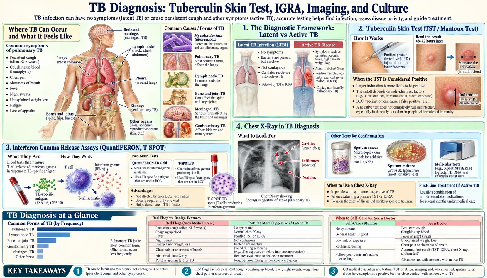
Diagnosing tuberculosis (TB) requires different tests for latent infection versus active disease, and no single test is perfect — understanding each test's strengths and limitations helps patients understand what results mean and why doctors sometimes order several tests before reaching a conclusion.
Table of Contents
- The Diagnostic Framework: Latent vs Active Tuberculosis
- Tuberculin Skin Test (TST / Mantoux Test)
- Interferon-Gamma Release Assays (QuantiFERON, T-SPOT)
- Chest X-Ray in Tuberculosis Diagnosis
- Sputum Smear Microscopy and AFB Culture
- GeneXpert MTB/RIF and Molecular Testing
- Bronchoscopy and Biopsy for Difficult Cases
- Special Diagnostic Situations
- Key Research Papers
- Connections
- Featured Videos
The Diagnostic Framework: Latent vs Active Tuberculosis
The most important concept in tuberculosis diagnostics is understanding that latent tuberculosis infection and active tuberculosis disease require completely different tests — and that confusing the two leads to misdiagnosis. These are not just different stages of the same condition; they are fundamentally different states requiring different diagnostic approaches.
Latent tuberculosis infection means the bacteria are present in the body but are being held in check by the immune system. The person has no symptoms, is not contagious, and the bacteria cannot be found in sputum or blood cultures. The only evidence of infection is the immune system's memory of having encountered tuberculosis antigens. This is why latent tuberculosis can only be detected through immune memory tests — the tuberculin skin test (TST) and interferon-gamma release assays (IGRAs). These tests detect the immune response, not the bacteria themselves.
Active tuberculosis disease means the bacteria are actively multiplying and causing tissue damage. In pulmonary tuberculosis, bacteria are shed into the airways and can often be found in sputum. This is when direct tests for bacteria become useful — sputum smear microscopy, culture, and molecular tests like GeneXpert can detect the actual organism. A positive TST or IGRA in a person with symptoms does not diagnose active tuberculosis; it only tells you that the person was previously infected. You still need direct microbiological evidence to confirm active disease.
The clinical algorithm runs as two separate tracks. If a person has symptoms consistent with active tuberculosis — chronic cough, night sweats, unexplained weight loss, fever — the workup should start with tests for active disease: chest X-ray, sputum examination, and GeneXpert. Do not start with a TST or IGRA in a symptomatic person; a negative result would be misleading because both tests can be falsely negative in active tuberculosis, especially in immunocompromised patients where the immune response may be blunted.
If a person has no symptoms but belongs to a high-risk group — a recent close contact of an active tuberculosis case, someone with human immunodeficiency virus (HIV), a healthcare worker, or a person who immigrated from a high-burden country — the workup should focus on latent tuberculosis with TST or IGRA. This is screening, not diagnosis of disease. A positive result in this scenario means treatment for latent tuberculosis is warranted, even without symptoms or radiographic abnormalities.
One critical nuance: a negative IGRA does not exclude active tuberculosis in immunocompromised people. Human immunodeficiency virus, chemotherapy, and biological immunosuppressants (tumor necrosis factor (TNF) inhibitors, corticosteroids) can blunt the interferon-gamma response enough to produce a false-negative result. In these patients, clinical suspicion and direct microbiological testing matter more than immune-based tests.
Tuberculin Skin Test (TST / Mantoux Test)
The tuberculin skin test, also called the Mantoux test, has been the backbone of tuberculosis screening for over a century. Despite its limitations, it remains widely used because it is inexpensive, requires no laboratory equipment, and has been validated in enormous populations worldwide.
The procedure is straightforward. A healthcare worker injects 0.1 mL of purified protein derivative (PPD) — a standardized extract of M. tuberculosis antigens — into the inner surface of the forearm, just below the skin (intradermally). A correctly placed injection creates a small pale wheal (raised bump) 6–10 mm in diameter immediately after injection. The patient returns 48–72 hours later for the reading. Waiting fewer than 48 hours risks a false negative; waiting more than 72 hours risks the reaction having faded.
What the healthcare worker measures at the reading is the induration — the firm, palpable, raised area caused by immune cells accumulating at the injection site. Redness (erythema) around the site is noted but does not count toward the measurement. The induration is measured across its widest point perpendicular to the forearm's long axis, in millimeters.
The interpretation of results is not a single fixed cutoff. Different threshold sizes are used for different populations based on their risk of true infection versus the probability of a false positive. This tiered approach reflects the fact that the test's positive predictive value depends on how common tuberculosis is in the population being tested.
A reaction of 5 mm or more is considered positive in the highest-risk groups: people with human immunodeficiency virus infection, people who have had recent close contact with an active tuberculosis case, people with organ transplants or on immunosuppressive therapy equivalent to 15 mg/day or more of prednisone for more than a month, and people with radiographic evidence of old healed tuberculosis. In these groups, even a small reaction should not be dismissed because the consequence of a missed latent tuberculosis that reactivates is serious.
A reaction of 10 mm or more is positive for people with intermediate risk factors: those born in or who lived in countries with high tuberculosis burden, current or former injection drug users, residents or employees of high-risk congregate settings (prisons, homeless shelters, nursing homes), healthcare workers who work with tuberculosis patients, children under 4, and people with medical conditions that increase the risk of reactivation (diabetes, silicosis, renal failure, head and neck cancers, low body weight).
A reaction of 15 mm or more is considered positive only for people with no identified risk factors for tuberculosis. In this population, the base rate of true latent tuberculosis infection is low, so a lower threshold would produce too many false positives.
The most important limitation of the TST is that it cannot distinguish between infection with M. tuberculosis and prior BCG vaccination. The BCG vaccine uses live attenuated Mycobacterium bovis, and because BCG shares many antigens with tuberculosis, people vaccinated with BCG will often mount a positive TST reaction even if they have never been infected with actual tuberculosis. This is a major problem in high-income countries screening immigrants from BCG-vaccinating countries — a positive TST in a BCG-vaccinated adult may be a false positive, but cannot be reliably distinguished from true infection without an IGRA.
The boosting effect is another important phenomenon in serial testing, particularly relevant in healthcare worker screening programs. In people with old tuberculosis infection whose immune response to PPD has waned, a first TST can stimulate the immune system and cause a stronger (falsely elevated) reaction to a second TST administered 1–3 weeks later. This can make it appear as though someone converted from negative to positive between two tests. Two-step testing — administering a second TST 1–3 weeks after the first — is used in healthcare workers at initial employment to establish a true baseline, so that later seroconversion (from a new infection) can be reliably identified as a real conversion rather than a boosted response.
The TST is also limited in immunocompromised patients. People with advanced human immunodeficiency virus, malnutrition, overwhelming active tuberculosis (miliary tuberculosis), or severe illness may be unable to mount a detectable skin reaction even if truly infected — a phenomenon called cutaneous anergy. A negative TST in these patients cannot be reassuring.
Interferon-Gamma Release Assays (QuantiFERON, T-SPOT)
Interferon-gamma release assays (IGRAs) are blood tests developed in the early 2000s that largely overcome the BCG-vaccination limitation of the TST. They represent a significant advance in latent tuberculosis diagnostics, although they have their own set of limitations.
The underlying principle is elegant. A blood sample is drawn and the white blood cells (T lymphocytes) are exposed to tuberculosis-specific antigens in the laboratory — specifically ESAT-6 and CFP-10, two proteins encoded by a region of the M. tuberculosis genome called the Region of Difference 1 (RD1). Crucially, RD1 is absent from the BCG vaccine strain and from most non-tuberculous mycobacteria species that cause false-positive TSTs. If the person's T cells have previously encountered real M. tuberculosis, they will recognize these antigens and respond by secreting interferon-gamma. The test measures how much interferon-gamma is produced.
Two IGRAs are commercially available. QuantiFERON-TB Gold Plus (QFT-Plus) uses an enzyme-linked immunosorbent assay (ELISA) to measure total interferon-gamma levels in the blood. It reports a quantitative result in international units per mL, with a cutoff of 0.35 international units (IU)/mL distinguishing positive from negative. Results also include an indeterminate category for samples where the positive control (mitogen-stimulated tube) fails or the negative control (nil tube) is too high, suggesting either immune suppression or procedural problems. T-SPOT.TB takes a different approach: it uses an ELISPOT method that counts the number of individual T cells secreting interferon-gamma in response to the antigens, reporting the result as the number of "spots" on a membrane.
The key advantages of IGRAs over the TST are substantial. Because ESAT-6 and CFP-10 are not present in BCG, a positive IGRA in a BCG-vaccinated person is much more likely to represent true M. tuberculosis infection. IGRAs require only a single visit — the patient doesn't have to return 48–72 hours later for a reading. This dramatically improves completion rates in populations that are difficult to follow up. IGRAs are also more specific in low-incidence settings, meaning they produce fewer false positives.
However, IGRAs have their own limitations that are important to understand. Indeterminate results occur in up to 10–15% of immunocompromised patients — a frustrating outcome because these are often the highest-risk patients where the result matters most. The blood sample must be processed within a specific time window (typically within 8–16 hours of collection for QFT-Plus), which requires coordinated laboratory logistics and limits their use in remote or resource-limited settings. IGRAs are considerably more expensive than TSTs. They are not recommended as the primary test in children under 5 because the T cell response is less mature and results are less reliable.
Head-to-head comparisons between IGRAs and TST show roughly equivalent sensitivity for detecting latent tuberculosis in immunocompetent adults, with IGRAs having higher specificity (fewer false positives) in BCG-vaccinated populations. Neither test has been validated against a true gold standard for latent tuberculosis — by definition, you cannot culture latent bacteria — so sensitivity estimates come from studies in people with known active tuberculosis, known recent exposure, or people who subsequently developed active tuberculosis despite preventive therapy.
Current guidance from the U.S. Centers for Disease Control and Prevention (CDC) and the World Health Organization (WHO) recommends IGRAs as the preferred test for BCG-vaccinated adults and for populations where follow-up for TST reading is unreliable. The TST remains acceptable and preferred for children under 5 years old, and either test is acceptable in most other situations. In high-risk populations, some guidelines recommend sequential testing (TST followed by IGRA, or vice versa) to reduce false negatives, treating a positive on either test as a positive overall.
Chest X-Ray in Tuberculosis Diagnosis
The chest X-ray is often the first investigation that raises suspicion of active tuberculosis, and it remains an essential part of the diagnostic workup. An experienced radiologist reading a chest X-ray can often identify patterns strongly suggestive of tuberculosis — but the chest X-ray cannot by itself confirm the diagnosis. Microbiological confirmation is always required.
Tuberculosis produces different radiographic patterns depending on whether this is a first-time infection (primary tuberculosis) or reactivation of latent disease (post-primary or reactivation tuberculosis), and depending on the patient's immune status.
Primary tuberculosis, which usually occurs in children or immune-naive adults experiencing their first tuberculosis exposure, typically appears as an area of air-space consolidation (an opacity indicating inflammation or fluid in the air sacs) in the lower or middle zones of the lungs, combined with enlarged hilar lymph nodes (the lymph nodes at the root of the lung where the airways branch). This combination — lung opacity plus prominent hilar lymphadenopathy — is called the primary complex or Ghon complex. Primary tuberculosis often resolves on its own in immune-competent people, leaving behind calcified scars (Ghon lesions) that can be seen on chest X-rays years later as evidence of old, healed infection.
Post-primary or reactivation tuberculosis in adults shows a dramatically different pattern that reflects the different immunological context. The hallmark is upper lobe predominance: infiltrates (areas of inflammation and consolidation) in the posterior segment of the upper lobes and the superior segment of the lower lobes. This upper lobe preference is thought to reflect the higher oxygen tension in the upper parts of the lungs, which favors M. tuberculosis growth. Thick-walled cavities are a classic and highly characteristic finding — these appear as areas of lucency (darker areas) within areas of consolidation, indicating that the center of the inflammatory mass has liquefied and drained into the airways. Satellite nodules — small nodular opacities surrounding a larger lesion — reflect local bronchogenic spread of disease.
Miliary tuberculosis produces a striking and alarming radiographic appearance: bilateral, symmetrical fine nodules measuring 1–2 mm distributed throughout both lungs, giving an appearance compared to millet seeds (hence the name). Miliary tuberculosis represents hematogenous dissemination of tuberculosis throughout the bloodstream and is a medical emergency requiring urgent treatment. It is more common in immunocompromised patients and young children.
Pleural effusion — fluid around the lung — can be the sole or predominant finding in tuberculosis pleuritis, particularly in younger patients. This typically causes blunting of one costophrenic angle on chest X-ray.
Computed tomography (CT) scanning of the chest is considerably more sensitive than plain chest X-ray for detecting early or subtle tuberculosis changes. Computed tomography can detect small cavities not visible on X-ray, hilar and mediastinal lymph node enlargement, and the "tree-in-bud" pattern — a branching nodular opacity in the peripheral airways that indicates endobronchial spread of infection and is highly suggestive of active infectious bronchiolitis, particularly tuberculosis.
A critical point that patients sometimes misunderstand: a completely normal chest X-ray does not rule out tuberculosis. Early pulmonary tuberculosis before significant tissue damage has occurred may have a normal X-ray. Extrapulmonary tuberculosis affecting lymph nodes, the spine, kidneys, or brain will not be visible on a chest X-ray at all. Human immunodeficiency virus-positive patients with very advanced immunosuppression can have active tuberculosis with a surprisingly unremarkable chest X-ray because the radiographic findings of tuberculosis (which are partly a product of the immune response to infection) are attenuated when the immune system is severely damaged. In all these situations, a normal chest X-ray should not terminate the diagnostic workup if clinical suspicion remains high.
Sputum Smear Microscopy and AFB Culture
Direct examination of sputum for acid-fast bacilli (AFB) has been the cornerstone of tuberculosis diagnosis in resource-limited settings for over 130 years. Despite being the oldest tuberculosis diagnostic technique, smear microscopy remains the most widely used test globally because it is fast, inexpensive, requires no electricity, and provides immediate actionable information about whether a person is infectious.
The Ziehl-Neelsen (ZN) staining technique exploits a unique property of mycobacteria: their cell walls contain a waxy lipid called mycolic acid that allows them to resist decolorization with acid alcohol after taking up a red dye (carbol fuchsin). This is why they are called "acid-fast" bacilli — they retain the red color after acid washing, while all other bacteria are decolorized and appear blue or colorless against the counterstain. The smear is positive when a trained microscopist sees the characteristic rod-shaped red bacilli under the microscope.
The major limitation of smear microscopy is its relatively low sensitivity. The test requires approximately 5,000–10,000 bacteria per milliliter of sputum to reliably detect them under the microscope. By contrast, liquid culture systems can detect as few as 10–100 organisms per mL. This means smear microscopy will miss approximately 40–60% of culture-positive pulmonary tuberculosis cases — the so-called smear-negative tuberculosis cases. This group includes early disease, patients co-infected with human immunodeficiency virus (who tend to have lower bacterial loads), and patients with less severe immune responses producing less cavitation and sputum bacterial load.
Smear microscopy also cannot distinguish M. tuberculosis from the many non-tuberculous mycobacteria (NTM) that also stain acid-fast. A positive smear means "acid-fast bacilli seen" — not "M. tuberculosis confirmed." Culture and molecular testing are required to identify the species.
Specimen collection matters enormously for smear accuracy. The World Health Organization recommends collecting two sputum specimens — early morning production tends to have the highest bacterial concentrations because secretions have accumulated overnight. If the patient cannot produce sputum spontaneously, induced sputum (inhaling nebulized hypertonic saline to trigger coughing) significantly improves yield. Three specimens were previously standard; current World Health Organization (WHO) guidance accepts two specimens in most settings without significant loss of diagnostic sensitivity when molecular tests are also being used.
Fluorescence microscopy using auramine-O stain on LED microscopes has largely replaced traditional ZN microscopy in well-resourced settings. The fluorescent stain makes AFB easier and faster to visualize (lower magnification covers more area per unit time) and increases sensitivity by approximately 10% compared to traditional ZN — it can detect organisms at lower concentrations. The World Health Organization recommends LED fluorescence microscopy as the preferred technique where available.
Culture is the gold standard for tuberculosis diagnosis and is required for all confirmed cases to determine drug susceptibility. Culture grows actual bacteria, allowing the laboratory to confirm M. tuberculosis as the species and to test which drugs will kill it. Two main culture methods exist. Solid media (Lowenstein-Jensen egg-based or Middlebrook agar) is inexpensive but slow — M. tuberculosis divides approximately every 16–24 hours, so colonies take 3–6 weeks to appear, and susceptibility testing on solid media takes another 3–4 weeks. Liquid media systems (BACTEC MGIT 960) detect bacterial growth by sensing oxygen consumption by a fluorescent sensor — growth appears in 2–3 weeks for smear-positive cases and 3–4 weeks for smear-negative cases, and drug susceptibility testing takes an additional 1–2 weeks. Liquid culture systems are more sensitive than solid media and are the preferred standard in high-income countries.
GeneXpert MTB/RIF and Molecular Testing
GeneXpert MTB/RIF, introduced in 2010 and endorsed by the World Health Organization in 2011, was arguably the most important advance in tuberculosis diagnostics in decades. It transformed the diagnostic landscape by delivering results in 2 hours that previously required weeks of culture — and simultaneously detecting rifampicin resistance.
GeneXpert is a cartridge-based, real-time polymerase chain reaction (PCR) system that requires minimal training. The healthcare worker places sputum into a liquid reagent, loads the mixture into a single-use cartridge, and inserts it into the GeneXpert machine. The machine performs all subsequent steps automatically: cell lysis to release DNA, PCR amplification of M. tuberculosis-specific sequences in the rpoB gene (which encodes RNA polymerase), and detection. The system simultaneously probes for mutations in the 81-base pair "hot spot" region of rpoB where >95% of rifampicin resistance-conferring mutations cluster. A result — positive/negative for tuberculosis and susceptible/resistant/indeterminate for rifampicin — is available in approximately 100 minutes.
The World Health Organization now recommends GeneXpert as the initial diagnostic test (replacing sputum smear microscopy) for all adults and children suspected of having tuberculosis in all settings, including low-income countries. This recommendation reflects the dramatic improvement in diagnostic accuracy that GeneXpert provides over smear microscopy.
Sensitivity of GeneXpert MTB/RIF is approximately 88% in smear-positive cases and 67% in smear-negative, culture-positive cases — compared to smear microscopy sensitivity of approximately 70% in smear-positive and essentially 0% in smear-negative cases (by definition). This means GeneXpert diagnoses a substantial proportion of patients who would have been missed by smear alone, reducing the period of undiagnosed infectiousness. Specificity is very high (>98%), meaning very few false positives.
GeneXpert can be applied to specimens beyond sputum. It has been validated for gastric aspirate (for children who cannot expectorate), lymph node biopsy tissue, cerebrospinal fluid, pleural fluid, urine, and other samples — making it invaluable in extrapulmonary tuberculosis where sputum is irrelevant. Sensitivity varies by specimen type and disease site, but even with lower sensitivity than in sputum, GeneXpert substantially outperforms smear microscopy on non-sputum specimens.
Rifampicin resistance detected by GeneXpert is a strong proxy for MDR-TB (multidrug-resistant tuberculosis, defined as resistance to both rifampicin and isoniazid). More than 95% of rifampicin-resistant strains are also isoniazid-resistant. A GeneXpert result showing rifampicin resistance should therefore trigger presumptive MDR-TB treatment while culture-based full drug susceptibility testing is awaited.
GeneXpert Ultra, the second-generation platform released in 2017, uses two sets of overlapping polymerase chain reaction targets and a larger detection chamber, increasing sensitivity to detect as few as 16 bacteria/mL — substantially lower than the original GeneXpert's limit of approximately 131 bacteria/mL. GeneXpert Ultra is particularly valuable for smear-negative, human immunodeficiency virus-associated, and pediatric tuberculosis, where bacterial loads tend to be lower. One trade-off is slightly lower specificity compared to the original GeneXpert, particularly in specimens from people with a history of previously treated tuberculosis where non-viable DNA fragments can persist and produce "trace" positive results.
Important limitations of molecular tests: GeneXpert detects DNA, not live bacteria. A person who has completed successful tuberculosis treatment may remain GeneXpert-positive for weeks to months because bacterial DNA fragments persist even after all live organisms are killed. This means GeneXpert cannot reliably be used to monitor treatment response. Additionally, GeneXpert only probes for rifampicin resistance; full resistance profiling for other drugs (isoniazid, fluoroquinolones, injectable agents, and newer drugs) requires either line probe assays or whole genome sequencing. For patients with rifampicin-resistant tuberculosis, comprehensive resistance testing is essential to guide appropriate regimen selection.
Line probe assays (LPA), such as the Hain GenoType MTBDRplus and MTBDRsl, extend molecular resistance detection to isoniazid, fluoroquinolones, and injectable second-line drugs. Whole genome sequencing (WGS) — now becoming available in some national reference laboratories — provides comprehensive resistance profiling for all clinically relevant drugs and simultaneously enables phylogenetic analysis for outbreak investigation and transmission tracking.
Bronchoscopy and Biopsy for Difficult Cases
When standard non-invasive investigations — chest X-ray, sputum smear, GeneXpert — fail to yield a diagnosis in a patient with high clinical suspicion of tuberculosis, more invasive procedures become necessary. Bronchoscopy, tissue biopsy, and pleural procedures each play a role in specific diagnostic scenarios.
Bronchoscopy with bronchoalveolar lavage (BAL) is indicated when a patient cannot produce adequate sputum — which is common in patients with early tuberculosis, human immunodeficiency virus-associated tuberculosis, or children, and in patients whose cough is non-productive. During bronchoscopy, a flexible tube is passed through the nose or mouth, down the trachea, and into the airways. Saline is instilled into a segment of lung and then aspirated back — this BAL fluid contains cells and organisms from the lower airways. GeneXpert, AFB smear, and mycobacterial culture can all be performed on BAL fluid, with sensitivity roughly comparable to induced sputum in most studies.
Transbronchial biopsy — using small forceps passed through the bronchoscope to take tiny pieces of lung tissue — can demonstrate the characteristic histological finding of tuberculosis: caseating granulomas. A granuloma is a cluster of immune cells (macrophages, epithelioid cells, Langerhans giant cells) organized around a focus of infection. "Caseating" refers to the central area of cell death that has a cheese-like consistency macroscopically — this pattern, while not unique to tuberculosis (sarcoidosis produces non-caseating granulomas), is highly characteristic when combined with clinical context. Tissue should be sent for both histopathology and mycobacterial culture simultaneously; never let the pathology laboratory use all available tissue for histology without reserving a portion for culture.
Endobronchial tuberculosis — tuberculosis involving the bronchial wall mucosa — can sometimes be directly visualized at bronchoscopy as mucosal irregularity, hyperemia, edema, or ulceration. Biopsies of visible abnormalities have high diagnostic yield in this situation.
Computed tomography-guided percutaneous needle biopsy is used for peripheral pulmonary nodules or masses not accessible by bronchoscopy. The radiologist uses computed tomography fluoroscopy to guide a needle through the chest wall into the lesion under local anesthesia. Core biopsies provide more tissue than fine-needle aspiration and should be sent for both histology and culture.
Lymph node tuberculosis (lymph node tuberculosis, or tuberculosis lymphadenitis) is the most common form of extrapulmonary tuberculosis globally. Cervical lymph nodes are most commonly affected. Lymph node biopsy — either excisional (surgical removal of the whole node) or core needle biopsy — shows caseating granulomas and may grow M. tuberculosis on culture. Fine-needle aspiration cytology can sometimes show caseating material and occasional AFB on smear, but a negative fine-needle aspirate does not exclude tuberculosis — the sensitivity is considerably lower than core biopsy or excisional biopsy. For mediastinal lymphadenopathy, endobronchial ultrasound-guided transbronchial needle aspiration (EBUS-TBNA) provides access to mediastinal nodes without surgical incision.
Pleural tuberculosis presents a particular diagnostic challenge because the pleural fluid itself is typically paucibacillary — very few bacteria are present. AFB smear on pleural fluid is positive in only approximately 5% of tuberculosis pleuritis cases. GeneXpert on pleural fluid has sensitivity of approximately 15–20%. Culture of pleural fluid is positive in 30–40% of cases. The best yield comes from closed pleural biopsy or video-assisted thoracoscopic surgery (VATS) pleural biopsy, which shows caseating pleural granulomas in over 80% of tuberculosis pleuritis cases. Pleural fluid adenosine deaminase (ADA) — an enzyme released by activated lymphocytes — is elevated in tuberculosis pleuritis (typically >40 U/L) and is a useful diagnostic marker when more invasive procedures are not immediately feasible.
Special Diagnostic Situations — Children, Extrapulmonary, and Point-of-Care
Several patient populations and disease presentations require modified diagnostic approaches because standard adult pulmonary tuberculosis algorithms do not translate directly to their situations.
Children present particular challenges because they rarely produce sputum — the cough in childhood tuberculosis tends to be non-productive and children swallow rather than expectorate secretions. Several alternative specimen collection strategies are used. Gastric aspirate is collected in the early morning before the child has eaten — a nasogastric tube is passed and stomach contents aspirated to recover swallowed overnight bronchial secretions. GeneXpert and culture on gastric aspirate have moderate sensitivity in childhood tuberculosis. Induced sputum uses nebulized 3–5% hypertonic saline followed by physiotherapy to promote productive coughing; this is feasible in children over 2 years in experienced centers. The string test (Entero-Test) involves the child swallowing a weighted capsule containing a nylon string; one end of the string hangs out of the mouth; 4–6 hours later the string is withdrawn and the bile-stained portion from the stomach is examined — this is less invasive than nasogastric aspiration for collecting gastric contents in children who cannot cooperate with tube insertion. Nasopharyngeal aspirate can also be used for GeneXpert and culture in very young children.
Tuberculosis meningitis is a medical emergency with high mortality if treatment is delayed, yet it is diagnostically challenging because the bacterial load in cerebrospinal fluid (CSF) is extremely low. CSF AFB smear is positive in only approximately 20% of cases. Cerebrospinal fluid GeneXpert has a sensitivity of approximately 40–80% depending on the volume of cerebrospinal fluid submitted and whether Ultra or standard cartridges are used — a higher volume of cerebrospinal fluid (at least 3–4 mL, ideally more) substantially improves yield. Cerebrospinal fluid culture, while more sensitive than smear, requires weeks to become positive. Cerebrospinal fluid biochemistry in tuberculosis meningitis typically shows lymphocyte-predominant pleocytosis (elevated white cell count with mostly lymphocytes), high protein, and low glucose. Adenosine deaminase in cerebrospinal fluid is less reliable than in pleural fluid but can provide supporting evidence.
Renal tuberculosis is often detected through early morning urine culture — the kidney can shed tuberculosis bacteria into urine when the renal parenchyma is infected. At least 3 early morning urine specimens should be collected for AFB smear, culture, and increasingly GeneXpert (though GeneXpert sensitivity on urine is variable). Intravenous urography or computed tomography urography may show calyceal destruction or ureteral strictures characteristic of renal tuberculosis.
Pericardial tuberculosis causes pericardial effusion (fluid around the heart) and can progress to constrictive pericarditis — scarring of the pericardium that restricts heart filling — if untreated. Pericardial fluid ADA is elevated and supports the diagnosis. Pericardiocentesis (needle drainage) provides fluid for AFB smear, culture, and GeneXpert, but bacterial yield is low. Pericardial biopsy at surgery or video-assisted procedures shows caseating granulomas in most cases.
Spinal tuberculosis (Pott's disease) typically affects the vertebral body and intervertebral disc with collapse creating the characteristic "gibbus" deformity (kyphotic angulation). Computed tomography-guided biopsy of the affected vertebra provides tissue for histology and culture.
Urine lateral-flow lipoarabinomannan (LAM) test represents the most promising point-of-care diagnostic development in recent years specifically for a defined patient population. LAM is a cell wall component of M. tuberculosis shed into urine when bacteria are breaking down, particularly in the context of immune suppression. The Alere Determine TB LAM Ag lateral-flow test (AlereLAM) produces a result in 25 minutes on urine — no laboratory equipment needed. The World Health Organization has conditionally recommended AlereLAM specifically for human immunodeficiency virus-positive adults with CD4 count below 100 cells/μL or those who are seriously ill, in settings of high tuberculosis prevalence. In this population the test has sensitivity of approximately 40–50% with high specificity. Critically, AlereLAM is not useful in human immunodeficiency virus-negative patients or in human immunodeficiency virus-positive patients with higher CD4 counts — sensitivity drops dramatically. Using it outside the recommended population leads to high rates of missed diagnoses.
The diagnostic algorithm for every special population ultimately follows the same principle: combine anatomically appropriate specimen collection, direct microbiological testing (smear, GeneXpert, culture on the relevant specimen), histopathology where tissue is available, and biochemical markers (ADA, protein, glucose in body fluids), assessed in the context of clinical presentation and epidemiological risk. No single test is sufficient — the diagnosis of tuberculosis in difficult presentations requires the integration of multiple imperfect pieces of evidence.
Key Research Papers
- — Systematic review demonstrating that both TST and IGRAs predict progression to active TB in latent TB infection, with IGRAs showing marginally better predictive value in BCG-vaccinated populations. — Search PubMed
- — Landmark meta-analysis in Lancet Infectious Diseases comparing IGRAs versus TST, establishing that IGRAs have greater specificity in BCG-vaccinated populations and equivalent sensitivity to TST in immunocompetent adults. — Search PubMed
- — Comprehensive review of advances in TB diagnostics including molecular tests, documenting the substantial gains in smear-negative TB detection achieved by GeneXpert over standard microscopy. — Search PubMed
- — Lancet review of current and emerging TB diagnostics detailing the limitations of culture-based methods and the role of molecular diagnostics in transforming case detection globally. — Search PubMed
- — Comprehensive Nature Reviews Disease Primers monograph on tuberculosis covering the full diagnostic spectrum from latent infection testing through molecular confirmation of drug resistance. — Search PubMed
- — Analysis of the global TB burden showing that smear-negative and extrapulmonary TB account for a substantial proportion of cases and TB mortality, underscoring the diagnostic gap that GeneXpert was designed to address. — Search PubMed
- — New England Journal of Medicine clinical overview of tuberculosis covering the current diagnostic and treatment landscape, including the role of host-directed therapy and the ongoing limitations of available tests. — Search PubMed
- — Pivotal New England Journal of Medicine multisite study of GeneXpert MTB/RIF demonstrating 92.2% sensitivity in smear-positive and 72.5% in smear-negative pulmonary TB, establishing the evidence base for WHO endorsement. — Search PubMed
- — European Respiratory Journal systematic review on the performance of the TST and IGRAs in diverse populations, providing updated meta-analytic estimates of sensitivity and specificity across different clinical contexts. — Search PubMed
- — American Thoracic Society statement on hepatotoxicity monitoring in TB treatment, addressing the intersection of diagnostic confirmation and treatment safety that makes accurate baseline diagnosis essential before committing patients to potentially hepatotoxic therapy. — Search PubMed