⚠ Evidence warning: Clinical trials or other evidence do not support the claims that reactive hypoglycemia reflects impaired alanine-mediated blood sugar recovery, that the alanine load test detects impairment earlier than standard liver markers, and that a 7-10 percent weight loss reverses early fibrosis. This page keeps it as a report of what people, websites or other media say.
Alanine for Liver Function (Alanine Aminotransferase, ALT)
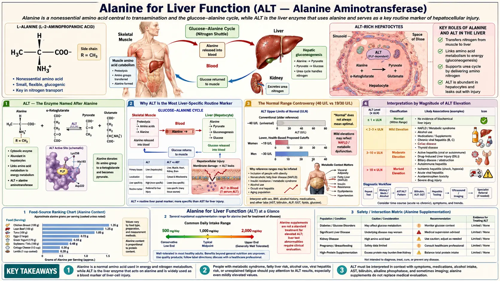
Alanine aminotransferase (ALT, historically SGPT) is the single most-ordered biomarker on every standard liver panel in clinical medicine worldwide. The enzyme is named after alanine because it catalyzes the central transamination between alanine and alpha-ketoglutarate to produce pyruvate and glutamate — the entry step into hepatic gluconeogenesis. Alanine aminotransferase is present at extraordinarily high concentrations inside hepatocytes (roughly 3,000-fold higher than serum levels) and at very low concentrations in all other tissues. The combination of liver-specific localization and high intracellular concentration makes serum alanine aminotransferase the most sensitive routine biochemical marker of hepatocyte injury: even modest liver-cell damage releases enough enzyme to produce measurable serum elevations. The 21st-century transformation of population liver disease — the global emergence of non-alcoholic fatty liver disease (NAFLD), now estimated at about 30% of adults worldwide — has elevated alanine aminotransferase from a hepatology workup tool to a routine population health screen for metabolic dysfunction. This page covers the biochemistry of alanine aminotransferase, the interpretation of alanine aminotransferase elevation patterns, the aspartate aminotransferase (AST):alanine aminotransferase ratio in distinguishing different liver pathologies, modern healthy-range cutoffs, non-alcoholic fatty liver disease epidemiology, drug-induced liver injury, and the practical workup of an isolated alanine aminotransferase elevation.
Table of Contents
- Alanine Aminotransferase — The Enzyme Named After Alanine
- Why Alanine Aminotransferase Is the Most Liver-Specific Routine Marker
- The Normal Range Controversy (40 U/L vs 19/30 U/L)
- Interpretation by Magnitude of Alanine Aminotransferase Elevation
- The Aspartate Aminotransferase:Alanine Aminotransferase Ratio — Alcoholic vs Non-Alcoholic Patterns
- Non-Alcoholic Fatty Liver Disease — The Global Liver Disease Epidemic
- Recent Drug Trials in Steatohepatitis With Liver Scarring
- Drug-Induced Liver Injury (DILI)
- Viral Hepatitis Pattern
- Practical Workup of an Isolated Alanine Aminotransferase Elevation
- The Alanine Load Test for Hepatic Function
- Cautions
- Key Research Papers
- Connections
- Featured Videos
Alanine Aminotransferase — The Enzyme Named After Alanine
Alanine aminotransferase (ALT, EC 2.6.1.2) is one of the two principal aminotransferases of clinical interest, the other being aspartate aminotransferase (AST, EC 2.6.1.1, historically SGOT). Both enzymes use pyridoxal-5-phosphate (the active form of vitamin B6) as cofactor and catalyze reversible transamination reactions. Alanine aminotransferase catalyzes:
L-alanine + alpha-ketoglutarate ↔ pyruvate + L-glutamate
This is the rate-limiting entry step for alanine's contribution to hepatic gluconeogenesis. In liver, alanine aminotransferase runs this reaction in the forward direction as written (consuming alanine to produce pyruvate for gluconeogenesis); in muscle, it runs in reverse (consuming pyruvate and glutamate to produce alanine for nitrogen export). The directional preference depends on cell-specific concentrations of the four reactants and on the cell's gluconeogenic vs gluconeogenic-suppressed state.
Alanine aminotransferase exists in two isozymic forms in humans: ALT1 (cytosolic, encoded by GPT) and ALT2 (mitochondrial, encoded by GPT2). The cytosolic ALT1 is the predominant form in liver and the form that is measured on standard liver panels. The mitochondrial ALT2 is more highly expressed in muscle, heart, and brain — muscle being where the alanine shuttle described above begins — and contributes only modestly to serum activity in healthy adults.
The historical name SGPT (serum glutamic-pyruvic transaminase) reflects the products of the reaction (glutamate and pyruvate) rather than the substrate (alanine). The shift to the alanine aminotransferase nomenclature occurred gradually through the 1980s and 1990s as the systematic IUPAC enzyme nomenclature became the international standard. Older patients and some European laboratories may still report SGPT; this is the same enzyme as alanine aminotransferase.
Why Alanine Aminotransferase Is the Most Liver-Specific Routine Marker
Alanine aminotransferase's clinical usefulness depends on two facts:
- Hepatocytes contain extremely high alanine aminotransferase concentrations — approximately 3,000-fold higher than serum concentrations. Even modest hepatocyte injury releases enough enzyme to produce measurable serum elevations within hours.
- Other tissues contain relatively little alanine aminotransferase — kidney, heart, and skeletal muscle contain detectable but much lower alanine aminotransferase activity. The serum alanine aminotransferase pool in a healthy adult therefore predominantly reflects hepatic origin.
Aspartate aminotransferase is the more revealing contrast. Aspartate aminotransferase is also abundant in liver, but is equally abundant in skeletal muscle, cardiac muscle, kidney, brain, and red blood cells. Hemolysis during phlebotomy can artifactually elevate aspartate aminotransferase. Vigorous exercise can elevate aspartate aminotransferase significantly. Acute myocardial infarction historically caused dramatic aspartate aminotransferase elevations (which is why aspartate aminotransferase was originally used as a cardiac marker before cardiac-specific troponins came along). All of these confounders are absent or markedly reduced for alanine aminotransferase.
The clinical inference is that an isolated alanine aminotransferase elevation almost always reflects hepatocyte injury, while an aspartate aminotransferase elevation is always interpreted in light of possible non-hepatic sources. This is the basis for the diagnostic primacy of alanine aminotransferase in hepatology workups and for the aspartate aminotransferase:alanine aminotransferase ratio as a discriminator (see below).
There are a few specific exceptions worth mentioning:
- Severe muscle injury (rhabdomyolysis) can elevate alanine aminotransferase modestly due to release from skeletal muscle. The creatine kinase (CK) elevation will be the dominant finding and will dwarf the alanine aminotransferase changes.
- Inflammatory myopathies (polymyositis, dermatomyositis) can produce chronic mild alanine aminotransferase elevations from muscle origin. Creatine kinase, aldolase, and clinical features distinguish from primary liver disease.
- Celiac disease can produce mild alanine aminotransferase elevations through poorly understood gut-liver axis mechanisms; the elevations resolve on a gluten-free diet.
- Strenuous exercise within 24-48 hours of blood draw can mildly elevate both alanine aminotransferase and aspartate aminotransferase through muscle origin. Repeat testing after exercise abstinence clarifies.
The Normal Range Controversy (40 U/L vs 19/30 U/L)
The standard laboratory "normal range" for alanine aminotransferase is typically reported as up to 40 U/L (some labs use up to 35 U/L). These ranges were originally derived in the 1970s from blood donor populations that, in retrospect, included many subjects with subclinical non-alcoholic fatty liver disease and chronic viral hepatitis (then unrecognized). The result is that the "normal range" was set too high — many subjects within the reported normal range actually had liver pathology.
The Prati 2002 Annals of Internal Medicine study redefined healthy alanine aminotransferase ranges using a much more rigorously screened reference population (blood donors with no clinical or laboratory evidence of liver disease, no body mass index (BMI) above 25, no metabolic syndrome). The Prati cutoffs are:
- Men: upper limit of normal 30 U/L (not 40)
- Women: upper limit of normal 19 U/L (not 30)
The study drew on 6,835 first-time blood donors and tested the new limits in donors with hepatitis C antibodies. The lower limits caught more of the people with hepatitis C virus in their blood (sensitivity 76.3% versus 55% with the older limits), at the price of flagging more people without it (specificity 88.5% versus 97.4%). The extra people caught mostly had minimal to mild liver changes on biopsy.
The Prati cutoffs have not been universally adopted by clinical laboratories, partly because the implications are uncomfortable: applying the stricter cutoffs to the general population reclassifies a substantial fraction of presumably-healthy adults as having mildly abnormal alanine aminotransferase. The National Health and Nutrition Examination Survey (NHANES) population data show that roughly 20-30% of US adults have alanine aminotransferase above the Prati upper limit, which corresponds well to the prevalence of non-alcoholic fatty liver disease established by hepatic ultrasound and biopsy studies.
By these updated figures, an alanine aminotransferase of 35 U/L — clearly within the standard 40 U/L reporting cutoff — is above the healthy range, a point raised especially for women and for people with metabolic risk factors. The 2017 American College of Gastroenterology (ACG) Clinical Guideline (Kwo et al.) gives its own figures, close to Prati's: a true healthy normal level of 29 to 33 IU/L for males and 19 to 25 IU/L for females, with levels above this to be assessed. Those figures are lower than the 40 U/L many laboratories report as "normal."
A common clinical rule of thumb built on these limits reads:
- Alanine aminotransferase below 19 (women) or 30 (men): within the Prati healthy range, with further testing usually driven by other indicators rather than by this number.
- Alanine aminotransferase 19-40 (women) or 30-40 (men): below the lab's reported upper limit but above the Prati cutoff. Clinicians commonly respond with a metabolic workup (lipids, hemoglobin A1c, body mass index, alcohol screen) and a re-check in 6 months.
- Alanine aminotransferase 40-150 U/L: mildly elevated by any standard. The usual workup is described in the practical workup section below.
- Alanine aminotransferase >150 U/L: moderately elevated and always abnormal; clinicians work this up, and a level above 500 is commonly sent to a liver specialist (hepatologist).
Interpretation by Magnitude of Alanine Aminotransferase Elevation
The magnitude of alanine aminotransferase elevation correlates roughly with the type of underlying liver pathology, providing a useful clinical heuristic:
- Mild elevation (1-2 times the upper limit of normal; alanine aminotransferase 40-80 U/L): Most commonly non-alcoholic fatty liver disease, mild drug-induced liver injury (statins, acetaminophen, anti-tuberculosis agents), chronic viral hepatitis (hepatitis B virus (HBV), hepatitis C virus (HCV)) in the inactive or low-replication phase, alcoholic liver disease (early), autoimmune hepatitis (mild), hemochromatosis, alpha-1 antitrypsin deficiency, Wilson disease (rarely).
- Moderate elevation (2-10 times the upper limit of normal; alanine aminotransferase 80-400 U/L): Active chronic viral hepatitis, moderate-severe non-alcoholic fatty liver disease/non-alcoholic steatohepatitis (NASH), autoimmune hepatitis with active inflammation, moderate drug-induced liver injury, alcoholic hepatitis (typically with aspartate aminotransferase > alanine aminotransferase), early acute viral hepatitis (rising phase).
- Marked elevation (10-25 times the upper limit of normal; alanine aminotransferase 400-1000 U/L): Acute viral hepatitis (peak phase), severe drug-induced liver injury, autoimmune hepatitis with severe inflammation, acute ischemic hepatitis (typically very high aspartate aminotransferase > alanine aminotransferase). This range commonly prompts a consultation with a liver specialist.
- Massive elevation (more than 25 times the upper limit of normal; alanine aminotransferase >1000 U/L): The differential narrows to three principal causes: (1) acute viral hepatitis (hepatitis A virus, hepatitis B virus, occasionally hepatitis E virus), (2) severe drug-induced liver injury (most classically acetaminophen overdose), and (3) ischemic hepatitis (shock liver). Acetaminophen toxicity in particular can produce alanine aminotransferase in the 5000-10000 U/L range, often the highest values seen in clinical practice. This range is handled as urgent, with a liver specialist consulted and the person assessed for fulminant hepatic failure (international normalized ratio (INR), encephalopathy).
The rate of change of alanine aminotransferase is also informative. Acute hepatitis typically rises over days and then falls predictably as the injury resolves. Chronic liver disease typically shows stable mildly-elevated values over months. A sudden rise in a previously stable alanine aminotransferase prompts workup for a new injury (drug, infection, ischemic event).
The Aspartate Aminotransferase:Alanine Aminotransferase Ratio — Alcoholic vs Non-Alcoholic Patterns
The ratio of aspartate aminotransferase to alanine aminotransferase (often called the De Ritis ratio after the Italian biochemist who first proposed it in 1957) provides important diagnostic information about the underlying liver process:
- Aspartate aminotransferase:alanine aminotransferase < 1 (more alanine aminotransferase than aspartate aminotransferase): The pattern of most non-alcoholic liver disease. Found in non-alcoholic fatty liver disease/non-alcoholic steatohepatitis (typically 0.7-1.0), chronic viral hepatitis (B and C in the chronic phase), acute viral hepatitis, drug-induced hepatitis, autoimmune hepatitis, hemochromatosis. This is the "default" hepatocellular pattern.
- Aspartate aminotransferase:alanine aminotransferase > 2 (twice as much aspartate aminotransferase as alanine aminotransferase): Strongly suggestive of alcoholic liver disease. The classic Cohen-Kaplan 1979 paper called a ratio above 2 highly suggestive of alcoholic hepatitis and cirrhosis: it occurred in 70% of those patients, compared with 26% with postnecrotic cirrhosis, 8% with chronic hepatitis, 4% with viral hepatitis and none with obstructive jaundice. The mechanism involves chronic alcohol-induced depletion of pyridoxal-5-phosphate (vitamin B6), which reduces both alanine aminotransferase and aspartate aminotransferase synthesis but reduces alanine aminotransferase more (alanine aminotransferase has a stricter B6 dependence) — so the ratio shifts toward aspartate aminotransferase dominance.
- Aspartate aminotransferase:alanine aminotransferase > 1 (but < 2): A "gray zone" that includes early cirrhosis from any cause (the aspartate aminotransferase:alanine aminotransferase ratio rises as cirrhosis progresses regardless of the original etiology), non-alcoholic steatohepatitis-related cirrhosis, and a mix of alcoholic and non-alcoholic disease.
- Aspartate aminotransferase:alanine aminotransferase > 4-5 (very high): Suggests acute Wilson disease with hemolysis, severe alcoholic hepatitis, or muscle injury contaminating the test (rhabdomyolysis would also show very high creatine kinase).
The Williams-Hoofnagle 1988 Gastroenterology paper expanded the diagnostic value of the aspartate aminotransferase:alanine aminotransferase ratio specifically for cirrhosis assessment: as liver disease progresses from chronic active inflammation to cirrhosis, the aspartate aminotransferase:alanine aminotransferase ratio rises regardless of the original etiology. Its 177 patients all had nonalcoholic chronic liver disease and a liver biopsy. Among 100 with chronic hepatitis B, the mean ratio was 0.59 without cirrhosis and 1.02 with cirrhosis, and it often rose above 1.0 when cirrhosis first became apparent. As an illustration not taken from that paper, a non-alcoholic steatohepatitis patient who starts with aspartate aminotransferase:alanine aminotransferase of 0.7 may have aspartate aminotransferase:alanine aminotransferase of 1.4 by the time non-alcoholic steatohepatitis-related cirrhosis develops. This makes serial aspartate aminotransferase:alanine aminotransferase trending useful for monitoring chronic liver disease progression.
The clinical use of the De Ritis ratio is therefore twofold: (1) initial differential diagnosis of acute or chronic liver disease (alcoholic vs non-alcoholic, principally), and (2) monitoring chronic liver disease progression toward cirrhosis. Both uses are based on the same biochemistry — the differential B6-cofactor dependence of alanine aminotransferase vs aspartate aminotransferase, modulated by the underlying disease's effect on hepatic protein synthesis.
Non-Alcoholic Fatty Liver Disease — The Global Liver Disease Epidemic
Non-alcoholic fatty liver disease (NAFLD), recently renamed metabolic-dysfunction-associated steatotic liver disease (MASLD) in the 2023 multi-society consensus, has become the leading cause of chronic liver disease worldwide. The Younossi 2016 Hepatology meta-analysis estimated global prevalence at approximately 25% of adults, with substantial regional variation: about 32% in South America, 27% in Asia, 24% in North America, 24% in Europe, and 31% in the Middle East. A 2023 update led by the same author pooled 92 studies and put the figure at 30.05% across 1990-2019, rising from 25.26% in 1990-2006 to 38.00% in 2016-2019. In the United States, the National Health and Nutrition Examination Survey data suggest that 30-40% of adults have hepatic steatosis on imaging, of whom an estimated 5-10% have progressed to the more severe non-alcoholic steatohepatitis with active inflammation and fibrosis.
Non-alcoholic fatty liver disease/metabolic dysfunction-associated steatotic liver disease is best understood as the hepatic manifestation of metabolic syndrome — the cluster of central obesity, insulin resistance, atherogenic dyslipidemia, hypertension, and type 2 diabetes that has become the dominant chronic disease pattern in developed and rapidly developing economies. The pathophysiology involves:
- Excess hepatic fatty acid delivery — from dietary fat, from de novo lipogenesis driven by chronic hyperinsulinemia, and from accelerated lipolysis of insulin-resistant visceral adipose tissue. The hepatocyte's capacity to package and export triglyceride as very-low-density lipoprotein (VLDL) is exceeded.
- Hepatic triglyceride accumulation — visible as macrovesicular steatosis on biopsy and as hepatic steatosis on ultrasound, computed tomography (CT), or magnetic resonance imaging (MRI).
- Lipotoxicity-induced hepatocyte injury — the accumulated free fatty acids, diacylglycerols, and ceramides activate inflammatory cytokine production and apoptotic pathways. Cell death releases alanine aminotransferase into the circulation.
- Hepatic stellate cell activation — the wound-healing response to chronic hepatocyte injury produces collagen deposition and progressive fibrosis. Over years to decades, this can progress to cirrhosis.
Alanine aminotransferase elevation is the most accessible biochemical signal of this process, but alanine aminotransferase has poor sensitivity for non-alcoholic fatty liver disease. The Mofrad 2003 study compared 51 biopsied patients who had non-alcoholic fatty liver disease and normal alanine aminotransferase with 50 who had raised values, and found the whole range of liver damage in both: 12 of the 51 had bridging fibrosis and 6 had cirrhosis. A figure of approximately 20% of patients is sometimes given for severe disease with normal values; that study reports no population percentage. The implication is that a normal alanine aminotransferase does not exclude non-alcoholic fatty liver disease — imaging (ultrasound, magnetic resonance imaging proton density fat fraction (MRI-PDFF), or transient elastography) is needed when the clinical suspicion is high (body mass index > 30, type 2 diabetes, metabolic syndrome features).
Management of non-alcoholic fatty liver disease is currently behavioral and pharmacological:
- Weight loss — the single most effective intervention. A 5% body weight loss is described as reversing steatosis in most patients, and 7-10% loss as able to reverse early fibrosis. In a 52-week study of 293 people with biopsy-proven non-alcoholic steatohepatitis, 58% of those who lost 5% or more of their weight had the steatohepatitis resolve; among those who lost 10% or more, 90% had it resolve and 45% had fibrosis regress. At 7-10%, that study reports lower disease-activity scores in people with few risk factors.
- Mediterranean diet — emphasizing whole foods, olive oil, fish, vegetables; modest carbohydrate restriction; minimization of fructose-containing sweetened beverages.
- Regular aerobic exercise — independent benefit beyond the weight loss effect.
- Pharmacological options — pioglitazone (an insulin sensitizer with proven non-alcoholic steatohepatitis benefit), glucagon-like peptide-1 (GLP-1) receptor agonists (semaglutide and tirzepatide both show significant non-alcoholic steatohepatitis improvement), and resmetirom (recently U.S. Food and Drug Administration-approved specifically for non-alcoholic steatohepatitis with fibrosis).
For more on non-alcoholic fatty liver disease, see our pages on Metabolic Syndrome and Liver Disease.
Recent Drug Trials in Steatohepatitis With Liver Scarring
Three trials report repeat liver biopsy results in adults with metabolic dysfunction-associated steatohepatitis (MASH), the newer name for non-alcoholic steatohepatitis, and liver scarring (fibrosis).
- Semaglutide (phase 3) — in the first 800 of 1,197 adults with moderate or advanced scarring, weekly semaglutide 2.4 mg resolved steatohepatitis without worsening scarring in 62.9% versus 34.3% on placebo at 72 weeks, and reduced scarring without worsening steatohepatitis in 36.8% versus 22.4%. Gut side effects were more common with semaglutide.
- Resmetirom (phase 3) — in 966 patients at 52 weeks, steatohepatitis resolved in 25.9% (80 mg) and 29.9% (100 mg) versus 9.7% on placebo, and scarring improved in 24.2% and 25.9% versus 14.2%. Diarrhea and nausea were more frequent with resmetirom.
- Tirzepatide (phase 2) — in 190 people with moderate or severe scarring at 52 weeks, steatohepatitis resolved in 44%, 56% and 62% at 5, 10 and 15 mg versus 10% on placebo, and scarring improved in 55%, 51% and 51% versus 30%. This is a smaller dose-finding trial; its authors state that larger and longer trials are needed.
Drug-Induced Liver Injury (DILI)
Drug-induced liver injury is the most common cause of acute liver failure in the developed world and a frequent cause of elevated alanine aminotransferase. Hundreds of medications and herbal products have been implicated. The patterns of injury fall into three principal categories:
- Hepatocellular pattern — alanine aminotransferase predominantly elevated relative to alkaline phosphatase. Suggests injury to hepatocytes. Classic offenders: acetaminophen overdose (highest alanine aminotransferase seen in clinical practice), isoniazid, statins, methotrexate, valproate, kava, green tea extract at high doses.
- Cholestatic pattern — alkaline phosphatase predominantly elevated relative to alanine aminotransferase. Suggests injury to biliary epithelium or impaired bile flow. Classic offenders: amoxicillin-clavulanate, anabolic steroids, chlorpromazine, erythromycin estolate, oral contraceptives.
- Mixed pattern — both alanine aminotransferase and alkaline phosphatase elevated, often with the R-value (alanine aminotransferase divided by its upper limit of normal, over alkaline phosphatase divided by its upper limit of normal) between 2 and 5. Classic offenders: phenytoin, sulfonamides, carbamazepine.
Specific medications worth knowing:
- Acetaminophen — the most common cause of drug-induced acute liver failure in the United States. The over-the-counter label warns that severe liver damage may occur with more than 4,000 mg in 24 hours, with other drugs containing acetaminophen, or with 3 or more alcoholic drinks every day while using it. In a U.S. series of 275 acetaminophen-related acute liver failure cases, the median dose taken was 24 g (Larson 2005). Toxicity above 2-3 g/day taken chronically is also described in heavy alcohol users. Alanine aminotransferase peaks at 48-72 hours after ingestion, often in the thousands to tens of thousands. N-acetylcysteine (NAC) is the antidote. In a national study of 2,540 patients, liver toxicity developed in 6.1% of those at probable risk when it was started within 10 hours and in 26.4% when started at 10-24 hours; started within 8 hours it was protective whatever the blood level, and high-risk patients treated at 16-24 hours (41%) still did better than historical controls. The authors concluded that treatment is still indicated at least as late as 24 hours after ingestion.
- Statins — mild alanine aminotransferase elevations in 1-3% of users. The Cohen-Anania-Chalasani 2006 hepatologist consensus established that mild statin-associated alanine aminotransferase elevation is not a contraindication to continued therapy in patients without underlying liver disease; it is a marker rather than a clinically meaningful injury. Severe statin hepatotoxicity is rare.
- Isoniazid — alanine aminotransferase elevation in up to 20% of treated patients, with clinical hepatitis in 0.5-2%. Monthly liver function test monitoring is standard during anti-tuberculosis therapy.
- Anabolic steroids — produce a distinctive cholestatic pattern with characteristic bile duct injury. Sometimes overlooked in young athletic men presenting with jaundice.
- Herbal hepatotoxins — kava, green tea extract (high-dose), comfrey, germander, chaparral, pyrrolizidine alkaloid-containing herbs (e.g., some bush teas). Underrecognized cause of unexplained alanine aminotransferase elevation; the 2017 ACG guideline includes a history of prescribed and over-the-counter medicines in the evaluation, and a detailed herbal and supplement history is how these products come to light.
The standard approach to suspected drug-induced liver injury is the modified Roussel-Uclaf Causality Assessment Method (RUCAM), which scores the temporal relationship, exclusion of other causes, risk factors, response to dechallenge, and prior knowledge of hepatotoxicity to estimate the probability that a specific agent caused the observed injury.
Viral Hepatitis Pattern
Viral hepatitis remains an important cause of alanine aminotransferase elevation worldwide. The clinical context and serology determine the workup:
- Hepatitis A — acute self-limited infection, fecal-oral transmission, peak alanine aminotransferase typically 1000-3000 U/L. Confirmed by hepatitis A virus immunoglobulin M (IgM) antibody. No chronic phase, no specific antiviral treatment, supportive care only. Vaccine-preventable.
- Hepatitis B — acute or chronic infection, blood/sexual transmission, alanine aminotransferase pattern varies by phase. Chronic hepatitis B may have alanine aminotransferase in the normal range during the immune-tolerant phase, mildly elevated during active replication, and very high during reactivation or hepatitis B e antigen (HBeAg) seroconversion. Confirmed by hepatitis B surface antigen (HBsAg; chronic carrier), antibody to the surface antigen (anti-HBs; immunity), and hepatitis B core immunoglobulin M antibody (anti-HBc; acute infection). Vaccine-preventable. Multiple effective antivirals available.
- Hepatitis C — usually chronic infection, blood-borne transmission, often mild alanine aminotransferase elevation or even normal alanine aminotransferase. Confirmed by hepatitis C virus antibody and a hepatitis C virus RNA polymerase chain reaction (PCR) test. Curable in >95% of cases with 8-12 weeks of direct-acting antiviral therapy (sofosbuvir-velpatasvir or glecaprevir-pibrentasvir).
- Hepatitis D — defective virus that requires hepatitis B surface antigen coexpression. It is tested for only when hepatitis B surface antigen is positive.
- Hepatitis E — fecal-oral or zoonotic transmission, acute self-limited in most cases but can be devastating in pregnancy. Confirmed by hepatitis E virus immunoglobulin M antibody and polymerase chain reaction.
- Epstein-Barr virus (EBV), cytomegalovirus (CMV), herpes simplex virus — can produce mild-to-moderate transient alanine aminotransferase elevations during acute viral syndromes. Usually presents with characteristic clinical features (fever, lymphadenopathy, atypical lymphocytosis).
The standard initial viral panel for an unexplained alanine aminotransferase elevation is hepatitis B surface antigen, hepatitis C virus antibody, and (in appropriate clinical settings) hepatitis A virus immunoglobulin M antibody and human immunodeficiency virus (HIV) antibody.
Practical Workup of an Isolated Alanine Aminotransferase Elevation
The Kwo 2017 ACG Clinical Guideline and the Newsome 2018 British Society of Gastroenterology (BSG) guideline both provide structured approaches to the patient with an isolated alanine aminotransferase elevation. A practical synthesis:
- Confirm and characterize — the liver function tests are repeated to show whether the elevation is real and not laboratory artifact. The aspartate aminotransferase:alanine aminotransferase ratio and the R-value (described above) are calculated. International normalized ratio and albumin are checked for synthetic function assessment.
- History — alcohol intake (the Alcohol Use Disorders Identification Test-Consumption questionnaire, AUDIT-C), medication and herbal supplement use, intravenous (IV) drug use risk factors, sexual risk factors, travel history, family history of liver disease, occupational exposures, prior episodes of jaundice, autoimmune disease history.
- Physical examination — body mass index and waist circumference (metabolic syndrome), stigmata of chronic liver disease (palmar erythema, spider angiomata, gynecomastia, ascites, splenomegaly), Kayser-Fleischer rings (Wilson disease), arthritis (autoimmune hepatitis association).
- First-tier laboratory workup — metabolic panel (fasting glucose, hemoglobin A1c, lipid panel for non-alcoholic fatty liver disease risk stratification), hepatitis B surface antigen, hepatitis C virus antibody, ferritin and transferrin saturation (hemochromatosis), antinuclear antibody (ANA) and anti-smooth muscle antibody (ASMA) (autoimmune hepatitis), tissue transglutaminase immunoglobulin A (IgA) (celiac).
- Imaging — abdominal ultrasound for hepatic steatosis, biliary tree, and any focal lesions. Magnetic resonance imaging proton density fat fraction if quantitative steatosis assessment is needed. Transient elastography if fibrosis assessment is needed (non-alcoholic fatty liver disease >F2 fibrosis is a hepatology referral trigger).
- Second-tier workup if first-tier negative — ceruloplasmin and 24-hour urinary copper (Wilson disease), alpha-1 antitrypsin level and Pi typing (alpha-1 antitrypsin deficiency), thyroid function (hypothyroidism-associated non-alcoholic fatty liver disease), human immunodeficiency virus testing, repeat viral hepatitis panel including hepatitis E virus in appropriate clinical contexts.
- Specialty referral — alanine aminotransferase more than 5 times the upper limit of normal despite negative initial workup; persistent alanine aminotransferase elevation >6 months without identified cause; any patient with cirrhosis or significant fibrosis (non-alcoholic fatty liver disease >F2); fulminant presentation (rising international normalized ratio, encephalopathy, jaundice).
- Liver biopsy — rarely needed in modern hepatology due to advances in non-invasive imaging and serological scoring. Indicated when the diagnosis remains unclear after the second-tier workup, or when management decisions require histological staging.
The most common ultimate diagnosis for an isolated mildly-elevated alanine aminotransferase in a developed-world primary care population is non-alcoholic fatty liver disease, followed by chronic viral hepatitis (B or C), alcoholic liver disease, drug-induced injury, and autoimmune hepatitis. The rarer diagnoses (Wilson disease, alpha-1 antitrypsin deficiency, hemochromatosis) are specifically looked for in younger patients without obvious metabolic or behavioral risk factors.
The Alanine Load Test for Hepatic Function
The alanine load test is a research-grade assessment of hepatic functional reserve that uses an oral alanine dose (15-30 g) to challenge the gluconeogenic capacity of the liver. The post-load plasma glucose and alanine kinetics provide a sensitive measure of hepatic gluconeogenic capacity that is impaired earlier than conventional liver function markers.
In healthy subjects, oral alanine produces a brisk rise in plasma alanine peaking at 60-90 minutes, with proportional plasma glucose rise and glucagon secretion. The plasma alanine clearance half-life is approximately 90 minutes. The test was originally developed in the 1970s from Genuth's 1973 work in fasting obese subjects and Genuth and Castro's 1974 work in normal and diabetic subjects, and subsequently adapted for research evaluation of hepatic function in cirrhosis, non-alcoholic steatohepatitis, and post-bariatric patients.
The alanine load test is rarely used in routine clinical hepatology because non-invasive imaging (magnetic resonance imaging elastography, transient elastography) and validated scoring systems (Fibrosis-4 index (FIB-4), non-alcoholic fatty liver disease Fibrosis Score, aspartate aminotransferase-to-platelet ratio index (APRI)) provide more practical assessments of hepatic function and fibrosis. The test remains useful in research settings where dynamic gluconeogenic capacity is the specific question of interest.
For patients curious about alanine's metabolic role in their own physiology, the clinical correlate is simpler: a standard glucose tolerance test (oral 75 g glucose with serial glucose and insulin measurements) implicitly assesses the post-load alanine response as part of the integrated postprandial metabolic state. Reactive hypoglycemia identified on a glucose tolerance test often reflects impaired alanine-mediated gluconeogenic counter-regulation.
Cautions
- An isolated mildly elevated alanine aminotransferase is common and often benign — 20-30% of US adults have alanine aminotransferase above the Prati cutoffs. Most have non-alcoholic fatty liver disease that improves substantially with weight loss and Mediterranean diet. Not every mildly elevated alanine aminotransferase requires aggressive workup; clinical context is everything.
- Normal alanine aminotransferase does not exclude significant liver disease — the Mofrad 2003 series found bridging fibrosis in 12 and cirrhosis in 6 of 51 biopsied patients with normal values, and a figure of up to 20% of patients with significant injury is sometimes given, though that study reports no population percentage. Imaging is more sensitive than alanine aminotransferase for steatosis.
- Pyridoxal-5-phosphate (vitamin B6) deficiency lowers alanine aminotransferase — chronic alcohol abuse, malnutrition, and isoniazid use can all deplete B6 and produce artifactually low alanine aminotransferase values. This is part of the mechanism behind the aspartate aminotransferase:alanine aminotransferase > 2 pattern of alcoholic liver disease.
- Pregnancy lowers alanine aminotransferase — physiological dilution of plasma proteins during pregnancy reduces alanine aminotransferase activity by approximately 25%. A pregnant woman's alanine aminotransferase is read against pregnancy-specific reference ranges.
- Acetaminophen safety — the over-the-counter label warns that severe liver damage may occur with more than 4,000 mg in 24 hours, with other drugs containing acetaminophen, or with 3 or more alcoholic drinks every day while using it; a lower limit of 2-3 g/day is commonly given for regular alcohol users. Many cold medicines and prescription pain medications contain acetaminophen and easily accumulate to toxic doses when stacked: in the 2005 U.S. series, 38% of people with unintentional overdoses had taken two or more acetaminophen preparations at once.
- Herbal supplement screening — a detailed history of all over-the-counter and herbal products is part of evaluating unexplained alanine aminotransferase elevation. Kava, green tea extract at high doses, comfrey, chaparral, and pyrrolizidine alkaloid-containing teas are well-documented hepatotoxins.
- Heavy exercise within 48 hours of blood draw — can artifactually elevate both alanine aminotransferase and aspartate aminotransferase through muscle origin. When exercise is suspected as the cause, clinicians commonly repeat the liver function tests after 1 week without exercise.
- L-alanine supplementation does not affect alanine aminotransferase in any clinically meaningful way — the enzyme is constitutively expressed at very high levels; substrate availability is not rate-limiting under normal physiological conditions. Routine L-alanine supplements are not a treatment for elevated alanine aminotransferase, nor a cause of it.
- Beta-alanine is a different molecule from L-alanine — a 2019 review of 101 human and 50 animal studies of the supplement beta-alanine found a small rise in blood alanine aminotransferase, with average values staying well within clinical reference ranges.
Key Research Papers
- Pratt DS, Kaplan MM (2000). Evaluation of abnormal liver-enzyme results in asymptomatic patients. NEJM 342(17):1266-1271. — DOI: 10.1056/NEJM200004273421707. — doi:10.1056/NEJM200004273421707
- Prati D, Taioli E, Zanella A, et al. (2002). Updated definitions of healthy ranges for serum alanine aminotransferase levels. Annals of Internal Medicine 137(1):1-10. — DOI: 10.7326/0003-4819-137-1-200207020-00006. — doi:10.7326/0003-4819-137-1-200207020-00006
- Kwo PY, Cohen SM, Lim JK (2017). ACG Clinical Guideline: Evaluation of abnormal liver chemistries. American Journal of Gastroenterology 112(1):18-35. — DOI: 10.1038/ajg.2016.517. — doi:10.1038/ajg.2016.517
- Newsome PN, Cramb R, Davison SM, et al. (2018). Guidelines on the management of abnormal liver blood tests. Gut 67(1):6-19. — DOI: 10.1136/gutjnl-2017-314924. — doi:10.1136/gutjnl-2017-314924
- Younossi ZM, Koenig AB, Abdelatif D, et al. (2016). Global epidemiology of nonalcoholic fatty liver disease — meta-analytic assessment. Hepatology 64(1):73-84. — DOI: 10.1002/hep.28431. — doi:10.1002/hep.28431
- Cohen JC, Horton JD, Hobbs HH (2011). Human fatty liver disease: old questions and new insights. Science 332(6037):1519-1523. — DOI: 10.1126/science.1204265. — doi:10.1126/science.1204265
- Mofrad P, Contos MJ, Haque M, et al. (2003). Clinical and histologic spectrum of nonalcoholic fatty liver disease associated with normal ALT values. Hepatology 37(6):1286-1292. — DOI: 10.1053/jhep.2003.50229. — doi:10.1053/jhep.2003.50229
- Williams ALB, Hoofnagle JH (1988). Ratio of serum aspartate to alanine aminotransferase in chronic hepatitis — relationship to cirrhosis. Gastroenterology 95(3):734-739. — DOI: 10.1016/s0016-5085(88)80022-2. — doi:10.1016/s0016-5085(88)80022-2
- Cohen JA, Kaplan MM (1979). The SGOT/SGPT ratio — an indicator of alcoholic liver disease. Digestive Diseases and Sciences 24(11):835-838. — DOI: 10.1007/BF01324898. — doi:10.1007/BF01324898
- Sookoian S, Pirola CJ (2012). Alanine and aspartate aminotransferase and glutamine-cycling pathway: their roles in pathogenesis of metabolic syndrome. World Journal of Gastroenterology 18(29):3775-3781. — DOI: 10.3748/wjg.v18.i29.3775. — doi:10.3748/wjg.v18.i29.3775
- Cohen DE, Anania FA, Chalasani N (2006). An assessment of statin safety by hepatologists. American Journal of Cardiology 97(8A):77C-81C. — DOI: 10.1016/j.amjcard.2005.12.014. — doi:10.1016/j.amjcard.2005.12.014
- Larson AM, Polson J, Fontana RJ, et al. (2005). Acetaminophen-induced acute liver failure: results of a US multicenter, prospective study. Hepatology 42(6):1364-1372. — DOI: 10.1002/hep.20948. — doi:10.1002/hep.20948
- Chalasani N, Younossi Z, Lavine JE, et al. (2018). The diagnosis and management of NAFLD: practice guidance from AASLD. Hepatology 67(1):328-357. — DOI: 10.1002/hep.29367. — doi:10.1002/hep.29367
- Genuth SM, Castro J (1974). Effect of oral alanine on blood beta-hydroxybutyrate and plasma glucose, insulin, free fatty acids, and growth hormone in normal and diabetic subjects. Metabolism 23(4):375-386. — PubMed PMID: 4206148
- Genuth SM (1973). Effects of oral alanine administration in fasting obese subjects. Metabolism 22(7):927-937. — PubMed PMID: 4710700
- Younossi ZM, Golabi P, Paik JM, et al. (2023). The global epidemiology of nonalcoholic fatty liver disease (NAFLD) and nonalcoholic steatohepatitis (NASH): a systematic review. Hepatology 77(4):1335-1347. — PubMed PMID: 36626630
- Vilar-Gomez E, Martinez-Perez Y, Calzadilla-Bertot L, et al. (2015). Weight loss through lifestyle modification significantly reduces features of nonalcoholic steatohepatitis. Gastroenterology 149(2):367-378. — PubMed PMID: 25865049
- Smilkstein MJ, Knapp GL, Kulig KW, Rumack BH (1988). Efficacy of oral N-acetylcysteine in the treatment of acetaminophen overdose. Analysis of the national multicenter study (1976 to 1985). NEJM 319(24):1557-1562. — PubMed PMID: 3059186
- Sanyal AJ, Newsome PN, Kliers I, et al. (2025). Phase 3 trial of semaglutide in metabolic dysfunction-associated steatohepatitis. NEJM 392(21):2089-2099. — PubMed PMID: 40305708
- Harrison SA, Bedossa P, Guy CD, et al. (2024). A phase 3, randomized, controlled trial of resmetirom in NASH with liver fibrosis. NEJM 390(6):497-509. — PubMed PMID: 38324483
- Loomba R, Hartman ML, Lawitz EJ, et al. (2024). Tirzepatide for metabolic dysfunction-associated steatohepatitis with liver fibrosis. NEJM 391(4):299-310. — PubMed PMID: 38856224
- Dolan E, Swinton PA, Painelli VS, et al. (2019). A systematic risk assessment and meta-analysis on the use of oral beta-alanine supplementation. Advances in Nutrition 10(3):452-463. — PubMed PMID: 30980076
PubMed Topic Searches
Connections
- All Amino Acids
- Alanine Overview
- Alanine Benefits Hub
- Alanine for Gluconeogenesis & Blood Sugar
- Beta-Alanine for Endurance
- Alanine for Immune Function
- Glutamic Acid (Transamination Partner)
- Glutamine
- Vitamin B6 (Alanine Aminotransferase Cofactor)
- Liver Disease
- Metabolic Syndrome (Non-Alcoholic Fatty Liver Disease)
- Diabetes (Non-Alcoholic Fatty Liver Disease)
- Obesity (Non-Alcoholic Fatty Liver Disease)
- Lab Tests
- N-Acetylcysteine (Acetaminophen Antidote)
- Liver Cleanse
- Milk Thistle
- History
- Sources